Immunoassay To Quantify Pneumococcal Capsular
Polysaccharide-Specific Antibodies
David L. Klein,aJoseph E. Martinez,aMichael H. Hickey,aF. Hassouna,aK. Zaman,band Mark Steinhoffc
Flow Applications, Inc., Okawville, Illinois, USAa
; International Centre for Diarrhoeal Disease Research, Bangladesh, Dhaka, Bangladeshb
; and Johns Hopkins University School of Public Health, Baltimore, Maryland, USAc
Enzyme-linked immunosorbent assay (ELISA), the traditional antibody quantification technique, has several limitations,
espe-cially when used to evaluate multivalent and/or infant vaccines. We have developed a multiplex bead-based antibody
quantifica-tion assay (MBIA) to measure antibody response to multiple pneumococcal (Pn) serotypes (St) in a single assay. MBIA was
com-pared with the WHO ELISA using a WHO panel of 12 international calibration sera for 7 Pn Sts. An agreement of 75 to 92% was
obtained for all 7 Sts. MBIA exhibited good robustness, with the assay variability at
<
16%. A major contributor to MBIA
vari-ability was the cell wall polysaccharide (CWPs) content in Pn St-specific capsular Ps. This necessitated careful CWPs (20
g/ml)
preadsorption of sera. MBIA is specific, robust, and reproducible and offers high throughput. The use of MBIA will greatly
re-duce the cost and time required to evaluate the immune response to multiple Pn Sts and could help promote the licensure of
fu-ture Pn and other multivalent vaccines.
A
number of immunoassays have been developed to measure
the concentration of serotype (St)-specific human IgG
anti-pneumococcal (anti-Pn) capsular polysaccharide (Ps) antibodies
following induced and natural immunization. The standard
mea-surement of Pn antibody in most laboratories today relies on the
World Health Organization (WHO) enzyme-linked
immunosor-bent assay (ELISA) (
3
,
17
). This standardized assay has several
limitations that result in substantial laboratory effort at a relatively
high cost. It is labor-intensive and can measure antibodies to only
one antigen at a time. The singleplex aspect of ELISA testing also
results in increased use of patient samples (
4
,
10
,
11
). Although an
increased volume of serum (10
l/St) is not troublesome to
achieve with adults, it is with infants. In such situations, a
multi-plex immunoassay that measures specific antibody to several
an-tigens simultaneously from a single small sample (i.e., 10
l) is
highly advantageous (
15
). The use of the multiplex assay to
mea-sure serum IgG reduces the time required per assay analyte by
80%, thereby markedly increasing throughput and greatly
reduc-ing the assay cost. In addition to increased automation and
effi-ciency, numerous studies have shown serological multiplexing
procedures to be sensitive, precise, and accurate (
1
,
5
,
7
).
Further-more, multiplex assays offer an efficient laboratory approach that
can serve as a primary antibody concentration assay for analyzing
imposing numbers of patient sera associated with large-scale
vac-cine efficacy trials and epidemiological studies.
In this study, we evaluated an in-house multiplex bead-based
immunoassay (MBIA) to quantitate anti-Pn Ps IgG to multiple
serotypes. The multiplex assay is based on Luminex’s xMAP
tech-nology (Luminex, Austin, TX). The primary objective of this study
was to characterize MBIA and elucidate the correlation between
MBIA and an in-house ELISA with the WHO reference panel of 12
pneumococcal calibration sera (
12
). Additional MBIA and
in-house ELISA validations were performed with paired maternal
sera (
n
⫽
50) from women vaccinated once with a 23-valent Pn Ps
vaccine during their third trimester of pregnancy. Assay
modifi-cation in terms of changes in cell wall Ps (CWPs) concentration in
the serum preadsorption buffer was necessary for a few Pn Sts.
MBIA exhibited excellent correlation with in-house ELISA for Pn
St-specific Ps IgG in the 12 WHO calibration sera and clinical
specimens.
MATERIALS AND METHODS
Standard and serum samples. The Food and Drug Administration’s (FDA) Pn human serological reference standard, 89SF, was used as the standard for all in-house ELISA and MBIA; it was never used as an un-known source. The WHO Pn calibration human serum panel (n⫽12) was used for primary assay correlation and validation studies. Secondary assay validations were carried out with paired sera (n⫽50) from pregnant women who received one dose of the 23-valent Pn Ps vaccine during their third trimester of pregnancy. Five in-house quality control (QC) sera generated from 23-valent Pn Ps-vaccinated laboratory technicians were also used in selected assay development experiments to conserve clinical sera.
Pn Ps IgG ELISA.The IgG ELISA procedure was performed according to the WHO consensus protocol, with modifications (9,17). Briefly, 89SF was prediluted (1:20) with phosphate-buffered Tween solution contain-ing 20g/ml CWPs (PBST⫹CWPs), subsequently adsorbed once with CWPs, and run in duplicate on each plate. Similarly, QC, calibration, and maternal sera were diluted (1:300) with PBST⫹CWPs that included 20 g/ml 22F Ps (PBST⫹CWPs⫹22F Ps). All samples were serially diluted 2-fold seven times in respective buffers in a non-ELISA 96-well plate. The diluted sera (100l) from each well were transferred to the corresponding well on an ELISA plate coated with 5g/ml Pn St-specific capsular Ps. The plates were incubated for 2 h at room temperature and washed four times with 100l PBST. Horseradish peroxidase-conjugated goat anti-human
Received11 November 2011Returned for modification30 December 2011 Accepted9 June 2012
Published ahead of print27 June 2012
Address correspondence to David L. Klein, [email protected].
Copyright © 2012, American Society for Microbiology. All Rights Reserved.
doi:10.1128/CVI.05535-11
on August 17, 2020 by guest
http://cvi.asm.org/
IgG (1:1,500 dilution in PBS with Tween 20) was added to each well (100 l) as a secondary antibody, followed by incubation and a washing pro-cedure. After 1 h of incubation at room temperature and four washes, 100 l/well TrueBlue peroxidase substrate (TMB Sureblue; KPL) was added to initiate a peroxidase-catalyzed color reaction. After 15 to 20 min, the reaction was stopped with hydrochloric acid (1 N HCl) and color intensity was measured in an ELISA reader (ELx 800; BioTek). KC Junior software (BioTek) was used for data acquisition and ELISA3.2 (CDC, Atlanta, GA) for data analysis.
MBIA for Pn Ps.Purified capsular Pn Ps (ATCC, Manassas, VA) and CWPs (SSI, Denmark) were activated and conjugated to fluorescent beads with minor modifications of previously published techniques (5,11). Pri-mary modifications pertained to the amount of Ps conjugated to the mi-crosphere (25g/ml to 50g/ml, St dependent) and conjugation time (overnight at room temperature for all Sts). Dilutions and preadsorption of standard, 89SF, QC, calibration, and maternal sera were carried out similarly to ELISA. After optimization studies with concentrations of CWPs that ranged from 20g/ml to 100g/ml, preadsorption buffer was supplemented with 20g/ml CWPs. Fifty microliters/well of the bead mix (5,000 beads/St/well) was transferred to appropriately labeled prewet multiscreen plates, and the plates were aspirated gently with a vacuum aspirator. Serum dilutions (50l/well) were transferred to a filter plate and incubated for 2 h at room temperature with agitation at 150 rpm. After incubation, the plates were washed 3 times with 100l/well assay buffer. Fifty microliters/well of phycoerythrin-labeled goat anti-human IgG reporter antibody (Jackson ImmunoResearch, West Grove, PA) was added and incubated for 1 h at room temperature with agitation at 150 rpm. After three washes with assay buffer, beads in the filter plate were resuspended in 75l/well assay buffer and read in the Luminex 100 reader. IgG concentration was calculated with the 89SF standard using the Masterplex QT 3.2 program (MiraiBio, San Francisco, CA).
Statistical methods.Statistical analyses were performed using Sig-maStat, version 3.5, software (Systat Software, Inc., Chicago, IL). Addi-tional descriptive statistics were determined using Excel 2010 (Microsoft Inc., Seattle, WA). Each sample was run in duplicate on each plate. Coef-ficient of variance (CV) between duplicate wells and between sample di-lutions was monitored to accept or reject a sample or plate for both ELISA and MBIA. Once the plate was run, we looked at the CV for concentra-tions determined between dilution wells, with a cutoff of 20%. Nonparal-lel dilutions, where the CV between the duplicates was⬎20% for un-knowns, were edited out. If a sample had⬎3 nonparallel dilutions, it was retested. In a test run, if the QC failed, the plate was summarily rejected and the test repeated. A nonparallel reference standard dilution was the major reason for plate rejection in the ELISA, and this wasⱕ5% for 6 Sts except St 6B, for which it was 20%. The reason for the higher plate rejec-tion rate for St 6B was improper plate coating. Addressing this with a new Ps batch brought down the plate rejection rate on par with the other Sts. Similarly, for MBIA, Sts 18C and 23F had higher plate failure rates (10% and 33%, respectively) due to Ps-bead conjugation errors. The rate of sample rejection, based on parallelism acceptance criteria, was 11% to 20% for both ELISA and MBIA. Among the Sts tested, the highest varia-tion was observed for Sts 14 and 23F (20%) with ELISA.
In the case of MBIA, Sts 19F and 23F (20%) exhibited higher variation, followed by Sts 4 and 14 (19%). We used the Westgard 13srule as a guideline for run acceptance and rejection decisions. In a Levey-Jennings chart, control limits were set at⫾2 standard deviations (SD) from the mean IgG concentration for the respective St. A run was rejected when a single control measurement exceeded the⫾2-SD control limit. The limit of detection (LOD) and limit of quantitation (LOQ) for MBIA were cal-culated using the method published by Lal et al. (5). Blank median fluo-rescent intensities (MFIs) for respective Sts were averaged from 30 test runs, and a mean⫹2 SD was calculated for each St. The St-specific IgG concentration (pg/ml), corresponding to the MFI (mean⫹2 SD), was set as the LOD for the respective St. The LOQ for individual Sts was set as 2⫻ the LOD.
Compliance with WHO and FDA guidelines.To ensure compliance with the FDA and WHO guidelines, and to demonstrate the accuracy of the MBIA, we tested it against a panel of 12 WHO reference or calibration sera with the 89SF standard and compared the results to a set of consensus IgG concentrations published previously. The WHO serum panel was tested for Pn St 4-, 6B-, 9V-, 14-, 18C-, 19F-, and 23F-specific IgG simul-taneously with MBIA (7-plex) and individually with ELISA. New assay methodology is considered acceptable by the FDA and WHO guidelines if ⱖ75% of the serum samples tested provide values that are within⫾40% of published target values (i.e., assigned IgG concentrations) for a specific St (9,12).
Correlation of MBIA with ELISA.Adult serum from immunized mothers was evaluated for anti-Pn Ps-specific IgG to Sts 4, 6B, 9V, 14, and 19F using both ELISA and MBIA. To help minimize the cost and labora-tory effort for providing a valid comparison between the ELISA and MBIA platforms, 5 rather than 7 Sts were selected for this analysis. Since the purpose of the study was to evaluate the level of agreement between the two assays, we randomly selected these five Pn Sts for testing. The rela-tionship between MBIA and ELISA was deduced andr2discussed.
Test for MBIA robustness.QC sera were prepared from plasma units obtained from adult donors 20 to 45 years of age. The donors were vacci-nated with a 23-valent Pn Ps vaccine, and serum was collected 30 days postvaccination. Five different QC sera were used in a run consisting of high, medium, and low antibody responders with a single QC sample on each plate. QC sera were used to monitor the day-to-day performance of the MBIA and assay acceptance. A Levey-Jennings chart was generated with Pn St-specific IgG concentrations on different days over a period of 25 days for QC1 and QC3.
Test for MBIA reproducibility.The reproducibility of the MBIA was determined by calculating the intraplate percent CV obtained for 89SF during a single week. A total of 200 samples were run as duplicates, with 8 dilutions in each plate assay. The mean percent CV was calculated for each of 9 Pn Sts during this period.
RESULTS
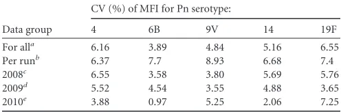
Pn Ps bead conjugation efficiency.
Serotype-specific Pn Pss were
effectively conjugated and revealed consistently high MFIs
(
⬎
10,000) for 89SF (1:20 dilution). The conjugation efficiency
was monitored based on the consistency and nondecay of signal in
terms of MFI and between-well CVs for 89SF over a 3-year period
(2008 to 2010). Data pertaining to five Pn Sts are presented in
Table 1
. The CVs were consistently low for all 5 Sts, with the most
recent batch of beads (year 2010) providing the lowest CVs for Sts
4, 6B, and 14.
Compliance with WHO and FDA guidelines.
A panel of 12
WHO reference or calibration sera (i.e., Goldblatt sera) were
tested for 7 Pn St-specific IgG using the MBIA (7-plex) and
com-TABLE 1Coefficient of variance for 89SF standard over a 3-year period in MBIA multiplex formatData group
CV (%) of MFI for Pn serotype:
4 6B 9V 14 19F
For alla 6.16 3.89 4.84 5.16 6.55
Per runb 6.37 7.7 8.93 6.68 7.4
2008c 6.55 3.58 3.80 5.69 5.76
2009d 5.52 4.54 3.55 4.88 3.65
2010e 3.88 0.97 5.25 2.06 7.25
an⫽259. b
Average of 6 plates (each sample was run in duplicate on each plate).
cn⫽128. d
n⫽100.
en⫽31.
on August 17, 2020 by guest
http://cvi.asm.org/
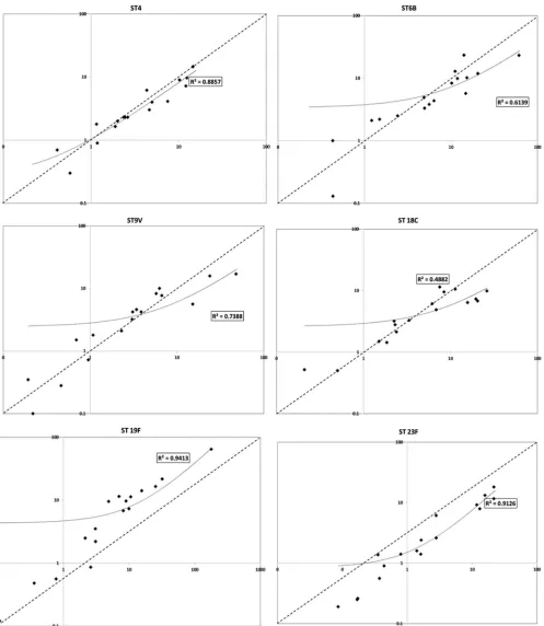
FIG 1Scatter plot to deduce the correlation between MBIA and WHO ELISA anti-pneumococcal polysaccharide IgG assignments for the WHO International pneumococcal calibration serum panel (12 sera).xaxis, log10of MBIA anti-pneumococcal serotype-specific IgG concentration (g/ml);yaxis, log10of WHO ELISA anti-pneumococcal serotype-specific IgG concentration (g/ml) assignments. The solid line represents the linear regression trend line and the dashed line the line of identity. Values for serotype 14 are not shown, as ther2wasⱖ0.99. Serotype 14 exhibited the highest correlation (r2⬎0.99), followed by serotypes 19F and 23F (r2⫽0.94 and 0.91, respectively). Serotype 18C exhibited poor correlation, with ther2at 0.488.
on August 17, 2020 by guest
http://cvi.asm.org/
pared to the results of a set of consensus IgG concentrations
pub-lished previously. Six of 7 Pn Sts tested demonstrated good
corre-lation between MBIA and the ELISA IgG assignments (
Fig. 1
). The
best agreement was recorded for St 14 (
r
2⬎
0.99), followed by St
19F and St 23F (
r
2⫽
0.94 and 0.91, respectively). Serotype 18C
exhibited poor correlation, with the
r
2at 0.488. Despite this
vari-ation in correlvari-ation between the MBIA and ELISA assignments,
MBIA as a technique passed the qualification criteria, with
ⱖ
75%
of WHO calibration sera falling within
⫾
40% of the assigned IgG
concentrations for all seven Sts (
Tables 2 to 5
).
Correlation of MBIA with ELISA.
Adult serum from
immu-nized mothers was evaluated for anti-Pn Ps-specific IgG to Sts 4,
6B, 9V, 14, and 19F to evaluate the correlation between our
in-house WHO ELISA and the bead-based multiplex assay.
Table 6
indicates a linear relationship in the Pn Ps St-specific IgG
concen-trations when measured by ELISA and MBIA, as well as
demon-strating a statistically significant positive relationship. The
r
2val-ues were consistently high (
ⱖ
0.84), with no significant difference
(
P
ⱖ
0.528) among the 5 Pn Sts. In addition, this positive
relation-ship provides the necessary statistical strength for MBIA to predict
the protective antibody levels similarly to ELISA as defined by the
WHO in test subjects (
Table 7
).
Test for MBIA robustness.
Robustness was tested on two
counts. Quality control 1 and QC 3 were tested every day for a
period of 25 consecutive days and the anti-Pn St 4, 6B, 9V, 14, and
19F IgG concentrations plotted in a Levey-Jennings chart (data
TABLE 2MBIA compliance with WHO assignments for pneumococcalserotypes 4 and 6Ba
WHO panel
Concn (g/ml) of IgG to:
Serotype 4 Serotype 6B
WHO assigned value MBIA WHO assigned value MBIA
A B C A B C
730 7.2 10.71 13.66 12.08 4.9 5.44 5.1 4.14
734 9 7.72 14.99 8.09 2.2 1.89 1.2 1.41
738 2.3 2.5 2 2.6 12.9 10.5 11.9 11.1
742 6.2 4.69 4.58 3.74 9.9 11.9 11.7 11.98
744 1.8 0.74 1.2 1.54 23.3 17.72 16.7 8.04
748 2.3 1.9 2.26 3.22 1 0.35 0.27 0.67
752 9.7 12.54 13.29 11.45 10.2 10.7 12.7 22.45
754 14.6 11.42 15.6 16.76 3.3 2.29 8.1 4.49
760 2.3 2.56 2.6 2.73 2.1 0.97 1.26 1.41
764 4 5.22 5.3 4.41 23.2 45.23 73.8 63.81
768 0.7 0.43 0.29 0.52 2.5 2.54 2.5 2.24
770 2 2.53 2.2 1.31 8.3 7.93 11.6 10.98
aMBIA values are from 3 individual runs (A, B, and C). For serotype 4, 83, 75, and 92%
of the values for runs A, B, and C, respectively, fell within⫾40% of the assigned value; for serotype 6B, 75, 75, and 75% of the values for runs A, B, and C, respectively, fell within⫾40% of the assigned value.
TABLE 3MBIA compliance with WHO assignments for pneumococcal serotypes 9V and 14a
WHO panel
Concn (g/ml) of IgG to:
Serotype 9V Serotype 14
WHO assigned value MBIA WHO assigned value MBIA
A B C A B C
730 1.5 0.76 0.65 0.68 66.10 14.59 9.7 11.39
734 7.7 6.8 6.3 7 322.10 392.67 450 371.34
738 3.2 2.81 4.3 2.05 18.40 65.4 16.5 18
742 2.1 2.36 2.3 2.24 7.40 6.88 4.4 4.41
744 10 3.29 8.7 6.95 3.70 2.54 3.31 3.66
748 4.2 5.28 3.7 2.65 10.60 9.9 12.3 9.95
752 17 18.8 60.3 65.16 27.80 39.4 98 85.06
754 15.8 10.84 21.7 39.37 160.80 102.4 156.8 214.99
760 1.8 1.02 0.89 1.35 19.20 21.21 30.8 19.75
764 8.3 4.99 7.2 5.11 17.2 10.91 12.2 11.99
768 4.6 2.77 3.9 3.61 14.00 7.77 10.2 8.95
770 4.2 4.1 2 3.19 115.60 103 134.86 146.46
a
MBIA values are from 3 individual runs (A, B, and C). For serotype 9V, 75, 75, and 75% of the values for runs A, B, and C, respectively, fell within⫾40% of the assigned value; for serotype 14, 75, 75, and 83% of the values for runs A, B, and C, respectively, fell within⫾40% of the assigned value.
TABLE 4MBIA compliance with WHO assignments for pneumococcal serotypes 18C and 19Fa
WHO panel
Concn (g/ml) of IgG to:
Serotype 18C Serotype 19F
WHO assigned value MBIA WHO assigned value MBIA
A B C A B C
730 3.2 2.54 2.23 1.99 9.7 9.1 11.5 6.47
734 6.8 7.68 34.7 19.39 11.3 7.4 15.49 8.98
738 6.1 5.4 7.2 5.9 2.5 2.1 2 2.35
742 11.5 7.22 8.1 7.35 11.5 6.2 6.9 8.06
744 9.5 7.16 9.1 9 9.5 2.97 3.2 8.5
748 10.6 10.3 10.89 12.87 16.5 17.8 34.6 23.96
752 9.9 9.9 36.7 32.35 64.1 131.1 215 190.52
754 4.9 4.37 9.9 6.38 14.1 8.55 19.2 19.1
760 3.3 2.82 4.4 2.84 6.8 7.7 7.6 9.28
764 6.4 23.34 5.45 18.41 21.7 30.19 30.1 35.97
768 1.5 1.78 1.15 1.62 3.5 3.2 2.6 3.43
770 2.8 3.41 1.69 1.83 7.3 10.12 9.38 10.46
aMBIA values are from 3 individual runs (A, B, and C). For serotype 18C, 92, 75, and
75% of the values for runs A, B, and C, respectively, fell within⫾40% of the assigned value; for serotype 19F, 75, 75, and 75% of the values for runs A, B, and C, respectively, fell within⫾40% of the assigned value.
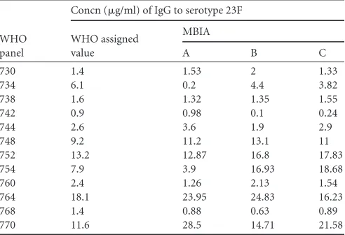
TABLE 5MBIA compliance with WHO assignments for pneumococcal serotype 23Fa
WHO panel
Concn (g/ml) of IgG to serotype 23F
WHO assigned value
MBIA
A B C
730 1.4 1.53 2 1.33
734 6.1 0.2 4.4 3.82
738 1.6 1.32 1.35 1.55
742 0.9 0.98 0.1 0.24
744 2.6 3.6 1.9 2.9
748 9.2 11.2 13.1 11
752 13.2 12.87 16.8 17.83
754 7.9 3.9 16.93 18.68
760 2.4 1.26 2.13 1.54
764 18.1 23.95 24.83 16.23
768 1.4 0.88 0.63 0.89
770 11.6 28.5 14.71 21.58
a
MBIA values are from 3 individual runs (A, B, and C). For all three runs, 75% of the values fell within⫾40% of the assigned value.
on August 17, 2020 by guest
http://cvi.asm.org/
not shown). The assay is robust for IgG values among all tested Sts
in QC 1 and QC 3 and, with one exception, fell within
⫾
2 SD of
the expected values. Additional robustness testing with different
operators and reagent lots is in progress as a part of an extensive
validation process.
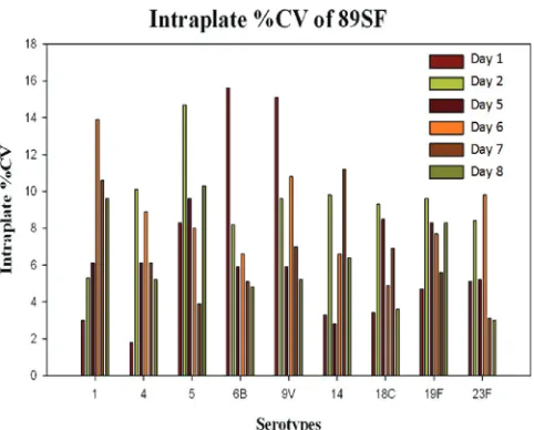
Test for assay reproducibility.
The reproducibility of the
MBIA was determined by calculating the intraplate CV obtained
for the 89SF MFI during a single week. The results are shown in
Fig. 2
. All CVs were less than 16% for any given day, with the mean
intraplate CV per St ranging from 6.2% for Sts 18C and 23F to
9.1% for St 5. Additional 89SF CV data were analyzed over a
pe-riod of 3 years for MFI. During this time, the CVs for each of the
nine serotypes measured were
⬍
9%.
DISCUSSION
This study describes the development and characterization of a
multiplex bead-based immunoassay (MBIA) to quantify Pn
St-specific IgG. The MBIA used in this clinical study was able to
provide a sensitive, efficient, and high-throughput analysis of a
large number of serum samples from both infants and adults. The
reproducibility of the multiplex assay values was evaluated daily
and over a 3-year period. The intra-assay variability for 89SF was
consistently low, with CVs of less than 8.93% for any given St on
any given day. These results suggest that the multiplex assay shows
good precision in measuring St-specific anti-Pn IgG. To further
assist in monitoring the day-to-day consistency of the multiplex
assay and to help normalize the results, a QC panel of 5 sera
con-taining low-, medium-, and high-titer anti-Pn antibody was run
daily on each plate. The fact that 3 different preparations of
con-jugate beads were used during the analysis period also provided
evidence that batch-to-batch variation during bead conjugation
was minimal, with no significant impact on assay results.
The MBIA platform introduces a number of improvements
compared to ELISA. The multiplex assay is valuable in situations
where small volumes of serum (5 to 10
l) are available for
ana-lyzing large multiples of Sts, as was the case in this study. These
volumes of serum are much less than what is needed to obtain
similar results using the ELISA (25 to 50
l) (
10
,
11
,
13
). In our
hands, the MBIA provided a 5-fold reduction in the required
amount of serum and a 29-fold reduction in the required amount
of Ps. Other studies have reported reductions as high as 25- and
200-fold for serum and Ps, respectively (
8
). Multiplexing of serum
samples for IgG antibody also reduces the assay time required for
each analyte by 80% and markedly increases sample throughput
(
4
,
5
,
7
). In contrast to ELISA, the multiplex technology allows for
the simultaneous analysis of as many as 100 distinct antigens in a
single microtiter well and the potential for high-throughput
screening of up to 1,000 sera per day (
16
). Finally, in terms of cost,
the multiplex assay is approximately seven times less expensive
than the ELISA for analyzing five Sts from each serum sample.
In our study, assessment of the maternal antibody response in
the multiplex assay indicated that serum samples with high
con-centrations of nonspecific, cross-reactive antibodies produced
ad-ditional fluorescent signals that were confounding and often
masked the true type-specific antibody response to capsular Ps. It
is possible that much of this background noise may be attributable
to the sensitivity of the multiplex assay. One approach for
elimi-nating this problem is to develop chemically assayed true
stan-dards. Another approach involves improving the quality of
com-mercially available Pn Ps antigen as close to reagent grade as
possible and eliminating the use of clinical-grade or vaccine grade
material as a target for
in vitro
immunoassays.
Due to the presence of variable amounts of CWPs and protein
contaminants associated with each Pn St as well as variations in
the production of anti-CWPs antibody by individual donors (
5
,
14
), it has been nearly impossible to create assay conditions in
TABLE 6Comparison of MBIA and ELISA anti-pneumococcal capsularpolysaccharide IgG assaysa
Serotype
Geometric mean concn of IgG (g/ml)
Pvalue r2value
Multiplex ELISA
4 4.50 (0.11–7.98) 4.14 (0.08–5.40) 0.707 0.84
6B 4.88 (0.13–9.23) 5.50 (0.09–6.30) 0.817 0.92
9V 6.17 (0.10–7.29) 6.49 (0.10–7.37) 0.591 0.86
14 27.99 (0.45–31.94) 24.50 (0.40–28.88) 0.528 0.89 19F 16.79 (0.39–27.99) 16.98 (0.34–24.62) 0.796 0.97 an⫽50. Adult pregnant women were vaccinated with a single dose of the 23-valent
polysaccharide vaccine during the third trimester of pregnancy. Values in parentheses are confidence intervals.Pvalues were determined by the Mann-Whitney rank sum test, andr2
values were determined by linear regression.
TABLE 7Percentage of vaccinated mothers with pneumococcal IgG antibody concentrations ofⱖ0.35g/mla
Serotype
% (n⫽50) as determined by:
MBIA ELISA
4 96.2 (92.8, 99.5) 94.3 (90.1, 98.2)
6B 90.6 (86.3, 93.7) 86.8 (81.2, 92.4)
9V 94.3 (90.1, 98.2) 92.4 (85.6, 97.7)
14 100.0 (92.7, 100.0) 98.1 (91.4, 99.6)
19F 96.2 (92.8, 99.5) 94.3 (90.1, 98.2)
a
Proposed WHO reference concentration for defining putative protective levels against invasive pneumococcal disease. Mothers were vaccinated once with a 23-valent pneumococcal polysaccharide vaccine during their third trimester of pregnancy. Values in parentheses are 95% confidence intervals.
FIG 2Intraplate CV for 89SF. Anti-pneumococcal serotype-specific IgG for 9 pneumococcal serotypes in 89SF standard serum was determined using MBIA during a single week. CVs wereⱕ16% for each serotype. Each box in the legend represents an assay day.
on August 17, 2020 by guest
http://cvi.asm.org/
which all of the confounding antibody effects have been removed
across all possible antibody levels and from all donors. The effect
of CWPs antibodies on a given serum IgG measure is dependent
on what St is being tested, the time allowed for CWPs adsorption,
and a patient’s CWPs antibody level. Because the concentration of
CWPs can be extremely high in some situations, the ability to
adsorb out all of the contaminants is often limited regardless of the
amount of time and adsorbent used in the procedure. Samples
with low levels of IgG antibody to Ps can be drastically reduced by
overly long CWPs adsorption, and samples with high CWPs
anti-body levels can demonstrate very high Ps IgG levels where the
CWPs are not completely removed by adsorption. In situations
where CWPs has been used to adsorb out nonspecific activity, it is
still possible to have up to 25% anti-CWPs remaining in the sera
(
11
,
17
). It is also possible that adsorption with high
concentra-tions of CWPs may have little effect on reducing nonspecific
an-tibody activity due to the presence of other contaminating cell wall
factors.
Validation of a new serological assay is often challenged by the
requirements to meet the assigned antibody titer obtained with an
alternate and highly regarded technique. While this is highly
plau-sible when comparing the reliability and usefulness of a technique
with similar chemistry and reaction dynamics, it becomes a real
challenge for techniques with clear differences in their operating
principles. This is further compounded by the need to meet
his-torical IgG assignments that were generated 15 to 20 years ago.
Realizing this challenge, previous studies attempted to
demon-strate a correlation between an in-house ELISA and an in-house
multiplex bead-based assay with a set of fresh in-house sera as a
surrogate to “true” validation. Despite these well-known
differ-ences, regulatory authorities insist on meeting the historical
ELISA assignments with the newly developed serological
tech-nique as the primary validation criteria for Pn Ps IgG
quantifica-tion (
3
,
12
). In this background, we have made an attempt to meet
the historical WHO Pn St-specific IgG assignments for 12
inter-national calibration sera and have been successful in our efforts.
MBIA has returned IgG concentrations for all seven Pn Sts within
the compliance criteria (
Tables 2 to 5
). In short, the MBIA data
presented here met the WHO qualification guidelines, with 75 to
92% of the serum panel showing strong agreement to
preestab-lished WHO values and falling within the acceptable 40% error
range. Many other assay protocols published in the literature have
failed to meet the qualification criteria for the same Pn Sts
evalu-ated in this study (
1
,
2
,
6
,
19
).
Recently, Whaley et al. (
18
) compared an MBIA-like assay with
two other multiplex assays. They examined several different
labo-ratory parameters, including assay accuracy, where the results
from each platform were compared against the assigned values of
the 12 WHO reference sera. All three assays performed in a similar
fashion as determined by comparing their individual mean IgG
concentrations to the WHO assigned reference serum values.
Fur-thermore, all three assays had significantly wide spreads for CVs,
and none of them reached total agreement with the
WHO-recom-mended values. In fact, only the MBIA-like assay had any Sts that
met the WHO requirement of at least 75% of the samples tested
returning values within the 40% error range. On the other hand,
Flow Applications, Inc., has developed a multiplex assay platform
that successfully demonstrated concurrence with the WHO
crite-ria for 7 Pn Sts associated with the first licensed Prevnar vaccine
(Wyeth Lederle, NY).
Other comparative results in our study showed a highly
signif-icant linear relationship and equivalence of the anti-Pn IgG
con-centrations when measured by the WHO in-house ELISA and
MBIA using 89SF as a standard. Additional correlation values
were observed between the two assays based on sera obtained from
pregnant women vaccinated with a 23-valent Pn Ps vaccine as part
of a randomized, double-blind, placebo-controlled clinical trial
(
20
). The results showed excellent agreement between the two
assays, with
r
2values ranging from 0.84 to 0.97 for the 5 Sts tested.
This relationship is critical when attempting to bridge an
un-known multiplex assay with a standardized and validated ELISA,
the latter being pivotal in providing serological data for the
licen-sure of the 7-valent Pn conjugate vaccine (
3
). The high level of
agreement between the ELISA and MBIA further suggests that
following additional characterization and complete validation of
the MBIA, it might be possible for the two assays to be used
inter-changeably when measuring the immune response to various Pn
vaccines in a clinical setting.
To conclude, the MBIA represents a comprehensive multiplex
assay system that can be applied to virtually any application that
requires analysis of anti-Ps antibody at both the basic and clinical
levels. Assay parameters such as the FDA 89SF, WHO
Interna-tional calibration serum panel, in-house QC sera, and pre- and
postvaccination immune sera indicate that the MBIA has good
precision, accuracy, and reproducibility, the three attributes
crit-ical for any assay system. The intra-assay variation for 89SF, when
measured over a 3-year period, was very low for all Sts (
⬍
9%) and
in some cases 1% or less, which again is indicative of a highly
reproducible assay. Most importantly, the MBIA met the
require-ments for assay qualification based on the FDA- and
WHO-pre-established criteria for concordance.
Many factors influence the success of a multiplex anti-Pn IgG
assay. These include (i) CWPs and protein contaminants and their
concentration in the target antigen; (ii) assay conditions; (iii)
se-rum antibody composition (e.g., specific versus nonspecific, IgG
versus IgM, and high versus low affinity) in each donor sample;
(iv) the Sts being tested; (v) the Ps conjugation procedure; (vi) the
extent to which confounding variables, such as CWPs antibodies
and antibodies to cell wall proteins, are removed from sera; and
(vii) the assay protocol and the data analysis process used in the
study. While the multiplex assay offers clear-cut advantages over
ELISA, careful optimization of the various above-cited factors is
required for its clinical use. Careful optimization, stringent
vali-dation, and continuous monitoring are the most critical factors
for the clinical exploitation of this unique multiplex,
high-throughput serological technique, MBIA.
ACKNOWLEDGMENTS
We thank Marla M. Martinez and Joseph Martinez, Jr. (Flow Applica-tions, Inc., Okawville, IL), for data management.
Mark Steinhoff receives research support from the Bill and Melinda Gates Foundation, USAID, the Thrasher Fund, Wyeth, GlaxoSmith Kline, Sanofi-Aventis, and Merck and lecture fees from Glaxo-SmithKline and Sanofi-Aventis. We claim no other potential conflict of interest relevant to this article.
REFERENCES
1.Biagini RE, et al.2003. Method for simultaneous measurement of anti-bodies to 23 pneumococcal capsular polysaccharides. Clin. Diagn. Lab. Immunol.10:744 –750.
2.Elberse KEM, Tsherniaeva I, Berbers GAM, Schouls LM.2010.
on August 17, 2020 by guest
http://cvi.asm.org/
mization and application of a multiplex bead-based assay to quantify se-rotype-specific IgG against Streptococcus pneumoniae polysaccharides: response to the booster vaccine after immunization with the pneumococ-cal 7-valent conjugate vaccine. Clin. Vaccine Immunol.17:674 – 682. 3.Jódar L, et al.2003. Serological criteria for evaluation and licensure of
new pneumococcal conjugate vaccine formulations for use in infants. Vaccine21:3265–3272.
4.Lal G, Balmer P, Joseph H, Dawson M, Borrow R.2004. Development and evaluation of a tetraplex flow cytometric assay for quantitation of serum antibodies to Neisseria meningitidis serogroups A, C, Y, and W-135. Clin. Diagn. Lab. Immunol.11:272–279.
5.Lal G, et al.2005. Development and validation of a nonaplex assay for the simultaneous quantitation of antibodies to nine Streptococcus pneu-moniae serotypes. J. Immunol. Methods296:135–147.
6.Marchese RD, et al.2006. Enzyme-linked immunosorbent assay for mea-suring antibodies to pneumococcal polysaccharides for the Pneumovax 23 vaccine: assay operating characteristics and correlation to the WHO inter-national assay. Clin. Vaccine Immunol.13:905–912.
7.Marchese RD, et al.2009. Optimization and validation of a multiplex, electrochemiluminescence-based detection assay for the quantitation of immunoglobulin G serotype-specific antipneumococcal antibodies in hu-man serum. Clin. Vaccine Immunol.16:387–396.
8.Musher DM, Watson DA, Baughn RE.1990. Does naturally acquired IgG antibody to cell wall polysaccharide protect human subjects against pneumococcal infection? J. Infect. Dis.161:736 –740.
9.Nahm M, and Goldblatt D.26 November 2002, posting date. Training manual for enzyme linked immunosorbent assay for the quantitation of Streptococcus pneumoniae serotype specific IgG. WHO Pneumococcal Serology Reference Laboratories, London, United Kingdom.http://www .vaccine.uab.edu.
10. Pickering JW, Martins TB, Schroder MC, Hill HR.2002. Comparison of a multiplex flow cytometric assay with enzyme-linked immunosorbent
assay for quantitation of antibodies to tetanus, diphtheria, and Haemo-philus influenzae type b. Clin. Diagn. Lab. Immunol.9:872– 876. 11. Pickering JW, et al.2002. A multiplexed fluorescent microsphere
immu-noassay for antibodies to pneumococcal capsular polysaccharides. Am. J. Clin. Pathol.117:589 –596.
12. Plikaytis BD, et al.2000. An analytical model applied to a multicenter pneumococcal enzyme-linked immunosorbent assay study. J. Clin. Mi-crobiol.38:2043–2050.
13. Schlottmann SA, Jain N, Chirmule N, Esser MT.2006. A novel chem-istry for conjugating pneumococcal polysaccharides to Luminex micro-spheres. J. Immunol. Methods309:75– 85.
14. Soininen A, van den Dobbelsteen G, Oomen L, Käyhty H.2000. Are the enzyme immunoassays for antibodies to pneumococcal capsular polysac-charides serotype specific? Clin. Diagn. Lab. Immunol.7:468 – 476. 15. Vignali DA.2000. Multiplexed particle-based flow cytometric assays. J.
Immunol. Methods21:243–255.
16. Waterboer T, Sehr P, Pawlita M.2006. Suppression of non-specific binding in serological Luminex assays. J. Immunol. Methods309:200 – 204.
17. Wernette CM, et al.2003. Enzyme-linked immunosorbent assay for quantitation of human antibodies to pneumococcal polysaccharides. Clin. Diagn. Lab. Immunol.10:514 –519.
18. Whaley MJ, et al.2010. Interlaboratory comparison of three multiplexed bead-based immunoassays for measuring serum antibodies to pneumo-coccal polysaccharides. Clin. Vaccine Immunol.17:862– 869.
19. Yu S, Sun Y, Frasch C, Concepcion N.1999. Pneumococcal capsular polysaccharide preparations may contain non-C polysaccharide contam-inants that are immunogenic. Clin. Diagn. Lab. Immunol.6:519 –524. 20. Zaman K, et al.2008. Effectiveness of maternal influenza immunization
in mothers and infants. N. Engl. J. Med.359:1555–1564. (Erratum,360: 648, 2009.)