Page 1 of 7 Original Research
Read online:
Scan this QR code with your smart phone or mobile device to read online. Authors:
Joseph M. Ochieng’ Oduor1,2
Nyamongo Onkoba1
Fredrick Maloba3
Atunga Nyachieo1
Affiliations:
1Institute of Primate Research
(IPR), Nairobi, Kenya
2School of Medicine, Kenyatta
University, Nairobi, Kenya
3School of Pure and Applied
Sciences, Kenyatta University, Nairobi, Kenya
Corresponding author: Joseph M. Ochieng’ Oduor, josemislredo@gmail.com Dates:
Received: 10 Mar. 2016 Accepted: 02 Aug. 2016 Published: 30 Sept. 2016 How to cite this article: Ochieng’ Oduor JM, Onkoba N, Maloba F, Nyachieo A. Experimental phage therapy against haematogenous multi-drug resistant
Staphylococcus aureus
pneumonia in mice. Afr J Lab Med. 2016;5(1), a435. http:// dx.doi.org/10.4102/ajlm. v5i1.435
Copyright:
© 2016. The Authors. Licensee: AOSIS. This work is licensed under the Creative Commons Attribution License.
Introduction
During lung infection with bacteria and viruses, the lung tissue becomes inflamed, deflated and occupied by pus and mucus, and the bronchial walls thicken due to accumulation of lymphocytes.1 Streptococcus pneumoniae, Haemophilus influenzae, Mycoplasma pneumoniae and Staphylococcus aureus all cause pneumonia.2 S. aureus pneumonia is usually associated with nosocomial infections common with patients in intensive care units.3
Community-acquired pneumonia, especially haematogenous (blood-borne) S. aureus pneumonia is rare; however, it has been documented to have high morbidity and mortality rates world-wide.4 Haematogenous pneumonia is common in children, immunocompromised persons and the elderly who have chronic skin and soft tissue infections.5 Globally, there has been an emergence of multidrug-resistant bacteria, including methicillin resistant S. aureus and multidrug-resistant S. aureus (MDRSA), which are difficult to treat with currently available antibiotics.6,7 These ‘super bugs’ have made the treatment of haematogenous pneumonia and other associated infections difficult.8,9,10,11 Thus, there is need for an alternative therapeutic measure, such as phage therapy, to combat super bugs.
Phage therapy refers to the use of bacteriophages (or phages) as antibacterial agents against bacterial pathogens.12 Currently, the use of phage therapy is common primarily in Georgia and Russia, despite once being practised globally before the advent of antibiotics.13,14,15 Its use ceased in western Europe, the United States and Canada in the 1940s due to poor understanding of the
Background: Community-acquired haematogenous Staphylococcus aureus pneumonia is a rare infection, though it can be acquired nosocomially. Currently, antibiotics used against S. aureus pneumonia have shown reduced efficacy. Thus, there is need for an alternative therapy against multidrug-resistant S. aureus (MDRSA) strains in the community.
Objective: We sought to determine the efficacy of environmentally-obtained S. aureus lytic phage against haematogenous MDRSA pneumonia in mice.
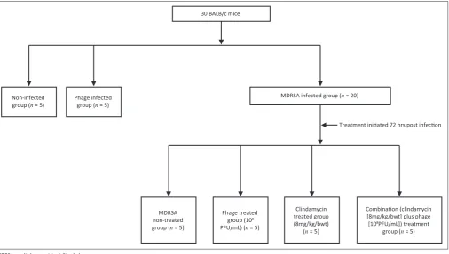
Methods: Phages and MDRSA were isolated from sewage samples collected within Nairobi County, Kenya. Isolated S. aureus bacteria were screened for resistance against ceftazidime, oxacillin, vancomycin, netilmicin, gentamicin, erythromycin, trimethroprim-sulfamethoxazole and cefuroxime. Thirty BALB/c mice aged six to eight weeks were randomly assigned into three groups: the MDRSA-infection group (n = 20), the phage-infection group (n = 5) and the non-infection group (n = 5). Mice were infected with either MDRSA or phage (108 CFU/mL) and treated after 72 hours with a single dose of clindamycin (8 mg/kg/bwt) or 108 PFU/mL of phage or a combination therapy (clindamycin and phage). The efficacy of phage, clindamycin or clindamycin with phage combination was determined using resolution of lung pathology and bacterial load in lung homogenates.
Results: The viable MDRSA count was 0.5 ± 0.2 log10 CFU/gm in the phage-treated group, 4.4 ± 0.2 log10 CFU/gm in the clindamycin-treated group and 4.0 ± 0.2 log10 CFU/gm in the combination-treated group. The efficacy of phage therapy was significantly different from other therapeutic modes (p = 0 < 0.0001). Histology showed that the mice treated with phage did not develop pneumonia.
Conclusion: Phage therapy is effective against haematogenous MDRSA infection. Thus, it can be explored as an alternative treatment method.
Experimental phage therapy against haematogenous
multi-drug resistant Staphylococcus aureus
pneumonia in mice
Read online:
nature, safety, therapeutics and pharmacology of phages.16 Phage therapy is cheap in terms of development and use as a therapeutic agent. Phages are abundant in nature and are capable of in vivo auto-dosing, that is to say, they are capable of multiplying into several copies at the infection site; hence, only a small dose is required for treatment.17 In Kenya, community-acquired MDRSA infections are commonly reported among HIV patients and at public hospitals that are located in informal settlements or slums.18 Thus, there is a need to characterise the profile of bacterial strains that are currently circulating in environmental sources. This will provide necessary information that can be used in management of infections caused by MDRSA bacteria. This study sought to determine the therapeutic potential of environmentally-obtained lytic phages in Nairobi, Kenya, against haematogenous MDRSA pneumonia in BALB/c mice.
Research method and design
Ethical considerations
All experimental protocols and procedures of the study were reviewed and approved by the Institutional Review Committee on Animals Ethics of the Institute of Primate Research (ref no: IRC/02/14) ethical committee on the use of laboratory animals for biomedical science in accordance with the international guidelines on animal care, handling and use for biomedical research. The experiments are reported in accordance with ARRIVE guidelines.19
Study animals
The study included 30 BALB/c mice of mixed sexes aged 6 to 8 weeks which were sourced from the Institute of Primate Research rodent house, Karen-Nairobi, Kenya, and maintained at their rodent facility. The mice were fed on antibiotic-free food rations (obtained from Unga Feeds, Nairobi, Kenya) and water was provided ad libitum.
Isolation of MDRSA
S. aureus bacterial strains were isolated from 20 sewage samples collected from informal settlements, sewage treatment plants and abattoirs within Nairobi County, Kenya. Specimens from all the samples were streaked on selective Mannitol salt agar (Liofilchem®, Roseto degli Abruzzi, Italy) supplemented with 4 µg of ciprofloxacin [Liofilchem®, Roseto degli Abruzzi, Italy]. Single colonies were amplified in nutrient agar (HiMedia, Mumbai, India). Staphylococcus strains were identified using microscopy, physiological tests and an Analytical Profile Index of Staphylococcus system [bioMérieux, Marcy l’Etoile, France]. We screened isolated S. aureus bacteria for antimicrobial resistance to the following drugs: ceftazidime 30 µg, oxacillin 1 µg, vancomycin 30 µg, netilmicin 30 µg, gentamicin 10 µg, erythromycin 15 µg, trimethroprim-sulfamethoxazole 25 µg and cefuroxime 30 µg (Liofilchem®, Roseto degli Abruzzi, Italy), according to the Clinical Laboratory Standard Institute protocol.20 Isolates were considered to be MDRSA, if they were non-susceptible to more than one class of antibiotics. Colonies identified as
MDRSA were re-suspended in 50% glycerol-nutrient broth and stored at -20 °C until use.
Phage isolation
We analysed half a litre of sewage samples collected from 10 sites (Nairobi City sewage treatment plant, informal settlements and an abattoir) within Nairobi County, Kenya. Twenty specific S. aureus lytic phages were isolated from these samples using the modified method of Anany et al.21 Briefly, equal volumes of ultra-filtered sewage samples and nutrient broth, plus 1 mL of 18-hour-old MDRSA culture were mixed and incubated overnight at 37 °C while shaking at 120 rpm (LAB-LINE® INCUBATOR-SHAKER, Waltham, Massachusetts, United States). The following day, the culture was centrifuged at 10 000 g for 10 minutes (Fisher Centrific® Centrifuge, Waltham, Massachusetts, United States), and the supernatant was filtered through a 0.22 µm filtration unit (Cambridge, Massachusetts, United States), then screened for the presence of phages using a double-layer plaque assay. Individual plaques were selected and sub-cultured in 2 mL of nutrient broth containing a sensitive bacterial host (106 CFU/ mL). The isolated phages were then tested against the previously-isolated MDRSA by spot assay. The virulence of the phages was determined by using an efficiency of plating analysis,22 whereby the strain with numerous plaques on MDRSA lawn was considered the most virulent. The virulent phage was selected and used as a therapeutic agent against MDRSA in mice.
Experimental design
Viable bacteria levels in mouse lung
homogenates
Upon necropsy, lungs were obtained aseptically and homogenised for bacterial culture as follows: 500 µL of homogenised tissue was diluted with normal saline at a ratio of 1:20 (homogenised lung tissue to saline). The diluted, homogenised tissue was then plated on 7.5% sodium chloride nutrient agar to select for MDRSA and incubated at 37 °C for 18–20 hours.
Histopathology
Phage safety and MDRSA pathogenicity were determined by euthanising the mice when they exhibited poor physical appearance and breathing difficulties in order to alleviate pain and suffering. Surviving mice were euthanised at 10 days post-infection. Lung samples were collected in 10% formaldehyde for histological analysis. Levels of inflammation were scored using categories as described by Schünemann et al.23 Briefly, the categories worst, worse, bad, good, better and best were assigned based on a scale of 5 to 0. The ‘worst’ category was assigned for severely inflammed septa, numerous collapsed alveoli, large pockets of pneumonia, mucus-congested alveoli, presence of perivascular fibrosis blood vessels and lack of ventilation (score = 5). The ‘worse’ category included all conditions in the worst category but with poor ventilation (score = 4). The ‘bad’ category was assigned for minor inflammation of septa, few pockets of pneumonia and moderate ventilation (score = 3). The ‘good’ category was assigned for minor inflammation of septa, few pockets of mucus-congested alveoli and good ventilation (score = 2). The ‘better’ category was assigned for no inflammed septa and a few pockets of mucus-congested alveoli (score = 1).
The ‘best’/normal category was assigned for no inflammation and good ventilation (score = 0).
Statistical analysis
Bacterial and phage counts were represented as mean ± standard error of the mean. The statistical significance of differences between groups was determined by one-way analysis of variance, followed by Tukey’s multiple comparison test, using Graph Pad Prism 5.0.1 (Graph pad software, San Diego, California, United States). A p-value of less than 0.05 was considered statistically significant.
Results
Multidrug-resistant S. aureus isolation
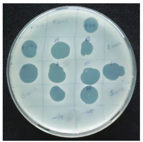
The environmentally-isolated S. aureus bacterium was resistant to β-lactam antibiotics (ceftazidime, cefuroxime and oxacillin). In addition, it was non-susceptible to glycopeptide (vancomycin), aminoglycoside (netilmicin and gentamicin) and macrolide (erythromycin) antibiotics. However, it was susceptible to nucleic acid synthesis inhibitors (co-trimoxazole [trimethroprim-sulfamethoxazole]) (Figure 2). Thus, the bacterial isolate was considered to be multidrug-resistant, as it was non-susceptible to more than one class of antibiotics.
Phage isolation and in vitro activities
Spot assays were used to identify 10 S. aureus lytic phages which produced large clear plaques on the MDRSA lawn (Figure 3) while other isolates failed. Of the 10 phage isolates,
Treatment iniated 72 hrs post infecon
MDRSA non-treated group (n = 5)
Phage treated group (108
PFU/mL) (n = 5)
Clindamycin treated group (8mg/kg/bwt)
(n = 5)
Combinaon (clindamycin [8mg/kg/bwt] plus phage [108PFU/mL]) treatment
group (n = 5) 30 BALB/c mice
Non-infected
group (n = 5) Phage infected group (n = 5)
MDRSA infected group (n = 20)
MDRSA, multidrug-resistant Staphylococcus aureus.
one created a larger patch on the MDRSA lawn. Its efficiency of plating analysis showed that it had more plaques than other phages.
Therapeutic effects in mice
Two groups of mice had 100% survivorship (n= 5), namely, the non-infected/non-treated group and the phage-infected mice. Only three mice survived in the MDRSA-infected group before treatment (72 hours post-infection) commenced.
All the mice that were treated with either clindamycin, phage or the combination therapy (clindamycin plus phage) survived at day 7 post-infection and each group had three mice. Mice that had phage infection (phage control group) were more active than mice in other treatment groups (clindamycin, phage and combination). Only one mouse in the MDRSA-infected control group survived at day 7 post-infection and it was weak (Table 1).
Viable bacteria levels in mouse lung
homogenates
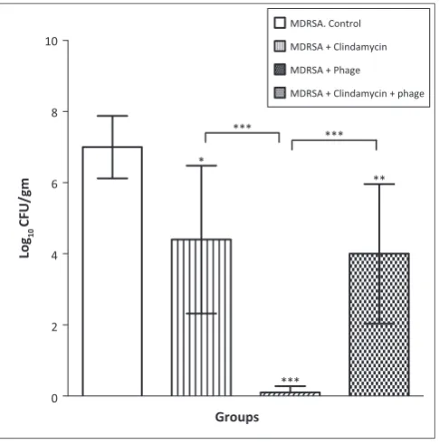
The number of viable MDRSA in mouse lung homogenates was significantly lower in mice treated with phage only (log10 CFU/gm = 0.5 ± 0.2) compared with those in the MDRSA-infected, non-treated control group (log10 CFU/gm = 8.0 ± 0.2) (p < 0.0001) (Figure 4). Compared with the MDRSA-infected, non-treated group, statistically-significantly differences were observed in the viable bacterial counts in the MDRSA-infected, clindamycin-treated (log10 CFU/gm = 4.4 ± 0.2;
p< 0.05) and MDRSA-infected, combination-treated groups (log10 CFU/gm = 4.0 ± 0.2; p < 0.001). The differences in counts between the phage-treated group and groups treated with clindamycin and the combination therapy were also statistically significant (p < 0.0001).
Histopathological examination
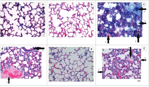
The lung tissues of mice in the phage-infected group were relatively normal, with minor focal congestion compared with tissue from mice in the MDRSA-infected non-treated group, which had deflated alveoli congested with mucus, lymphocyte-infiltrated septa and pockets of serous fluid. The FIGURE 2: Antibiogram test of S. aureus isolate. The isolate was resistant to: ceftazidime, 30 µg (1); oxacillin, 1 µg (2); vancomycin, 30 µg (4); netilmicin, 30 µg (6); gentamicin, 10 µg (7); erythromycin, 15 µg (8); and cefuroxime, 30 µg (5). However, it was sensitive to trimethroprim-sulfamethoxazole, 25 µg (3).
lung tissue of MDRSA-infected, non-treated mice were similar to lung tissue from clindamycin-treated mice. However, lung tissue from the clindamycin-treated group (n = 3) overall was graded as bad, since they had less congestion, fewer pockets of serous fluid and deflated alveoli compared with the non-treated MDRSA infected mice. Mice that received the combination therapy (n = 3) showed inflammation of the septa and blood vessel walls, moderately deflated alveoli and several serous fluid pockets compared with the non-infected (n = 5) and phage-infected mice (n = 5) (p < 0.0001) (Figure 5).
Discussion
This study found that S. aureus isolates collected from sewage water and found to be resistant to multiple drug therapies caused haematogenous pneumonia in mice, which was characterised by infiltration of immune cells and accumulation of serous fluid in the lungs. This confirmed that the isolated bacterium was pathogenic and able to cause sepsis, then was further confirmed by isolation of viable MDRSA bacteria from the lung
homogenates. The accumulation of serous fluid and pus in the lungs indicated that the mice induced an innate immune response toward multiplying MDRSA bacteria.24 This inflammatory immunological phenomenon may have led to the inflammation observed in the bronchi, bronchioles, alveoli and septa, as well as blood vessels. Our findings agree with other studies in which mice were infected intravenously with methicillin-resistant S. aureus, then administered a dose of antibiotic on a daily basis for a week.25,26,27 However, in our study we used a single dose of S. aureus-specific lytic phage.
The use of phage as a treatment method attained 100% efficacy, an indication that the single dose administered was enough to clear the bacteria from the infected mice. This is probably due to the auto-dosing properties of phages, whereby they increase in numbers at the site of an infection.28 The mode of phage administration, concentration, dosage and timing of treatments and resistance to phages by the pathogenic bacteria are some of the factors that determine the efficacy and safety of phage therapy.29 These factors may be responsible for the differences between our findings and those of other studies,30,31 in which methicillin-resistant S. aureus-infected mice were treated immediately or six hours after infection with phage to achieve a 100% efficacy. The timing of the initiation of treatment may be the crucial factor in achieving 100% efficacy. However, we cannot rule out the role of immunity in clearing the bacteria, considering that Yao et al.32 established that within 72 hours of infection the mouse immune system has the capacity to clear bacteria from the system. In our study, the combination therapy (clindamycin plus phage) reduced the efficacy of the phage to about 60%, indicating that clindamycin may have an antagonist effect on phage lytic activity. Clindamycin is a bacteriostatic antibiotic; thus, it may interfere with phage protein synthesis, which would interrupt the phage auto-dosing mechanism.
Limitations of the study
Our study results were generated in mice. While our findings provide vital information on the antimicrobial resistance profile in Kenya, they cannot be translated directly to humans. However, comparative animal models can be utilised to ascertain the safety and therapeutic potential of environmentally-available phages against infections caused by multidrug-resistant bacteria.
TABLE 1: Number of surviving mice at 72 hours post-infection and 7 days post-treatment, Nairobi, Kenya.
Groups Initial number
of mice Number of mice 72 hours post-infection during treatmentNumber of mice post-infection (end point)Number of mice 7 days
All MDRSA infected mice 20 12 12 10
Non-treated 5 3 3 1
Clindamycin-treated 5 3 3 3
Phage-treated 5 3 3 3
Combination-treated 5 3 3 3
Phage-infected mice 5 5 5 5
Non-infected mice 5 5 5 5
Total 30 22 22 20
MDRSA, multidrug-resistant Staphylococcus aureus.
0 2 4 6 8 10
*** *
**
*** ***
Groups
Lo
g10
CFU/gm
MDRSA + Clindamycin + phage MDRSA + Clindamycin
MDRSA + Phage MDRSA. Control
MDRSA, multidrug-resistant Staphylococcus aureus.
Conclusion
In conclusion, we report for the first time that a single dose of MDRSA-specific lytic phage is efficacious against haematogenous pneumonia caused by multidrug-resistant S. aureus in mice.
Acknowledgements
We acknowledge the Institute of Primate Research staff for providing logistical support during experimentation; Dr Geoffrey Omuse and Dr Allan Njoroge (Aga Khan University Hospital, Nairobi, Kenya) for donating ATCC 43300; Prof W. Jaoko and Mr J. Oloo (University of Nairobi, Medical School, Kenya) for donating ATCC 29213; and Prof. B. Weber-Dabrowska and Mr M. Lusiak-Szelachowska (Institute of Immunology and Experimental Therapy, Wroclaw, Poland) for providing phage isolation protocols.
This study was approved by the ethics committees of the Institute of Primate Research (ref no: IRC/02/14). All institutional and national guidelines for the care and use of animals were followed.
Competing interests
The authors declare that they have no financial or personal relationship(s) that may have inappropriately influenced them in writing this article.
Sources of support
None.Authors’ contributions
J.M.O.O. made conceptual contributions. J.M.O.O. and A.N. designed the study and the experimental protocols. J.M.O.O. collected the samples, performed the experimentation, and collected and analysed the data. F.M. assisted in reading the histology slides. J.M.O.O. and N.O. wrote the article. All authors read and approved the final article for publication.
References
1. Alcón A, Fàbregas N, Torres A. Pathophysiology of pneumonia. Clin Chest Med. 2005;26(1):39–46. http://dx.doi.org/10.1016/j.ccm.2004.10.013
2. Köksal I, Oslü T, Bayraktar O, et al. Etiological agents of community-acquired pneumonia in adult patients in Turkey; a multicentric, cross-sectional study. Tuberk Toraks. 2010;58(2):119–127.
3. Kyaw MH, Kern DM, Zhou S, et al. Healthcare utilization and costs associated with S. aureus and P. aeruginosa pneumonia in the intensive care unit: a retrospective observational cohort study in a US claims database. BMC Health Serv Res. 2015;15:241. http://dx.doi.org/10.1186/s12913-015-0917-x
4. Rubinstein E, Kollef MH, Nathwani D. Pneumonia caused by methicillin-resistant Staphylococcus aureus. Clin Infect Dis. 2008;46(Suppl 5):S378–S385. doi: 10.1086/533594
5. Dicpinigaitis PV, Levy DE, Gnass RD, et al. Pneumonia due to Staphylococcus aureus in a patient with AIDS: review of incidence and report of an atypical roentgenographic presentation. South Med J. 1995;88(5):586–590. http://dx.doi. org/10.1097/00007611-199505000-00017
6. Monecke S, Coombs G, Shore AC, et al. A field guide to pandemic, epidemic and sporadic clones of methicillin-resistant Staphylococcus aureus. PLoS One. 2011;6(4):e17936. http://dx.doi.org/10.1371/journal.pone.0017936
a
b
c
d
e
f
1 2
3 4
2
1
3
1
5
40X
MDRSA, multidrug-resistant Staphylococcus aureus.
7. Yu F, Lu C, Liu Y, et al. Emergence of quinupristin/dalfopristin resistance among livestock-associated Staphylococcus aureus ST9 clinical isolates. Int J Antimicrob Agents. 2014;44(5):416–419. http://dx.doi.org/10.1016/j.ijantimicag.2014.06.020. Epub 2014 Aug 17.
8. Rasmussen RV, Fowler VG, Skov R, et al. Future challenges and treatment of Staphylococcus aureus bacteremia with emphasis on MRSA. Future Microbiol. 2011;6(1):43–56. http://dx.doi.org/10.2217/fmb.10.155
9. Westwood J, Burnett M, Spratt D, et al. The hospital microbiome project: meeting report for the UK science and innovation network UK-USA workshop ‘beating the superbugs: hospital microbiome studies for tackling antimicrobial resistance’, October 14th 2013. Stand Genomic Sci. 2014;9:12. http://dx.doi.org/10.1186/ 1944-3277-9-12
10. Ferrara AM. Treatment of hospital-acquired pneumonia caused by methicillin-resistant Staphylococcus aureus. Int J Antimicrob Agents. 2007;30(1):19–24. Epub 2007 May 1. http://dx.doi.org/10.1016/j.ijantimicag.2007.02.011
11. Khan SN, Khan AU. Breaking the spell: combating multidrug resistant ‘superbugs’. Front Microbiol. 2016;7:174. http://dx.doi.org/10.3389/fmicb. 2016.00174. eCollection 2016.
12. Clokie MR, Millard AD, Letarov AV, et al. Phages in nature. Bacteriophage. 2011;1(1):31–45. http://dx.doi.org/10.4161/bact.1.1.14942
13. Abedon ST, Kuhl SJ, Blasdel BG, et al. Phage treatment of human infections. Bacteriophage. 2011;1(2):66–85. http://dx.doi.org/10.4161/bact.1.2.15845 14. Matsuzaki S, Uchiyama J, Takemura-Uchiyama I, et al. Perspective: the age of the
phage. Nature. 2014;509(7498):S9. http://dx.doi.org/10.1038/509S9a 15. Reardon S. Phage therapy gets revitalized. Nature. 2014;510(7503):15–16. http://
dx.doi.org/10.1038/510015a
16. Sulakvelidze A, Alavidze Z, Morris JG. Bacteriophage therapy. Antimicrob Agents Chemother. 2001;45(3):649–659. http://dx.doi.org/10.1128/AAC.45.3. 649-659.2001
17. Miedzybrodzki R, Fortuna W, Weber-Dabrowska B, et al. Phage therapy of staphylococcal infections (including MRSA) may be less expensive than antibiotic treatment. Postepy Hig Med. Dosw (Online). 2007;61:461–465.
18. Ouko TT, Ngeranwa JN, Orinda GO, et al. Oxacillin resistant Staphylococcus aureus among HIV infected and non-infected Kenyan patients. East Afr Med J. 2010;87(5):179–186.
19. Kilkenny C, Browne W, Cuthill IC, et al. Animal research: reporting in vivo experiments: The ARRIVE guidelines. Br J Pharmacol. 2010;160(7):1577–1579. http://dx.doi.org/10.1111/j.1476-5381.2010.00872.x
20. Clinical Laboratory Standards Institute. M100-S17: Performance standards for antimicrobial Susceptibility testing; Seventeenth informational supplement. Wayne, PA: CLSI; 2007.
21. Anany H, Lingohr EJ, Villegas A, et al. A Shigella boydii bacteriophage which resembles Salmonella phage ViI. Virol J. 2011;8:242. http://dx.doi.org/10.1186/ 1743-422X-8-242
22. Khan Mirzaei M, Nilsson AS. Isolation of phages for phage therapy: a comparison of spot tests and efficiency of plating analyses for determination of host range and efficacy. PLoS One. 2015;10(3):e0118557. http://dx.doi.org/10.1371/journal. pone.0118557. eCollection 2015.
23. Schünemann HJ, Jaeschke R, Cook DJ, et al. An official ATS statement: grading the quality of eevidence and strength of recommendations in ATS guidelines and recommendations. Am J Respir Crit Care Med. 2006;174(5):605–614. http://dx. doi.org/10.1164/rccm.200602-197ST
24. Wardenburg JB, Patel RJ, Schneewind O. Surface proteins and exotoxins are required for the pathogenesis of Staphylococcus aureus pneumonia. Infect Immun. 2007;75(2):1040–1044. http://dx.doi.org/10.1128/IAI.01313-06 25. Yanagihara K, Ohnishi Y, Morinaga Y, et al. Efficacy of ME1036 against
meticillin-resistant Staphylococcus aureus and vancomycin-insensitive S. aureus in a model of haematogenous pulmonary infection. Int J Antimicrob Agents. 32(5):401–404. http://dx.doi.org/10.1016/j.ijantimicag.2008.04.030 Epub 2008 Aug 19.
26. Oduor JMO, Onkoba WN, Maloba F, et al. Safety and therapeutic efficacy of staphylococcus aureus specific lytic phage against multidrug-resistant S. aureus (MDRSA) in BALB/c mice: A prospective study. Int J Infect Dis. 2016;45(Suppl 1):108.
27. Chhibber S, Kaur T, Kaur S. Co-therapy using lytic bacteriophage and linezolid: effective treatment in eliminating methicillin resistant Staphylococcus aureus (MRSA) from diabetic foot infections. PloS One. 2013;8(2):e56022. http://dx.doi. org/10.1371/journal.pone.0056022. Epub 2013 Feb 13.
28. Chhibber, S. & Kumari, S. in Bacteriophages (ed. Kurtoboke, I.) 139–158 (InTech, 2012).
29. Ly-Chatain MH. The factors affecting effectiveness of treatment in phages therapy. Front Microbiol. 2014;5:51. http://dx.doi.org/10.3389/fmicb.2014.00051 30. Matsuzaki S, Yasuda M, Nishikawa H, et al. Experimental protection of mice
against lethal Staphylococcus aureus infection by novel bacteriophage phi MR11. J Infect Dis. 2003;187(4):613–624. Epub 2003 Feb 7. http://dx.doi.org/10.1086/ 374001
31. Thiel K. Old dogma, new tricks—21st century phage therapy. Nat Biotechnol. 2004;22(1):31–36. http://dx.doi.org/10.1038/nbt0104-31