Abstract
Lawhon, Sara Dyann. Genetic and environmental regulation of virulence genes in
Salmonella enterica serovar Typhimurium. (Under direction of Craig Altier and Paul Orndorff)
The purpose of this research has been to assess the effect of the genetic regulator, CsrA, and environmental conditions on the expression of virulence genes in Salmonella enterica serovar Typhimurium. CsrA is an RNA binding protein that alters messenger RNA stability in E. coli and regulates virulence genes located on the Salmonella pathogenicity island 1 (SPI-1). This work demonstrates that CsrA in S. typhimurium like its counterpart in E. coli regulates expression of genes required for flagellar synthesis and suggests a role for CsrA in the positive regulation of utilization of propanediol and ethanolamine, vitamin B12 synthesis, and expression of maltoporin, which transports maltose and
maltodextrins across the bacterial cell membrane. Propanediol, ethanolamine, and maltodextrins are byproducts of digestion likely present in the intestinal tract. Vitamin B12 is required for utilization of propanediol and ethanolamine. The sensor kinase BarA and its response regulator SirA control levels of CsrA
indirectly through the expression of an untranslated RNA CsrB, which binds CsrA. SirA regulates expression of Salmonella virulence genes and is required for
short chain fatty acids (SCFA) acetate, propionate, and butyrate are present in the ileum and colon at differing total concentrations and relative percentages. SCFAs representing the ileum and acetate alone are able to restore SPI-1 invasion gene expression to a barA mutant but not to a sirA mutant. Additionally, ileal SCFAs increase the expression of SPI-1 virulence genes required for invasion of epithelial cells and increase the expression of genes required for survival within epithelial cells and macrophages. SCFAs representing the colon decrease expression of genes required for epithelial cell invasion and decrease the expression of genes required for flagellar synthesis and maltose transport while increasing the
Genetic and environmental regulation of virulence genes in Salmonella enterica serovar Typhimurium.
by
Sara Dyann Lawhon
A dissertation submitted to the Graduate Faculty of North Carolina State University
in partial fulfillment of the requirements for the Degree of
Doctor of Philosophy
Comparative Biomedical Sciences
Raleigh 2002
APPROVED BY:
Stephen Libby Geraldine Luginbuhl
Craig Altier Paul Orndorff
Dedication
Biography
Acknowledgements
I would particularly like to recognize Craig Altier for his patience, guidance, his incisive observations, and above all meticulous analysis, Mitsu Suyemoto for her many hours of support, her expertise with experiment design and execution, and above all her friendship, Wondwossen Gebreyes for his wise counsel, his enthusiasm, and his sense of humor, and Russ Maurer, who patiently offered advice and insightful explanations of biochemistry. I would also like to thank Jonathan G. Frye, Steffen Porwollik, and Michael McClelland who provided the Salmonella LT2 microarray, performed many of the microarray experiments and provided expert mentoring in the area of microarray analysis, John Roth who generously provided strains, Brian Ahmer who shared unpublished results, David Klapper of the UNC Protein Sequencing Core Facility who sequenced outer membrane proteins, and Michael Dykstra of the Laboratory for Advanced Electron and Light Optical Methods at the North Carolina State University College of
Veterinary Medicine. I would also like to thank Patty Spears and John Horton who never failed to share their time, advice, and countless reagents.
Finally, I would like to thank members of my committee, Paul Orndorff, Steve Libby, and Gerry Luginbuhl, for their insight, their guidance, sharing their experiences, and most of all their open doors.
Table of Contents
List of Tables... vi
List of Figures ... vii
Literature Review ...1
Salmonella Infection ...1
Salmonella Pathogenicity Island I ...4
Salmonella Pathogenicity Island 2 ...6
Flagellar regulation and chemotaxis ...7
The intestinal environment ...9
Short chain fatty acids ...11
Maltose ...11
Vitamin B 12...15
Regulation of genes associated with vitamin B12 synthesis ...15
Regulation of genes associated with vitamin B12 utilization...16
Conclusion...17
References ...19
Intestinal short chain fatty acids alter Salmonella typhimurium invasion gene expression and virulence through BarA/SirA ...33
Summary ...33
Introduction ...34
Results ...40
Discussion...49
Experimental Procedures ...55
References ...60
Global regulation by CsrA in Salmonella typhimurium...82
Summary ...82
Introduction ...83
Results ...87
Discussion...96
Experimental Procedures ... 101
References ... 107
Regulation of virulence gene expression by intestinal short chain fatty acids and BarA/SirA... 122
Summary ... 122
Introduction ... 123
Results ... 127
Discussion... 142
Experimental Procedures ... 155
List of Tables
Intestinal short chain fatty acids alter Salmonella typhimurium invasion gene expression and virulence through BarA/SirA
Table 1 Virulence of S. typhimurium strains in BALB/c mice...69 Table 2 Mutations used...70
Global regulation by CsrA in Salmonella typhimurium
Table 1. Regulation of invasion genes by csrA... 113 Table 2. Regulation of flagellar synthesis and chemotaxis by csrA... 114 Table 3. Regulation of vitamin B12 synthesis and of metabolic pathways requiring
B12 by csrA... 115 Table 4. Regulation of the maltose operon by csrA ... 116 Table 5 Strains and plasmids used ... 117
Regulation of virulence gene expression by intestinal short chain fatty acids and BarA/SirA
Table 1 Summary of gene regulation by intestinal short chain fatty acids (SCFA) and barA/sirA... 164 Table 2 Regulation of Salmonella typhimurium SPI-1 invasion genes by intestinal
short chain fatty acids (SCFA) and barA/sirA... 165 Table 3 Regulation of Salmonella enterica serovar Typhimurium SPI-2 invasion
genes by ileal short chain fatty acids (SCFA)... 166 Table 4 Regulation of Salmonella typhimurium flagellar synthesis and chemotaxis
by intestinal short chain fatty acids (SCFA) and barA/sirA... 167 Table 5 Regulation of genes required for vitamin B-12 synthesisand utilization by
intestinal short chain fatty acids (SCFA) and barA/sirA in Salmonella
typhimurium... 168 Table 6 Regulation of the maltose transport system by intestinal short chain fatty
acids (SCFA) and barA/sirA in Salmonella typhimurium... 170 Table 7 Regulation of the metabolism by intestinal short chain fatty acids (SCFA)
List of Figures
Intestinal short chain fatty acids alter Salmonella typhimurium invasion gene expression and virulence through BarA/SirA
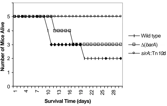
Figure 1. Time to death of mice...71
Figure 2. Acetate restores invasion gene expression in a ∆(barA) muta nt ...72
Figure 3. Invasion of HEp-2 cells ...74
Figure 4. Propionate and butyrate inhibit invasion gene expression...76
Figure 5. Gastrointestinal levels of short-chain fatty acids (SCFA) regulate invasion gene expression...78
Figure 6. Intracellular acetate restores e xpression of CsrB in a ∆(barA) mutant ..80
Figure 7. A model for the regulation of invasion gene expression by acetate and BarA ...81
Global regulation by CsrA in Salmonella typhimurium Figure 1. CsrA is required for production of flagella and for motility... 118
Figure 2. Loss of csrA alters the expression of Salmonella outer membrane proteins ... 119
Figure 3. CsrA regulates the utilization of ethanolamine and propanediol and vitamin B12 synthesis ... 120
Figure 4. Regulation of hydrogen sulfide production by csrA... 121
Regulation of virulence gene expression by intestinal short chain fatty acids and BarA/SirA Figure 1. Regulation of HilD by intestinal SCFA... 175
Figure 2. Regulation of SseB by intestinal SCFA... 176
Figure 3. Colonic SCFAs regulate the utilization of propanediol... 177
Figure 4. Regulation of CsrB by intestinal SCFA... 178
Literature Review
Salmonella Infection
Salmonella enterica serovar Typhimurium causes enteritis and systemic disease in susceptible host species and can be carried asymptomatically by food animals, including poultry and swine. Human infection is typically marked by self-limiting enteritis but may progress to systemic infection characterized by fever, nausea, vomiting, diarrhea, neutropenia, and in severe cases death. In addition to its effects on human hosts, S. typhimurium causes systemic disease in mice reminiscent of human typhoid or enteric fever caused by S. typhi. Because of this relationship, infection of mice with S. typhimurium has been widely employed as a model of human infection with S. typhi. Both diseases result from fecal-oral
transmission of the bacteria. Once ingested by a new host, Salmonella traverses the gastrointestinal tract where it encounters a series of complex environments and reaches the ileum, the primary site of Salmonella infection (Carter and Collins, 1974). In the ileum, Salmonella can infect epithelial cells and M-cells. Preferential infection of the ileum is not due solely to the presence of specialized cells such as M-cells because Salmonella can colonize other regions of the small intestine and can infect colonic epithelium in mice treated with streptomycin (Meynell and
Subbaiah, 1963). As the colon lacks M-cells, it is likely that the antibiotic alters the environment of the colon, thereby making infection possible. One way that
fatty acids (SCFAs) which inhibit the growth of Salmonella (Meynell and Subbaiah, 1963; Meynell, 1963; Bohnhoff et al., 1964a; Bohnhoff et al., 1964b). In the ileum,
Salmonella induces its uptake by the intestinal epithelial cells through the action of a type III secretion system encoded at centisome 63 on the Salmonella
chromosome (Galan and Curtiss, 1989; Galan et al., 1992; Behlau and Miller, 1993; Mills et al., 1995). This 40 kb region of the chromosome is designated the
Salmonella Pathogenicity Island 1 (SPI-1). The SPI-1 type III secretion system consists of the secretion apparatus, transcriptional regulators, and secreted effector proteins that are translocated to the host enterocyte cytoplasm. SPI-1 is required for invasion of the intestinal epithelium but is not required for systemic infection (Murray and Lee, 2000).
Epithelial cells infected by Salmonella produce interleukin 1 (Il-1), Il-8, pathogen-elicited epithelial chemoattractant (PEEC), prostaglandin E2 (PGE2), and chemokines. Il-1, IL-8, PEEC, and PGE2 attract polymorphonuclear cells (PMN) to the intestinal epithelium (Eckmann et al., 1993; Jung et al., 1995;
McCormick et al., 1995a; McCormick et al., 1995b; McCormick et al., 1998). The recruited PMN migrate between the epithelial cells (enterocytes) and into the intestinal lumen by expressing tight junction proteins (Parkos et al., 1992;
the bacteria cross the basolateral membrane of the enterocytes and enter the lamina propria of the intestine. Once internalized by the macrophages, Salmonella
is protected from destruction by the immune system. Within the macrophage,
Salmonella withstands the acidic environment of the phagolysosome and
replicates (Carrol et al., 1979; Alpuche-Aranda et al., 1994; Rathman et al., 1996). Phagocytosed bacteria are disseminated throughout the host by macrophages and go on to cause systemic disease, including infection of the liver and spleen. In addition to the well-studied infection of and dissemination by macrophages,
another population of CD18+ cells, dendritic cells, has also recently been identified as a mechanism for Salmonella dissemination throughout the host (Vazquez-Torres et al., 1999). Dendritic cells are recruited to the intestinal epithelium in response to the presence of bacteria (Rescigno et al., 2001). Once present, the dendritic cells express tight junction proteins (just as the PMNs do), enabling them to form tight junctions with the enterocytes. This allows the dendritic cells to extend into and sample the intestinal lumen without disrupting the epithelial border
(Rescigno et al., 2001). The dendritic cells are specialized cells whose function is not completely understood, although they are known to sample antigens at
2001). For these reasons, dendritic cells have been proposed to offer an additional site where Salmonella may be protected from the adaptive immune system.
Salmonella Pathogenicity Island I
Engulfment of Salmonella by the normally non-phagocytic enterocytes requires the expression of invasion genes that comprise the type III secretion system encoded at SPI-1. The expression of the SPI-1 type III secretion system is regulated by genetic regulators both within SPI-1 such as HilC/HilD, HilA, and InvF, and outside SPI-1 such as PhoP/PhoQ, BarA/SirA, and CsrA/CsrB.
Additionally, environmental conditions similar to those encountered in the intestinal tract, such as near-neutral pH, high osmolarity, and reduced oxygen tension, are known to regulate SPI-1 invasion genes (Galan and Curtiss, 1990; Francis et al., 1992; Lee et al., 1992). Genetic and environmental control of invasion gene expression is coordinated within the pathogenicity island at the level of HilA (Lee et al., 1992; Bajaj et al., 1996). HilA is a transcriptional regulator of the
OmpR/ToxR family (Bajaj et al., 1995). Both HilC and HilD are positive regulators of hilA expression (Schechter et al., 1999; Lucas and Lee, 2001). InvF regulates expression of secreted effector proteins both dependently and independently of
been identified. Genetic evidence suggests that SirA interacts with BarA (Altier et al., 2000a). Regulation of invasion genes by CsrA/CsrB is complex. Csr stands for carbon storage regulator. Originally identified in E. coli, the csr system is a global regulatory system that functions at the level of mRNA stability and regulates glycogen synthesis and gluconeogenesis in E. coli (Romeo et al., 1993; Liu et al., 1995; Romeo, 1996; Liu and Romeo, 1997; Romeo, 1998; Wei et al., 2001). The
csr system consists of CsrA, a 61 amino acid protein, and CsrB, an untranslated RNA. CsrB regulates the level of CsrA by binding the protein (Liu et al., 1997). CsrA is a post-transcriptional regulator of mRNA stability that can act either to enhance degradation of mRNA, as in the case of glgC, or to stabilize it, as in the case of flhDC (Wei et al., 2001; Baker et al., 2002). Loss of CsrA results in
increased levels of enzymes involved in glycogen storage and decreased ability of
E. coli to grow on acetate as a sole carbon source (Liu et al., 1995; Baker et al., 2002). In S. typhimurium, CsrA regulates SPI-1 invasion gene expression and invasion of epithelial cells (Altier et al., 2000b). Additionally, both loss and over expression of csrA decrease invasion gene expression, suggesting that the level of csrA must be tightly controlled for optimal invasion (Altier et al., 2000b). Loss of CsrB also decreases invasion gene expression and epithelial cell invasion (Altier et al., 2000a)
The type III secretion system translocates proteins encoded within SPI-1 including AvrA, SipABCD, and proteins encoded elsewhere on the Salmonella
enterocyte, SipA, a secreted effector protein, binds to the host cell’s actin cytoskeleton preventing its depolymerization and inducing membrane-ruffling (Zhou et al., 1999). The host cell membrane then surrounds and engulfs the bacteria (Takeuchi, 1967). SipA also functions as an activator of the signaling cascade, leading to production of PEEC by enterocytes, which in turn induces PMN migration to the intestinal lumen (Lee et al., 2000). SipB activates caspase-1, which also functions as interleukin-1 converting enzyme (ICE) thereby inducing apoptosis within the epithelial cells (Hersh et al., 1999). The activation of this enzyme results in the production and secretion of interleukin-1 (Il-1) by the
infected epithelial cells. SopE activates Rho GTPases including Cdc42 and Rac, which influence cytoskeletal rearrangements (Hardt et al., 1998). SopE2 is a homolog of SopE and has similar functions (Bakshi et al., 2000; Stender et al., 2000). SopB is an inositol phosphate phosphatase that induces an increase in inositol phosphate, thereby blocking chloride channel closure thus inducing secretory diarrhea (Norris et al., 1998). Loss of SopB decreases fluid secretion and the influx of PMNs, but sopB mutants still cause diarrhea in calves when the dose is high (Galyov et al, 1997, Watson et al., 1998, Tsolis et al., 1999).
Salmonella Pathogenicity Island 2
Falkow, 1997; Pfeifer et al., 1999). In vitro, SPI-2 is induced in Salmonella grown in minimal media with low levels of magnesium or calcium or by phosphate
starvation, conditions thought to replicate the environment of the phagolysosome or intracellular vesicles (Valdivia and Falkow, 1997; Cirillo et al, 1998; Deiwick et al. 1999). Genetic regulators of SPI-2 include PhoP/PhoQ, which responds to low levels of magnesium, EnvZ/OmpR, which may respond to low osmolarity in the phagolysosome, and SsrA/SsrB, which responds to an as yet undetermined signal (Garcia-Vescovi et al., 1996; Deiwick et al. 1999; Lee et al., 2000). SPI-2 gene expression is dependent on SsrA/SsrB, which in turn is regulated by EnvZ/OmpR (Cirillo et al., 1998; Deiwick et al., 1999; Lee et al., 2000). SsrB has significant homology to UvrY of E. coli and SirA of Salmonella, while the predicted structure of SsrA is that of a phospho-relay type sensor kinase similar to BarA or ArcA (Deiwick et al., 1999). Recent work has shown that SsrA/SsrB regulate genes outside SPI-2, specifically pipB, which is encoded at SPI-5 (Knodler et al., 2002). Also encoded at SPI-2 is the anaerobic tetrathionate reductase, ttrABC and an associated two component regulatory system, ttrRS (Hensel et al., 1997; Hensel et al., 1999). This will be discussed in detail below.
Flagellar regulation and chemotaxis
Flagellar synthesis in E. coli and S. typhimurium requires the coordinated expression of seventeen operons. These operons are expressed in stages early, middle and late. The early genes consist primarily of the central regulatory genes
response to a variety of environmental and global regulatory signals (Reviewed Chilcott and Hughes, 2000). Regulators of flagellar synthesis include cyclic AMP-cAMP receptor protein (AMP-cAMP-CRP) (Yokota and Gots, 1970; Silverman and Simon, 1974), heat shock proteins (Shi et al., 1992), DNA supercoiling (Shi et al., 1993; Li et al., 1993; Kutsukake, 1997), phosphatidylethanolamine and
phosphatidylglycerol synthesis (Shi et al., 1993; Mizushima et al., 1994), acetyl phosphate through OmpR (Shin and Park, 1995), and the carbon storage regulator, CsrA (Wei et al., 2001).
FlhDC regulate the transcription of the middle genes that encode the hook-basal body of the flagella. The middle genes include fliA, which encodes the alternative transcription factor, σ28
(Ikebe et al., 1999). The expression of the late flagellar genes, which includes genes that encode the external filament and the chemotaxis proteins, requires σ28. During synthesis of the hook-basal body, σ28
is prevented from binding the promoters of the late genes by FlgM (Reviewed
Chilcott and Hughes, 2000). Once the basal body is complete, FlgM is secreted by the cell, thereby freeing σ28
and initiating transcription of the late genes. Middle genes include those that encode structure and assembly of the hook-basal body of the flagella including flgAMN, flgBCDEFGHIJKL, flhBAE, fliAZY, fliDST, fliE,
ó28, allowing it to initiate transcription of the late genes. The late genes are those required for the late assembly stage and for motility, chemotaxis, and aerotaxis and include flgMN, flgKL, fliC, fliDST, fljBA, motAB, cheAW, cheRBYZ, tar, tsr, and
aer.
In addition to its role in coordinated movement of the bacteria, flagella also play a significant role in invasion and the host immune response. Flagellar
mutants have decreased ability to invade epithelial cell monolayers and induce less fluid secretion in bovine ileal loops than their wild type counterparts (Schmitt et al., 1996; Schmitt et al., 2001). FliA also regulates SPI-1 invasion gene
expression (Eichelberg and Galan, 2000). Salmonella produces two forms of the structural protein flagellin FliC and FljB, which are subject to phase variation. Flagellin induces proinflammatory mediators in host epithelial cells by traversing the epithelial cells and binding to Toll-like receptor 5 (TLR5) on the basolateral surface of the enterocytes (Eaves-Pyles et al., 2001 Gewirtz et al., 2001; Reed et al., 2002). FliE mutants are able to interact with the apical surface of intestinal epithelial cells but are unable to induce the cells to produce IL -8 and have reduced signaling through NF-κΒ (Reed et al., 2002). Flagellin also induces recruitment of
dendritic cells by stimulating epithelial cell secretion of CCL20, also called MIP -3 alpha and the ligand for dendritic cell receptor CCR6 (Sierro et al., 2001).
The intestinal environment
epithelium of the small intestine. A number of pathogens and parasites have exploited this unique environment, and the ability of S. typhimurium to sense and respond to the intestinal environment enhances its ability to survive and replicate and to find new hosts. The intestinal tract is awash in breakdown products of protein, fat, and carbohydrate metabolism. Among these are amino acids,
triglycerides, glucose, maltose and maltodextrins, and SCFAs. Additional SCFAs are produced by bacterial fermentation in the colon. Retrograde leakage of the ileocecal valve probably accounts for the increased presence of SCFAs in the ileum as compared to the rest of the small intestine. The ileum is also unique in its ability to absorb vitamin B12. Vitamin B12 is released from food during digestion. It is first bound to haptocorrin. Then, in the small intestine, it is transferred to
Short chain fatty acids
Short chain fatty acids (SCFAs) are produced primarily by anaerobic bacteria found in the cecum and colon of mammalian species as a result of carbohydrate fermentation (Macfarlane et al., 1992). The predominant SCFAs found in the gastrointestinal tract are acetate, propionate, and butyrate. These vary in both total concentration and relative proportion to each other depending upon the location within the gastrointestinal tract. In the small intestine,
particularly the distal ileum, acetate is the predominant SCFA, comprising 85% of the total SCFA concentration, while propionate and butyrate in equal proportions make up the remainder (Argenzio and Southworth, 1974a; Argenzio and
Southworth, 1974b). In the cecum and colon the total level of SCFA increases from the 30 to 40 mM present in the ileum to approximately 200 mM depending on diet (Argenzio and Southworth, 1974a; Argenzio and Southworth, 1974b;
Cummings et al., 1987). In general, SCFAs are considered to inhibit the growth of bacteria. However, Salmonella utilizes the products of ackA-pta and the
prpBCDE operon to phosphorylate acetate and propionate and to convert them to acetyl-CoA and CoA respectively. Ultimately acetyl-CoA and propionyl-CoA are converted to pyruvate for use in either the TCA cycle or gluconeogenesis.
Maltose
are linked together with either α 1-6 bonds or α 1-4 bonds. Salivary and pancreatic amylase cleaves starch at α 1-4 linkages yielding maltose and maltotriose.
Maltose is converted to glucose, which has its own transport system, by maltase. The uptake of maltodextrins by S. typhimurium and E. coli utilizes a maltose transport system that consists of an outer membrane pore, a translocation complex, and a maltose binding protein (MBP). The translocation complex is encoded by malFGK2 , the maltose binding protein (MBP) by malE, and lamB
encodes a specific pore for maltodextrins that also serves as the receptor for phage λ(Reviewed in Boos and Shuman, 1998). The system also includes
several enzymes required for the catabolism of maltodextrins. Among these are
malP and malQ, which encode essential enzymes for maltodextrin metabolism, maltodextrin phosphorylase and amylomaltase respectively and malS, which encodes a nonessential maltodextrin metabolizing enzyme, periplasmic α
MBP binds are transported into the cell. mal gene expression is low in cells actively growing on rich media such as Luria Bertani broth and probably reflects concentration of internal cAMP and catabolite repression (Reviewed in Boos and Shuman, 1998). High levels of mal expression in E. coli grown in the absence of maltose or maltodextrins suggest endogenous production of a MalT inducer. To date the only known inducer of malT in vitro is maltotriose (Raibaud and Richet, 1987). Endogenous maltotriose is synthesized either from glycogen or from trehalose. Glycogen is thought to be converted to maltotriose through the action of glgX, whose amino acid sequence shares homology with amylases (Romeo et al., 1988). malQ mutants are constitutive for expression of the maltose system unless they carry secondary mutations in glgA, which encodes glycogen synthase or glgC, which encodes ADP-glucose pyrophosphorylase. The presence of these secondary mutations results in loss of constitutive expression of the maltose system and the mutant regains its ability to be induced by maltose (Decker et al., 1993). In the absence of glycogen, trehalose is able to induce the mal genes (Klein and Boos, 1993). Trehalose is metabolized into glucose and glucose-1-phosphate (Rimmele and Boos, 1994). The presence of free glucose induces the
phosphoenolpyruvate dependent sugar phosphotransferase system (PTS) and
malK and is not present in glucose limiting conditions or in trehalose metabolism (Reviewed in Boos and Shuman, 1998). Thus control of the mal operon is
achieved by integrating recognition of internal free glucose levels into control of expression of malT.
Loss of the ability to utilize maltose has been associated with decreased virulence of enteropathogenic E. coli (EPEC) and Vibrio cholerae. EPEC
infections are characterized by close association of the bacteria with the host cell, but invasion does not occur. EPECs carrying mutations in espB, an EPEC secretory protein, are defective in expression of maltoporin (Kumar et al., 2001). Expression of maltoporin is restored by complementing the espB mutation. Complementation also restores the ability of the mutant to induce cytopathic changes in cultured cells to wild type levels. Purified maltoporin itself is not cytopathic and its role in virulence is not fully understood, although it is possible that maltoporin offers an alternate route of protein secretion. EspB is translocated to the host cell cytoplasm and to the cell membrane, and maltoporin may provide an alternate means of secreting this protein. Maltoporin has also been associated with secretion of additional proteins required for virulence. One example of this is the decreased secretion of cholera toxin and decreased production of
Vitamin B 12
Regulation of genes associated with vitamin B12 synthesis.
Unlike E. coli, Salmonella is able to synthesize vitamin B12 de novo. B12 synthesis occurs under anaerobic conditions and requires the expression of the
cob operon, which is located at 44 min on the Salmonella chromosome and includes cob and cbi genes (Roth et al., 1993; Reviewed Roth et al., 1996). There are two B12 co-enzymes, adenosylcobalamin and methylcobalamin.
Adenosylcobalamin is involved in conversion of methylmalonyl-CoA to succinyl CoA and as such is important in catabolism of odd chain fatty acids. Synthesis of vitamin B12 requires expression of genes associated with threonine synthesis, glycine synthesis, and synthesis of the adenosyl cobalamide precursor. Threonine is utilized as part of the aminopropanol side chain (Lowe and Turner, 1970;
Kurumaya and Kajiwara, 1990). Threonine is also converted to glycine. Glycine is used in the synthesis of dimethylbenzimidazole, which is attached to the corrin ring as part of the nucleotide loop of B12 (Reviewed Roth et al., 1996). The glycine cleavage system converts tetrahydrofolate to 5,10 methelene tetrahydrofolate. The methyl group from methelene tetrahydrofolate is donated to cobalamin, generating methylcobalamin, which in turn donates the methyl group to homocysteine in the synthesis of methionine. In addition to synthesis of B12,
Once in the periplasmic space, B12 binds to BtuF, which in turn interacts with an inner membrane translocation system formed by BtuC and BtuD (Reviewed Roth et al., 1996). As stated earlier, the distal ileum is the primary site of vitamin B12 absorption in most mammalian species and is well studied in humans. As such it is most likely present in highest concentration at this site, suggesting a possible role for it as a signal for Salmonella. Additionally, vitamin B12 is required by
Salmonella for use of two additional carbon sources that are likely present in abundance in the intestinal tract, ethanolamine and propandiol, to be discussed in greater detail below.
Regulation of genes associated with vitamin B12 utilization.
Utilization of ethanolamine and propanediol as carbon and energy sources requires B12 (Rondon and Escalante-Semerena, 1992; Lawrence and Roth, 1995; Walter et al., 1997). Both ethanolamine and propanediol are carbon sources present in the gastrointestinal tract. Ethanolamine is a component of both
procaryotic and eucaryotic cell membranes, while propanediol is a product of the breakdown of rhamnose, a component of plant cell walls, and fucose, a
glycoconjugate found on the surface of intestinal epithelial cells (Badia et al., 1985; Obradors et al., 1988; Bry et al., 1996). Ethanolamine is utilized as a carbon source by both Salmonella and E. coli and is converted to acetyl-CoA, which can enter the TCA cycle and the glyoxalate shunt (Reviewed Roth et al. 1996).
which joins oxaloacetate via the 2-methyl-citrate pathway and is converted to succinate and pyruvate (Horswill and Escalante-Semerena, 1999; Tsang et al., 1998). B12 is synthesized anaerobically, while early work demonstrated only aerobic metabolism of ethanolamine and propanediol. Recent work has demonstrated that the utilization of tetrathionate as an electron acceptor allows metabolism of ethanolamine and propanediol in the absence of oxygen (Price-Carter et al. 2001). A proposed mechanism of reduction of tetrathionate suggests reduction of tetrathionate to thiosulfate by enzymes encoded by the ttr operon (Barrett and Clark, 1987; Hensel et al., 1999; Price-Carter et al. 2001). Thiosulfate is then reduced to sulfite and hydrogen sulfide by enzymes encoded by the phs
operon (Heinzinger et al., 1995).
Conclusion
The intestinal tract provides an interface between the host and the virtually unlimited variety of molecules that result from the breakdown of protein, fats, and carbohydrates. In the ileum, these include SCFAs, maltodextrins, vitamin B12, ethanolamine, and propanediol. The ability of Salmonella and other pathogens to exploit these and other byproducts of metabolism either as signals or as energy sources enhances their ability to colonize the intestinal tract and potentially to spread to additional hosts. Ongoing work studying the effect of exposure to acid, bile, and bicarbonate on gene expression in Salmonella and other enteric
References
Ahmer, B.M.M., van Reeuwijk, J., Watson, P.R., Wallis, T. S., and Heffron, F. (1999) Salmonella SirA is a global regulator of genes mediating enteropathogenesis. Mol Microbiol 31: 971-982.
Alpuche-Aranda, C.M., Racoosin, E.L., Swanson, J.A., and Miller, S.I. (1994)
Salmonella stimulate macrophage macropinocytosis and persist within spacious phagosomes. J Exp Med 179: 601-608.
Altier, C., Suyemoto, M., Ruiz, A.I., Burnham, K.D., and Maurer, R. (2000a) Characterization of two novel regulatory genes affecting Salmonella
invasion gene expression. Mol Microbiol 35: 635-646.
Altier, C., Suyemoto, M., and Lawhon, S.D. (2000b) Regulation of Salmonella enterica serovar Typhimurium invasion genes by csrA. Infect Immun 68: 6790-6797.
Argenzio, R.A., and Southworth, M. (1974a) Sites of organic acid production and absorption in gastrointestinal tract of the pig. American Journal of
Physiology 228: 454-460.
Argenzio, R.A., and Southworth, M. (1974b) Sites of organic acid production and absorption in the equine gastrointestinal tract. American Journal of
Physiology 228: 454-460.
Badia, J., Ros, J., and Aguilar, J. (1985) Fermentation mechanism of fucose and rhamnose in Salmonella typhimurium and Klebsiella pneumoniae. J Bacteriol 161: 435-437.
Bajaj, V., Hwang, C., and Lee, C.A. (1995) hilA is a novel ompR/toxR member that activates the expression of Salmonella typhimurium invasion genes. Mol Microbiol 18: 715-727.
Bajaj, V., Lucas, R. L., Hwang, C., and Lee, C.A. (1996) Coordinate regulation of
Salmonella typhimurium invasion genes by environmental and regulatory factors is mediated by control of hilA expression. Mol Microbiol 22: 703-714.
Baker, C.S., Morozov, I., Suzuki, K., Romeo, T., and Babitzke, P. (2002) CsrA regulates glycogen biosynthesis by preventing translation of glgC in
Bakshi, C.S., Singh, V.P., Wood, M.W., Jones, P.W., Wallis, T.S., and Galyov, E.E., (2000) Identification of SopE2, a Salmonella secreted protein, which is highly homologous to SopE and involved in bacterial invasion of epithelial cells. J Bacterial 182: 2341-2344.
Barrett, E., and Clark, M. (1987) Tetrathionate reduction and production of hydrogen sulfide from thiosulfate. Microbiol Rev 51: 192-205. Baumler, A.J., Kusters JG, Stojiljkovic I, and Heffron F. (1994) Salmonella
typhimurium loci involved in survival within macrophages. Infect Immun 62: 1623-1630.
Behlau, I., and Miller, S.I. (1993) A PhoP-repressed gene promotes Salmonella typhimurium invasion of epithelial cells. J Bacteriol 175: 4475-4484. Bernhard, D., Ausserlechner, M.J., Tonko, M., Löeffler, M., Hartmann, B.L.,
Csordas, A., and Kofler, R. (1999) Apoptosis induced by the histone
deacetylase inhibitor sodium butyrate in human leukemic lymphoblasts. The FASEB Journal 13: 1991-2001.
Böhmig, G.A., Krieger, P.-M., Säemann, M.D., Ullrich, R., Karimi, H., Wekerle, T., Mühlbacher, F., and Zlabinger, G.J. (1999) Stable prodrugs of n-butyric acid: suppression of T cell alloresponses in vitro and prolongation of heart allograft survival in a fully allogeneic rat strain combination. Transplant Immunol 7: 221-227.
Bohnhoff M., Miller, C.P., and Martin, W.R. (1964b) Resistance of the mouse’s intestinal tract to experimental Salmonella infection. II. Factors responsible for its loss following streptomycin treatment. J Exp Med 120: 817-828. Bohnhoff, M., Miller, C.P., and Martin, W.R. (1964a) Resistance of the mouse’s
intestinal tract to experimental Salmonella infection. I. Factors which interfere with the initiation of infection by oral inoculation. J Exp Med 120: 805-816.
Boos, W. and Shuman, H. (1998) Maltose/maltodextrin system of Escherichia coli: transport, metabolism, and regulation. Microbiol Mol Biol Rev 62: 204-229. Bordonard, M., Lazarova, D.L., Augenlicht, L.H., and Sartorelli, A.C. (2002) Cell
Bry, L., Falk, P.G., Midtvedt, T., and Gordon, J.I. (1996) A model of host-microbial interactions in an open mammalian ecosystem. Science 273: 1380-1383. Carter, P.B., and Collins, F.M. (1974) The route of enteric infection in normal mice.
J Exp Med 139: 1189-1203.
Carrol, M.E., Jackett, P.S., Aber, V.R., and Lowrie, D.B. (1979) Phagolysosome formation, cyclic adenosine 3':5'-monophosphate and the fate of Salmonella typhimurium within mouse peritoneal macrophages. J Gen Microbiol. 110: 421-429.
Chapon, C. (1982) Role of the catabolite activator protein in the maltose regulon of
Escherichia coli. J Bacteriol. 150:722-729.
Chapon, C., and Kolb, A. (1983) Action of CAP on the malT promoter in vitro. J Bacteriol 156: 1135-1143.
Cherrington, C.A., Hinton, M., Mead, G.C., and Chopra, I. (1991) Organic acids: chemistry, antibacterial activity and practical applications. Adv Microb Physiol 32: 87-108.
Chilcott, G.S., and Hughes, K.T. (2000) Coupling of flagellar gene expression to flagellar assembly in Salmonella enterica Serovar Typhimurium and Escherichia coli. Microbiol Mol Biol Rev 64: 694-708.
Cirillo, D.M., Valdivia, R.H., Monack, D.M., and Falkow, S. (1998) Macrophage-dependent induction of the Salmonella pathogenicity island 2 type III secretion system and its role in intracellular survival. Mol Microbiol 30: 175-188.
Collazo, C.M., and Galán, J.E. (1997) The invasion-associated type III system of
Salmonella typhimurium directs the translocation of Sip proteins into the host cell. Mol Microbiol 24:747-756.
Cuisset,L., Tichonicky, L., Jaffray, P., and Delpech, M. (1997) The effects of sodium butyrate on transcription are mediated through activation of a protein phosphatase. J Biol Chem 272: 24148-24153.
Dardonville, B., and Raibaud, O. (1990) Characterization of malT mutants that constitutively activate the maltose regulon of Escherichia coli. J Bacteriol 172:1846-1852.
Darwin, K.H., and Miller, V.L. (1999) InvF is required for expression of genes encoding proteins secreted by the SPI1 type III secretion apparatus in
Salmonella typhimurium. J Bacteriol 181: 4949-4954.
Death, A., and Ferenci, T. (1993) The importance of the binding-protein-dependent Mgl system to the transport of glucose in Escherichia coli growing on low sugar concentrations. Res Microbiol 144: 529-537.
Debarbouille, M., and Schwartz, M. (1979) The use of gene fusions to study the expression of malT the positive regulator gene of the maltose regulon. J Mol Biol 132: 521-534.
Decker, K., Peist, R., Reidl, J., Kossmann, M., Brand, B., and Boos, W. (1993) Maltose and maltotriose can be formed endogenously in Escherichia coli
from glucose and glucose-1-phosphate independently of enzymes of the maltose system. J Bacteriol 175: 5655-5665.
Deiwick J, Nikolaus T, Erdogan S, Hensel M. (1999) Environmental regulation of
Salmonella pathogenicity island 2 gene expression. Mol Microbiol. 31:1759-1773.
Eaves-Pyles, T., Murthy, K., Liaudet, L., Virag,L., Ross, G., Soriano, F.G., Szabo, SC., and Salzman, A.L. (2001) Flagellin, a novel mediator of Salmonella -induced epithelial activation and systemic inflammation: I kappa B alpha degradation, induction of nitric oxide synthase, induction of proinflammatory mediators, and cardiovascular dysfunction. J Immunol 166:1248-1260. Eckmann, L., Kagnoff, M.F., and Fierer, J. (1993) Epithelial cells secrete the
chemokine interleukin-8 in response to bacterial entry. Infect Immun. 61: 4569-4574.
Eckmann, L., Rudolf, M.T., Ptasznik, A., Schultz, C., Jiang, T., Wolfson, N., et al. (1997) D-myo-Inositol 1,4,5,6-tetrakisphosphate produced in human intestinal epithelial cells in response to Salmonella invasion inhibits phosphoinositide 3-kinase signaling pathways. Proc Natl Acad Sci U S A. 94: 14456-14460.
encoded transcriptional activators InvF and HilA. Infect Immun 67: 4099-4105.
Eichelberg, K., and Galán, J.E. (2000) The flagellar sigma factor FliA (σ28 ) regulates the expression of Salmonella genes associated with the centisome 63 type III secretion system. Infect Immun 68: 2735-2743. Eichelberg, K., Hardt, W.-D., and Galán, J.E. (1999) Characterization of SprA, an
AraC like transcriptional regulator encoded with the Salmonella typhimurium
pathogenicity island 1. Mol Microbiol 33: 139-152.
Francis, C.L., Starnbach, M.N., and Falkow, S. (1992) Morphological and
cytoskeletal changes in epithelial cells occur immediately upon interaction with Salmonella typhimurium grown under low-oxygen conditions. Mol Microbiol 6: 3077-3087.
Galán, J.E. and Curtiss, III, R. (1989) Virulence and vaccine potential of phoP
mutants of Salmonella typhimurium. Microb Pathog 6: 433-443. Galán, J.E. and Curtiss, III, R. (1990) Expression of Salmonella typhimurium
genes required for invasion is regulated by changes in DNA supercoiling. Infect Immun 58: 1879-1885.
Galán, J.E., Ginocchio, C., and Costeas, P. (1992) Molecular and functional characterization of the Salmonella invasion gene invA: homology of InvA to members of a new protein family. J Bacteriol. 174: 4338-4349.
Galyov, E.E., Wood, M.W., Rosqvist, R., Mullan, P.B., Watson, P.R., Hedges, S., and Wallis, T.S. (1997) A secreted effector protein of Salmonella dublin is translocated into eukaryotic cells and mediates inflammation and fluid secretion in infected ileal mucosa. Mol Microbiol 25: 903-12.
Garcia-Vescovi, E., Soncini, R.C., and Groisman, E.A. (1996) Mg2+ as an
extracellular signal: environmental regulation of Salmonella virulence. Cell 84: 165-174.
Gilbert, K.M., Wahid, R., Fecher, N. P., Freeman, J.P., and Fifer, E.K. (2000) Potential clinical use of butyric acid derivatives to induce antigen-specifiic T cell inactivation. J Pharm and Exp Therapeutics 294: 1146-1153.
Hardt, W.-D., Chen, L.-M., Schuebel, K.E., Bustelo, X.R., and Galán, J.E. (1998)
Salmonella typhimurium encodes an activator of Rho GTPases that induces membrane ruffling and nuclear responses in host cells. Cell 93: 815-826. Heinzinger, N.K., Fujimoto, S.Y., Clark, M.A., Moreno, M.S., and Barrett, E.L.
(1995) Sequence analysis of the phs operon in Salmonellatyphimurium and the contribution of thiosulfate reduction to anaerobic energy metabolism. J Bacteriol 177: 2813-2820.
Hensel, M., Shea, J.E., Baumler, A.J., Gleeson, C., Blattner, F., and Holden, D.W. (1997) Analysis of the boundaries of Salmonella pathogenicity island 2 and the corresponding chromosomal region of Escherichia coli K-12. J Bacteriol 179: 1105-1111.
Hensel, M., Shea, J.E., Waterman, S.R., Mundy, R., Nikolaus, T., Banks, G., Vazquez-Torres, A., Gleeson, C., Fang, F.C., and Holden, D.W. (1998) Genes encoding putative effector proteins of the type III secretion system of
Salmonella pathogenicity island 2 are required for bacterial virulence and proliferation in macrophages. Mol Microbiol 30: 163-174.
Hensel, M., Hinsley, A., Nikolaus, T., Sawers, G., and Berks, B. (1999) The genetic basis of tetrathionate respiration in Salmonella typhimurium. Mol Microbiol 31: 489-498.
Hersh, D., Monack, D.M., Smith, M.R., Ghori, N., Falkow, S., and Zychlinsky, A. (1999)The Salmonella invasin SipB induces macrophage apoptosis by binding to caspase-1. Proc Natl Acad Sci U S A. 96: 2396-2401.
Horswill, A.R., and Escalante-Semerena, J.C. (1999) Salmonella typhimurium LT2 catabolizes propionate via the 2-methylcitric acid cycle. J Bacteriol. 181: 5615-5623.
Ikebe, T., Iyoda, S., and Kutsukake, K. (1999) Promoter analysis of the class 2 flagellar operons of Salmonella. Genes Genet Syst 74: 179-183. Iwasaki, A., and Kelsall, B.L. (1999) Mucosal immunity and inflammation. I.
Jones, B.D., and Falkow, S. (1994) Identification and characterization of a
Salmonella typhimurium oxygen-regulated gene required for bacterial internalization. Infect Immun 62: 3745-3752.
Jones, B.D., Lee, C.A., and Falkow, S. (1992) Invasion by Salmonella typhimurium
is affected by the direction of flagellar rotation, Infect Immun 60: 2475-2480. Jung, H.C., Eckmann, L., Yang, S.-K., Panja, A., Fierer, J., Morzycka-Wroblewska, R., and Kagnoff, M.F. (1995) A distinct array of proinflammatory cytokines is expressed in human colon epithelial cells in response to bacterial invasion. J Clin Invest 95: 55-65.
Klein, W., and Boos, W. (1993) Induction of the lambda receptor is essential for effective uptake of trehalose in Escherichia coli. J Bacteriol 175: 1682-1686. Knodler, L.A., Celli, J., Hardt, W.D., Vallance, B.A., Yip, C., and Finlay, B.B. (2002)
Salmonella effectors within a single pathogenicity island are differentially expressed and translocated by separate type III secretion systems. Mol Microbiol 43: 1089-1103.
Kumar, S.S., Sankaran, K., Haigh, R., Williams, P.H., and Balakrishnan, A. (2001) Cytopathic effects of outer-membrane preparations of enteropathogenic
Escherichia coli and co-expression of maltoporin with secretory virulence factor, EspB. J Med Microbiol 50:602-12
Kurumaya, K., and Kajiwara, M, (1990) Studies on the biosynthesis of corrinoids and porphyrinoids. III. The origin of amide nitrogen of vitamin B12. Chem Pharm Bull 38: 2589-2590.
Kutsukake, K. (1997) Autogenous and global control of the flagellar master operon, flhD, in Salmonella typhimurium. Mol Gen Genet 254: 440-448. Lang, H., Jonson, G., Holmgren, J., and Palva, E.T. (1994) The maltose regulon of Vibrio cholerae affects production and secretion of virulence factors. Infect. Immun. 62: 4781-4788.
Lawrence, J.G. and Roth, J.R. (1995) The cobalamin (coenzyme B12) biosynthetic genes of Escherichia coli. J Bacteriol 177: 6371-6380.
Lee, C.A., Silva, M., Siber, A.M., Kelly, A.J., Galyov, E., and McCormick, B.
(2000) A secreted Salmonella protein induces a proinflammatory response in epithelial cells, which promotes neutrophil migration. PNAS 97:12283-12288.
Li, C., Louise, C.J., Shi, W., and Adler, J. (1993) Adverse conditions which cause lack of flagella in Escherichia coli. J Bacteriol 175: 2229-2235.
Liu, M.Y., Yang, H., and Romeo, T. (1995) The product of the pleiotropic
Eschericia coli gene csrA modulates glycogen biosynthesis via effects on mRNA stability. J Bacteriol 177: 2663-2672.
Liu, M.Y., and Romeo, T. (1997) The global regulator CsrA of Escherichia coli is a specific mRNA-binding protein. J Bacteriol 179: 4639-4642.
Liu, M.Y., Gui, G., Wei, B., Preston, III, J.F., Oakfor, L., Yüksel, Ü., and Romeo, T. (1997) The RNA molecule CsrB binds to the global regulatory protein CsrA and antagonizes its activity in Eschericia coli. J Biol Chem 272: 17502-17510.
Lostroh, C. P., Bajaj, V. and Lee, C.A. (2000) The cis requirements for
transcriptional activation by HilA, a virulence determinant encoded on SPI-1. Mol Microbiol 37:300-315.
Lowe, D.A., and Turner, J.M. (1970) Oridin of the D-1-aminopropan-2-ol fragment of vitamin B12. J Gen Microbiol 64:119-122.
Lucas, R.L., Lee, C.A. (2001) Roles of hilC and hilD in regulation of hilA
expression in Salmonella enterica serovar Typhimurium. J Bacteriol 183: 2733-45.
Macfarlane, G. T., Gibson, G.R., and Cummings, J.H., (1992) Comparison of fermentation reactions in different regions of the human colon. J Appl Bacteriol 72: 57-64.
McCormick BA, Colgan SP, Delp-Archer C, Miller SI, Madara JL. (1993)
Salmonella typhimurium attachment to human intestinal epithelial
monolayers: transcellular signaling to subepithelial neutrophils. J Cell Biol. 123: 895-907.
epithelia imprints the subepithelial matrix with gradients chemotactic for neutrophils. J Cell Biol. 131:1599-608.
McCormick BA, Miller SI, Carnes D, Madara JL. (1995b) Transepithelial signaling to neutrophils by salmonellae: a novel virulence mechanism for
gastroenteritis. Infect Immun 63: 2302-2309.
McCormick, B.A., Hofman, P.M., Kim, J., Carnes, D.K., Miller, S.I. and Madara, J.L. (1998) Apical secretion of a pathogen-elicited epithelial
chemoattractant activity in response to surface colonization of intestinal epithelia by Salmonella typhimurium. J Immunol 160: 455-466.
Meynell, G.G. (1963) Antibacterial mechanisms o the mouse gut. II: The role of EH and volatile fatty acids in the normal gut. Brit. J Exp Path 44: 209-219. Meynell, G.G., and Subbaiah, T.V. (1963) Antibacterial mechanisms of the mouse
gut. I. Kinetics of infection by Salmonella typhimurium in normal and streptomycin-treated mice studied with abortive transductants. Brit. J Exp Path 44: 197-208.
Miller, S.I., Kukral, A.M., and Mekalanos, J.J. (1989) A two component regulatory system (phoP/phoQ) controls Salmonella typhimurium virulence. Proc Natl Acad Sci 86:5054-5058.
Mills, D.B., Bajaj, V., and Lee, C.A. (1995) A 40 kilobase chromosomal fragment encoding Salmonella typhimurium invasion genes is absent from the corresponding region of the Escherichia coli K-12 genome. Mol Microbiol 15: 749-759.
Mizushima, T., Tomura, A., Shhinpuku, T., Miki, T., and Sekimizu, K. (1994) Loss of flagellation in dnaA mutants of Escherichia coli. J Bacteriol 176: 5544-5546.
Murray, R.A. and Lee, C.A. (2000) Invasion genes are not required for Salmonella enterica serovar Typhimurium to breach the intestinal epithelium: evidence that Salmonella Pathogenicity Island has alternative functions during infection. Infect Immun 68:5050-5055.
Niedergang, F., Sirard, J.C., Blanc, C.T., and Kraehenbuhl, J.P. (2000) Entry and survival of Salmonella typhimurium in dendritic cells and presentation of recombinant antigens do not require macrophage-specific virulence factors. Proc Natl Acad Sci U S A. 97:14650-14655.
Norris, F.A., Wilson, M.P., Wallis, T.S., Galyov, E.E., and Majerus, P.W. (1998) SopB, a protein required for virulence of Salmonella dublin, is an inositol phosphate phosphatase. Proc Natl Acad Sci USA 95: 14057-14059. Obradors, N., Badia, J., Baldomà, L., and Aguilar, J. (1988) Anaerobic metabolism
of the L-rhamnose fermentation product 1, 2-propanediol in Salmonella typhimurium. J Bacteriol 170: 2159-2162.
Ochman, H., Soncini, F.C., Solomon, F., and Groisman, E.A. (1996) Identification of a pathogenicity island required for Salmonella survival in host cells. Proc Natl Acad Sci U S A. 93:7800-7804.
Parkos CA, Colgan SP, Delp C, Arnaout MA, Madara JL. (1992) Neutrophil
migration across a cultured epithelial monolayer elicits a biphasic resistance response representing sequential effects on transcellular and paracellular pathways. J Cell Biol 117: 757-64.
Pegues, D.A., Hantman, M.J., Behlau, I., and Miller, S.I. (1995) PhoP/PhoQ transcriptional repression of Salmonella typhimurium invasion genes: evidence for a role in protein secretion. Mol Microbiol 17: 169-181. Price-Carter, M., Tingey, J., Bobik, T., and Roth, J.R. (2001) The alternative
electron acceptor tetrathionate supports B12-dependent anaerobic growth of
Salmonella enterica serovar Typhimurium on ethanolamine or 1,2-propanediol. J Bacteriol 183: 2463-2475.
Raibaud, O. and Richet, E. (1987)Maltotriose is the inducer of the maltose regulon of Escherichia coli. J Bacteriol. 169:3059-3061.
Rathman M, Sjaastad MD, Falkow S. (1996) Acidification of phagosomes
containing Salmonella typhimurium in murine macrophages. Infect Immun. 64: 2765-2773.
Reed, K.A., Hobert, M.E., Kolenda, C.E., Sands, K.A., Rathman, M., O’Connor, M., Lyons, S., Gewirtz, A.T., Sansonetti, P.J., and Madara, J.L. (2002) The
Rescigno, M., Rotta, G., Valzasina, B., and Ricciardi-Castagnoli, P. (2001) Dendritic cells shuttle microbes across gut epithelial monolayers. Immunobiol 204: 572-581.
Richet, E. and Raibaud, O. (1989) MalT, the regulatory protein of the Escherichia coli maltose system, is an ATP-dependent transcriptional activator. EMBO J 8:981-987.
Rimmele, M., and Boos, W. (1994) Trehalose-6-phosphate hydrolase of
Escherichia coli. J Bacteriol. 176: 5654-5664.
Romeo, T., Kumar, A., and Preiss, J. (1988) Analysis of the Escherichia coli
glycogen gene cluster suggests that catabolic enzymes are encoded among the biosynthetic genes. Gene 70: 363-376.
Romeo, T., Gong, M., Liu, M.Y., and Brun-Zinkernagel, A.M. (1993) Identification and molecular characterization of csrA, a pleiotropic gene from Escherichia coli that affects glycogen biosynthesis, gluconeogenesis, cell size, and surface properties. J Bacteriol. 175: 4744-55.
Romeo, T. (1996) Post-transcriptional regulation of bacterial carbohydrate
metabolism: evidence that the gene product CsrA is a global mRNA decay factor. 14th forum in Microbiology.
Romeo, T. (1998) Global regulation by the small RNA-binding protein CsrA and the non-coding RNA molecule CsrB. Mol Microbiol 29: 1321-1330.
Rondon, R.M., and Escalante-Semerena, J.C. 1992. The poc locus is required for 1,2-propanediol-dependent transcription of the cobalamin biosunthetic (cob) and propanediol utilitzation (pdu) genes of Salmonella typhimurium. J Bacteriol 174: 2267-2272.
Roth, J.R., Lawrence, J.G., Rubenfield, M., Kieffer-Higgins, S., Church, G.M.
(1993) Characterization of the cobalamin (vitamin B12) biosynthetic genes of
Salmonella typhimurium. J Bacteriol 175: 3303-3316.
Roth, J.R., Lawrence, J.G., and Bobik, T.A. (1996) Cobalamin (Coenzyme B12): Synthesis and biological significance. Annu Rev Microbiol 50: 137-181. Schechter, L.M., Damrauer, S.M., and Lee, C.A. (1999) Two AraC/XylS family
Schmitt, C.K., Darnell, S.C., and O'Brien, A.D. (1996) The attenuated phenotype of a Salmonella typhimurium flgM mutant is related to expression of FliC flagellin. J Bacteriol 178:2911-2915.
Schmitt, C.K., Ikeda, J.S., Darnell, S.C., Watson, P.R., Bispham, J., Wallis, T.S., Weinstein, D.L., Metcalf, E.S., and O'Brien, A.D. (2001) Absence of all components of the flagellar export and synthesis machinery differentially alters virulence of Salmonella enterica serovar Typhimurium in models of typhoid fever, survival in macrophages, tissue culture invasiveness, and calf enterocolitis. Infect Immun 69:5619-5625.
Shi, W., Bogdanov, M., Dowhan, W., and Zusman, D.R. (1993) The pss and psd
genes are required for motility and chemotaxis in Escherichia coli. J Bacteriol 175: 7711-7714.
Shi, W., Zhou, Y., Wild, J., Adler, J., and Gross, C.A. (1992) DnaK, DnaJ, and GrpE are required for flagellum synthesis in Escherichia coli. J Bacteriol 174: 6256-6263.
Shin, S. and Park, C. (1995) Modulation of flagellar expression in Escherichia coli
by acetyl phosphate and the osmoregulator OmpR. J Bacteriol 177: 4696-4702.
Shin, S., Song, S.G., Lee, D.S., Pan, J.G., and Park, C. (1997) Involvement of iclF
and rpoS in the induction of acs, the gene for acetyl coenzyme A
synthetase of Escherichia coli, K-12. FEMS Microbiol Lett 146: 103-108. Sierro, F., Dubois, B., Coste, A., Kaiserlian, D., Kraehenbuhl, J.P., and Sirard, J.C.
(2001)Flagellin stimulation of intestinal epithelial cells triggers CCL20-mediated migration of dendritic cells. Proc Natl Acad Sci U S A. 98: 13722-13727.
Silverman, M., and Simon, M. (1974) Characterization of Escherichia coli flagellar mutants that are insensitive to catabolite repression. J Bacteriol 120: 1196-1203.
Takeuchi, A. (1967) Electron microscope studies of experimental Salmonella
infection. I. Penetration into the intestinal epithelium by Salmonella typhimurium. Am J Pathol 50: 109-136.
Tsang, A.W., Horswill, A.R., and Escalante-Semerena, J.C. (1998) Studies of regulation of expression of the propionate (prpBCDE) operon provide
insights into how Salmonella typhimurium LT2 integrates its 1,2-propanediol and propionate catabolic pathways. J Bacteriol 180: 6511-6518.
Tsolis, R.M., Adams, L.G., Ficht, T.A., and Baumler, A.J. (1999) Contribution of
Salmonella typhimurium virulence factors to diarrheal disease in calves. Infect Immun 67: 4879-4885.
Uchiya, K., Barbieri, M.A., Funato, K., Shah, A.H., Stahl, P.D., and Groisman, E.A. (1999)A Salmonella virulence protein that inhibits cellular trafficking. EMBO J. 18: 3924-3933.
Valdivia, R.H., and Falkow, S. (1997) Fluorescence-based isolation of bacterial genes expressed within host cells. Science. 277:2007-2011.
Vazquez-Torres, A., Jones-Carson, J., Baumler, A.J., Falkow, S., Valdivia, R., Brown, W., Le, M., Berggren, R., Parks, W.T., and Fang, F.C. (1999) Extraintestinal dissemination of Salmonella by CD18-expressing phagocytes. Nature. 401: 804-808.
Walter, D., Ailion, M., and Roth, J. (1997) Genetic characterization of the pdu
operon: use of 1,2-propanediol in Salmonella typhimurium. J Bacteriol 179:1013-1022.
Watson, P.R., Galyov, E.E., Paulin, S.M., Jones, P.W., and Wallis, T.S. (1998) Mutation of invH, but not stn, reduces Salmonella-induced enteritis in cattle. Infect Immun 66: 1432-1438.
Wei, B., Shin, S., LaPorte, D., Wolfe, A. J., and Romeo, T. (2000) Global
regulatory mutations in csrA and rpoS cause severe central carbon stress in
Escherichia coli in the presence of acetate. J Bacteriol 182: 1632-1640. Wei, B.L., Brun-Zinkernagel, A.M., Simecka, J.W., Pruss, B.M., Babitzke, P., and
Yokota, T., and Gots, J.S. (1970) Requirement of adenosine 3’, 5’-cyclic phosphate for flagellum formation in Escherichia coli and Salmonella typhimurium. J Bacteriol 103: 513-516.
Intestinal short chain fatty acids alter Salmonella typhimurium invasion gene
expression and virulence through BarA/SirA
Summary
Salmonellatyphimurium causes enteric and systemic disease by invading the intestinal epithelium of the distal ileum, a process requiring the invasion genes of Salmonella pathogenicity island 1 (SPI-1). BarA, a sensor kinase postulated to interact with the response regulator SirA, is required for the expression of SPI-1 invasion genes. We found, however, that a barA null mutation had little effect on virulence using the mouse model for septicemia. This confounding result led us to seek environmental signals present in the distal ileum that might supplant the need for BarA. We found that acetate restored the expression of invasion genes in the
barA mutant, but had no effect on a sirA mutant. Acetate had its effect only at a pH that allowed its accumulation within the bacterial cytoplasm and not with the deletion of ackA and pta, the two genes required to produce acetyl-phosphate. These results suggest that the rising concentration of acetate in the distal ileum provides a signal for invasion gene expression by the production of
acetyl-phosphate in the bacterial cytoplasm, a pathway that bypasses barA. We further found that a ∆(ackA-pta) mutation alone had no effect on virulence, but in
combination with ∆(barA) it increased the oral LD50 by 24-fold. Thus, the
those of acetate: neither restored invasion gene expression in the barA mutant, and both, in fact, reduced expression in the wild type strain. Further, a
combination of SCFA found in the distal ileum restored invasion gene expression in the barA mutant, while colonic conditions failed to do so and also reduced expression in the wild type strain. These results suggest that the concentration and composition of SCFA in distal ileum provide a signal for productive infection by Salmonella, while those of the large intestine inhibit invasion.
Introduction
Salmonella enterica serovar typhimurium is the leading cause of human death due to foodborne illness in the United States. In humans, S. typhimurium
causes gastroenteritis that can develop into systemic disease. S. typhimurium
Kaniga et al., 1996; Collazo and Galán, 1997; Fu and Galán, 1998; Hardt et al., 1998a, Hardt et al., 1998b; Kubori et al., 1998; Norris et al., 1998; Zhou et al., 1999). These rearrangements result in ruffling of the epithelial cell membrane and cause engulfment of the bacteria by the host cell. Two additional pathogenicity islands are required for the enteric phase of pathogenesis, 4 and 5. SPI-4 is a 27 kb fragment present at centisome 92 that may encode a type 1 secretion system (Wong et al., 1998). SPI-4 is also the site of a Tn10 insertion that renders
Salmonella unable to replicate within macrophages (Baumler et al., 1994; Wong et al., 1998). SPI-5 encodes SopB, a Salmonella secreted protein. SopB is an inositol phosphatase that is translocated into the eucaryotic host cell by the SPI-1 TTSS (Galyov et al., 1997; Hong and Miller,1998; Norris et al., 1998; Wood et al., 1998). Once present in the eucaryotic cell, SopB causes an increase in inositol phosphate that results in closure of the chloride channels, affecting electrolyte transport and causing fluid secretion (Norris et al., 1998, Eckmann et al., 1997). Loss of SopB reduces but does not completely abrogate fluid accumulation and polymorphonuclear leukocyte (PMN) migration in the bovine ligated loop model (Galyov et al., 1997). Genes in SPI-4 and SPI-5 are controlled by HilA, a transcriptional regulator encoded within SPI-1, and by SirA, a regulator of hilA
(Ahmer et al., 1999).
required for invasion gene expression and enteropathogenesis (Ahmer et al., 1999, Altier et al., 2000a). Loss of either barA or sirA reduces SPI-1 invasion gene expression (Ahmer et al., 1999; Altier et al., 2000a). Overproduction of BarA does not suppress a sirA mutation, but overproduction of SirA does suppress a barA
mutation (Altier et al., 2000a), indicating that BarA requires SirA for its function, but that SirA, at least when overexpressed, can act independently of BarA. The csr
system is comprised of csrB, an untranslated RNA, and CsrA, a small protein that has been shown in E. coli to regulate gene expression post-transcriptionally by modulating the half-life of target messages (Liu et al., 1995; Liu and Romeo, 1997; Wei et al., 2001). csrB binds CsrA, titrating it, and thereby reducing the level of free CsrA protein (Liu et al., 1997). In S. typhimurium, loss of CsrA causes a severe reduction in the expression of SPI-1 genes, at least in part through its effects on hilC and hilD (Altier et al., 2000b). Overexpression of CsrA also reduces expression of invasion genes, suggesting that levels of CsrA must be tightly controlled for maximal invasion gene expression. Other organisms such as
Erwinia carotovora and Pseudomonas fluorescens also utilize homologs of the csr
system to regulate genes involved in pathogenesis. In E. carotovora, rsmA and
Induction of invasion genes requires coordinated response to the varied environmental signals present in the gastrointestinal tract. Environmental conditions control expression of SPI-1 invasion genes through induction and
repression of hilA (Bajaj et al., 1996). Conditions that induce hilA expression and therefore SPI-1 invasion gene expression include near-neutral pH, low oxygen tension, and increased osmolarity (Galán and Curtiss, 1990; Lee and Falkow, 1990; Lee et al., 1992; Bajaj et al., 1996). These conditions are likely present in the distal ileum, the primary site of Salmonella infection (Carter and Collins, 1974). The pH of the intestinal tract varies with diet, but the small intestine and colon
typically range between 6 and 7 (Bohnhoff et al., 1964a; Argenzio and Southworth 1974a; Cummings et al, 1987). The lumen of the small intestine and colon is typically considered anaerobic while the brush border of the small intestine is considered microaerophilic. Osmolarity in the small intestine is high at greater than 300 mOsm (Fordtran and Ingelfinger, 1968). In addition to its effects on hilA, conditions of high osmolarity induce changes in DNA supercoiling that affect invasion gene transcription (Galán and Curtiss, 1990). In addition to these conditions, hilA is repressed by PhoP/PhoQ (Bajaj et al., 1996). PhoP/PhoQ is activated by low-cation concentrations, implying that hilA is repressed by these conditions (Garcia-Vescovi et al., 1996; Groisman, 1998).
bacteria, particularly members of the species Lactobacillus and Bacteroides. SCFA can provide a significant source of energy for the host, and high levels of SCFA inhibit growth of some pathogenic bacteria including Salmonella (Bohnhoff et al., 1964a; Meynell, 1963). Mice treated with streptomycin have reduced
colonic flora and resultant decreases in levels of SCFA, making them more susceptible to S. typhimurium infection (Bohnhoff et al., 1964b; Meynell and Subbaiah, 1963; Meynell, 1963). Altering the dietary levels of cellulose and carbohydrates to alter SCFA levels has long been proposed as a method for changing the intestinal flora of food-producing animals to prevent colonization by pathogens (Bailey et al., 1991; Durant et al., 1999; Fukata et al., 1999; Naughton et al., 2001). SCFA vary in concentration through the gastrointestinal tract. Levels in the small intestine, the site of Salmonella invasion, are low at between 20 to 40mM total SCFA, while levels in the colon are high, ranging from 130 to 300 mM, depending on animal species and diet (Bohnhoff et al., 1964a; Argenzio and Southworth, 1974a; Argenzio and Southworth, 1974b; Macfarlane et al., 1992; Cummings et al., 1987). In contrast to the inhibitory effects of SCFA on
colonization of the gastrointestinal tract by Salmonella, recent studies in poultry have shown that alteration in pH and SCFA levels can increase Salmonella
gastrointestinal tract by S. typhimurium, low levels of SCFA may induce invasion gene expression and increase host susceptibility.
Here we investigate the role of SCFA and BarA/SirA on Salmonella
invasion and virulence. We report that acetate, the predominant SCFA in the distal ileum, is able to compensate for the loss of barA, but not sirA, in invasion gene expression in the form of acetyl-phosphate. We also find that SCFA in concentrations and proportions that mimic the ileum and colon have differing effects. Ileal conditions restore invasion gene expression in the barA mutant while colonic conditions do not supplant the barA defect and in fact decrease invasion gene expression in wild type bacteria. These results are consistent with a
mechanism in which SCFA serve as a signal for bacterial invasion of the ileum but reduce invasion in the colon, and suggest an explanation for the long-held
observation that Salmonella preferentially invades the distal ileum.
Results
BarA is not required for virulence in mice
S.typhimurium requires the expression of invasion genes, found in
we inoculated BALB/c mice orally with wild type S. typhimurium (ATCC 14028s), and with an isogenic ∆(barA) mutant. While mice do not develop gastroenteritis,
they are susceptible to Salmonella and develop septicemia that first requires bacterial penetration of the intestinal epithelium. We found that loss of barA had little effect on lethality in mice, increasing the LD50 only 3.9-fold (Table 1). Mice that died in the group inoculated with the barA mutant had only a slightly longer time until death than did mice inoculated with the wild type (Figure 1). We similarly tested the virulence of a sirA::Tn10 mutant. When administered to mice in the same concentration as the wild type strain, none of the mice died (three groups of five mice each, using 10-fold differences in bacterial numbers), producing an LD50 at least 9.3-fold higher than the wild type (Figure 1 and Table 1). These results indicate that the loss of barA has little if any effect on virulence in mice and suggest that the requirements for BarA and SirA are different in an animal host.
Acetate suppresses the effects of a barA mutation on invasion gene expression
and invasion
To explain our findings that BarA is required for invasion in vitro, but apparently not in vivo, we speculated that environmental conditions in the mouse gastrointestinal tract might induce the expression of SPI-1 genes independent of
1974a; Argenzio and Southworth, 1974b; Macfarlane et al., 1992; Cummings et al., 1987). We therefore tested the hypothesis that SCFA in physiologically relevant concentrations could restore invasion gene expression in the barA
mutant, using lacZY operon fusions to the SPI-1 genes hilA, invF, and sipC. We found that the loss of barA reduced the expression of these three genes: hilA by 7-fold, invF by 14-fold, and sipC by 18-fold (Figure 2A-C). However, the addition of 30 mM sodium acetate in medium buffered to pH 6.7 restored the expression of all three genes to a level greater than that of the wild type. This effect was not due simply to an increase in medium osmolarity, since an equivalent concentration of sodium chloride produced no increase in expression. The loss of sirA reduced expression of the SPI-1 genes from 5- to 21-fold, but in contrast to the response of the barA mutant, the addition of acetate failed to restore expression in the sirA