Abstract
BALL, TAKIYAH ASHA. Implementing and Sustaining Antimicrobial Resistance Surveillance Programs in Developing Countries. (Under the Direction of Dr. Paula Cray and Dr. Maria Correa)
The antimicrobial surveillance programs which have been sustained over a period of time
are important as they provide data which illustrate changes in antimicrobial resistance (AMR)
and allow for trend analysis to be conducted; they are also useful as they may help identify
newly emerging or re-emerging resistant pathogens. A surveillance system can also aid in
informing possible threats and illness burdens with populations by detecting various shifts in
susceptibility in organisms. One example of these types of surveillance systems is the National
Antimicrobial Resistance Monitoring Program (NARMS) here in the US. The ecosystem of
AMR involves many components including food animals and associated meatstuffs, the
environment, and the human population; this makes AMR a critical and relevant One Health and
One World Issue. When assessing surveillance data that may be available on resistance in the
food chain and food-producing animals, major gaps in data are noted. In this study, we evaluate
the process it takes for a laboratory in Uganda to implement an AMR surveillance program
successfully in countries that have the economic and educational capacity to sustain. The specific
objectives of this project are to: evaluate laboratory capacity and provide recommendations for
the development and implementation of an AMR surveillance system in food animals at the
University of Makerere in Kampala, Uganda; conduct a pilot study examining the prevalence and
phenotype of Salmonella and E. coli isolated from cattle and chicken farms in the Wakiso district
of Uganda two times a year; and genotypically characterize Salmonella and E. coli isolated from
cattle and chicken farms in the Wakiso district of Uganda. Our overall goal is to assist in the
such as Uganda, will encourage successful, sustainable and harmonized programs that will result
Implementing and Sustaining Antimicrobial Resistance Surveillance Programs in Developing Countries
By
Takiyah Asha Ball
A thesis submitted to the Graduate Faculty of North Carolina State University
in partial fulfillment of the requirements for the degree of
Doctor of Philosophy
Comparative Biomedical Sciences
Raleigh, North Carolina
2018
APPROVED BY:
______________________________ ______________________________ Dr. Paula Cray Dr. Maria Correa
______________________________ ______________________________ Dr. Awa Aidara-Kane Dr. Megan Jacob
External member
ii
Dedication
iii
Biography
iv
Acknowledgments
v
Table of Contents
List of Tables ... vii
List of Figures ... viii
Chapter 1: Introduction ... 1
Introduction ... 2
Chapter 2: Literature Review ... 4
Literature Review ... 5
Gram-Negative Bacteria ... 11
Surveillance Programs... 16
Program Evaluation ... 18
Public Health Impact of Surveillance Systems for Antimicrobial Resistance ... 19
Uganda ... 20
References ... 29
Chapter 3: Phenotypic Characterization of Salmonella and E. coli from Cattle and Chicken Farms in the Wakiso District, Uganda ... 34
Abstract ... 35
Introduction ... 36
Methods ... 38
Results ... 40
Discussion ... 43
Conclusion ... 47
Acknowledgments ... 48
References ... 49
Chapter 4: Genotypic Characterization of Salmonella from Cattle and Chicken Farms in the Wakiso District in Uganda ... 57
Abstract ... 58
Introduction ... 59
Methods ... 60
vi
Discussion ... 64
Conclusion ... 67
Acknowledgments ... 67
References ... 68
Chapter 5: Molecular Characterization of Extended-Spectrum β-Lactamase Escherichia coli from Cattle and Chicken Farms in the Wakiso District of Uganda ... 72
Abstract ... 73
Introduction ... 74
Methods ... 76
Results ... 78
Discussion ... 79
Conclusion ... 83
References ... 84
Chapter 6: Antimicrobial Resistance Project: Evaluation of Makerere University College of Veterinary Medicine Laboratory Surveillance Capability ... 88
Executive Summary ... 89
Background ... 91
Program Description ... 94
Logic Model ... 95
Stakeholders ... 97
Literature review ... 99
Evaluation... 100
Strengths and Limitations... 114
Recommendations ... 116
Conclusions ... 119
References ... 120
Chapter 7: Discussion ... 121
vii
List of Tables
Table 2.1 Demographics of Uganda……….21
Table 2.2 Top 10 Causes of Death in Uganda (2015)………..22
Table 2.3 Introduction of antimicrobials to Uganda………28
Table 3.1 Salmonella serotype distribution among chicken isolates (N=51)………...51
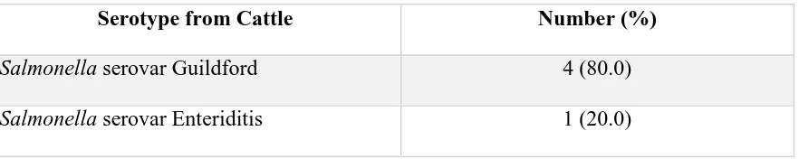
Table 3.2 Salmonella serotype distribution among cattle isolates (N=5)……….…51
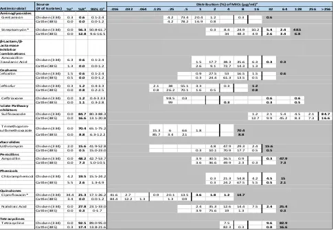
Table 3.3 Distribution of MICs and Resistance by Animal Source among E. coli, 2016………52
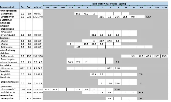
Table 3.4 Distribution of MICs and Resistance of Chicken* among Salmonella, 2016……...53
Table 3.5 MDR E. coli from cattle and chicken, 2016……….54
Table 3.6 MDR Salmonella from cattle and chicken, 2016………...54
Table 3.7 Top 10 Resistance patterns of E. coli from cattle (N=385) ……….55
Table 3.8 Top 10 Resistance patterns of E. coli from chicken (N=334)………..55
Table 3.9 Top Resistance patterns of Salmonella from chicken (N=51)……….56
Table 4.1 Prevalence of plasmid replicon types (%) from chicken and cattle Salmonella Isolates (n=56)…..………70
Table 4.2 Antimicrobial resistance profiles and associated Inc replicon types of Salmonella from chicken and cattle (N=56)………...……….70
viii
List of Figures
Figure 2.1 Land Use in Uganda (2012)………....23
Figure 2.2 Livestock Numbers in Uganda (2012-2014)………...24
Figure 2.3 Cattle Distribution in Uganda……….24
Figure 4.1 PFGE Dendrogram of chicken and cattle (N=56)……….…..71
Figure 5.1 Summary of phenotypic and genotypic characteristics of ESBL E. coli isolates from Cattle and Chicken Farms in the Wakiso District of Uganda...……….86
2 Introduction
The antimicrobial surveillance programs which have been sustained over a period of time
are important as they provide data which illustrate changes in antimicrobial resistance (AMR)
and permit trend analysis to be conducted; they are also useful as they may help identify newly
emerging or re-emerging resistant pathogens (Masterton, 2008). Examples of emerging
pathogens include Shiga.toxin Escherichia coli (Griffin & Karmali, 2017) and Salmonella
4,[5],12:i:- (Arnott et al., 2018). A surveillance system can also aid in informing possible threats
and illness burdens with populations by detecting various shifts in susceptibility in organisms
(Bax et al., 2001; A. P. Johnson, 2015). When using surveillance data, it may also be possible to
develop mitigation strategies to control AMR. Not only does surveillance provide invaluable
information on resistance, but it also provides information on pathogen incidence and prevalence
(Masterton, 2008). Use and in particular misuse of antimicrobials are just some contributing
factors for resistant to develop (Bronzwaer et al., 2002). Monitoring the usage of antimicrobial
drugs can be one means to help control the increasing problem of AMR (Bronzwaer et al., 2002).
Although surveillance programs seem like the ideal way to manage AMR, many factors must be
considered before implementing a surveillance program. Factors include the populations to be
studied, program funding, political and economic public policies, sampling methods and
associated costs, the organisms to be studied, the methodology of susceptibility testing, and
dissemination of results (Bax et al., 2001).
When assessing surveillance data available on resistance in the food chain and
food-producing animals, major gaps in data are seen. Data sharing is one aspect that can impact
analytics of surveillance data which in turn can impact the health and well being of animals and
3 program to compare data from both humans and animals, which includes data sharing, will be an
important tool in combatting the AMR problem (WHO, 2014).
The ecosystem of AMR involves many components including food animals and
associated meatstuffs, the environment, and the human population; this makes AMR a critical
and relevant One Health and One World Issue (Robinson et al., 2016). In this study, we evaluate
a process for a laboratory in Uganda to develop a framework to implement an AMR surveillance
program successfully. The objectives of this project are to:
• Evaluate laboratory capacity and provide recommendations for the development and implementation of an AMR surveillance system in food animals at the University of
Makerere in Kampala, Uganda
• Conduct a pilot study examining the prevalence and phenotype of Salmonella and E. coli isolated from cattle and chicken farms in the Wakiso district of Uganda over a
one year period
• Genotypically characterize Salmonella and E. coli isolated from cattle and chicken farms in the Wakiso district of Uganda
The overall goal is to assist in the implementation of antimicrobial resistance surveillance
programs in developing countries, such as Uganda, which will encourage successful, sustainable
and harmonized programs that will result in international integrated data systems to monitor
5 Literature Review
Antimicrobials and Development of Resistance
Antimicrobials
The terms ‘antibiotic’ and ‘antimicrobial’ are often used interchangeably in the literature.
However, between the two there are important differences. Antibiotics are naturally produced
from molds or bacteria. Antimicrobials, however, can be chemically synthesized (Quinn,
Markey, Carter, Donnelly, & Leonard, 2002; Salyers & Whitt, 1994). In addition to killing
(bactericidal) or inhibiting (bacteriostatic) fungi and bacteria, (Quinn et al., 2002; Salyers &
Whitt, 1994) they can also kill some viruses particularly when chemical sanitizers are used. For
this thesis, the term ‘antimicrobial’ is used.
Alexander Fleming reported the first antibiotic, penicillin, a compound naturally
produced from a mold. Florey and Chain later purified penicillin for clinical use (Quinn et al.,
2002). Interestingly, most antibiotics used in human medicine occur naturally. Chemically
synthesized antimicrobials include sulfonamide drugs, which were discovered in the 1930’s,
quinolones in the 1960’s, and oxazolidinone in the 2000’s (Walsh, 2003).
Since their discovery, antimicrobials were/are categorized as either broad or narrow
spectrum antimicrobials. They can also be categorized further into classification schemes such
as penicillins, macrolides, cephalosporins, and fluoroquinolones (B. Berger-Bachi, 2002; Salyers
& Whitt, 1994). Broad-spectrum antimicrobials work on Gram-negative and Gram-positive
bacteria; Gram-positive bacteria are typically more sensitive to their action. While
broad-spectrum antimicrobials can kill pathogens, they can also disrupt resident microflora resulting in
6 antimicrobials are typically directed toward a target bacterium and minimize disturbance against
the microflora of the body (Yao et al., 2016).
Antimicrobials are sorted into classes based on their mechanism of action against
bacteria. Antimicrobial classes include β-Lactams, glycopeptides, aminoglycosides,
tetracyclines, macrolides and lincosamides, quinolones, and trimethoprim and sulfonamides (B.
Berger-Bachi, 2002; Salyers & Whitt, 1994).
Beta-Lactams are bactericidal antimicrobials that inhibit cell wall synthesis. They consist
of penicillins, cephalosporins, carbapenems, and monobactams. All contain a β-Lactam ring and
inhibit the last step in peptidoglycan synthesis of microorganisms. They account for
approximately one.half of all antimicrobials used, considered broad spectrum, and are effective
against Gram-negative and Gram-positive bacteria (B. Berger-Bachi, 2002; Salyers & Whitt,
1994). Cephalosporins are traditionally divided into four generations which have
pharmacokinetic differences. The first generation is known to be more effective against
Gram-positive organisms, while the second generation is more effective against Gram-negative
organisms. Third generation cephalosporins are effective against Gram-negative, but not effective against β-Lactamase enzymes in Gram-negative organisms. Fourth generation act as
the same as the third generation, but are also effective against Gram-positive organisms and are more stable against β-Lactamase enzymes (Scholar, 2007).
Glycopeptides are antimicrobials that inhibit peptidoglycan synthesis and include
vancomycin and teicoplanin. Their primary targets are Gram-positive bacteria including MRSA
(B. Berger-Bachi, 2002; Salyers & Whitt, 1994). Vancomycin is very effective against
life.threatening Gram-positive bacteria, where others are less toxic and not effective, for
7 Aminoglycosides are produced by strains of Streptomyces, Micromonospora, and
Bacillus species and account for about three percent of all antimicrobials used (Quinn et al.,
2002; Salyers & Whitt, 1994). Common aminoglycosides include kanamycin, gentamicin,
streptomycin, and neomycin (Salyers & Whitt, 1994). These antimicrobials inhibit protein
synthesis by preventing the 30S subunit of the bacterial ribosome from binding to the 50S
subunit (Quinn et al., 2002; Salyers & Whitt, 1994) They are active against Gram-negative
bacteria and are typically bacteriocidal. Aminoglycosides are usually held in reserve until
treatment failure of other antimicrobials occurs (Salyers & Whitt, 1994).
Tetracyclines are broad-spectrum antimicrobials that inhibit the 30S subunit of bacterial
ribosomes by distorting the tRNA’s A site where it cannot align with the mRNA codon. They are
typically derived from individual tetracyclines include chlortetracycline and oxytetracycline,
commonly used in animal production to promote growth. Tetracyclines are bacteriostatic and
most often used in human medicine to treat acne and Lyme disease (Salyers & Whitt, 1994).
Macrolides include erythromycin, oleandomycin, spiramycin, and tylosin and account for
11% of antimicrobials used. Erythromycin is often used for treatment in people with allergies to
penicillin. They prevent the elongation of the 50S subunit and the translocation of the ribosome
and are derived from a strain of actinomycetes. Macrolides are bacteriostatic and bacteriocidal
against Gram-positive bacteria (B. Berger-Bachi, 2002; Salyers & Whitt, 1994).
Quinolones are bactericidal antimicrobials that include ciprofloxacin, nalidixic acid,
enrofloxacin and all newer fluoroquinolones. Quinolones inhibit nucleic acid synthesis by inhibiting the β subunit of the DNA gyrase from supercoiling in DNA replication (B.
Berger-Bachi, 2002; Quinn et al., 2002; Salyers & Whitt, 1994). They also inhibit topoisomerase IV,
8 completely degrade when excreted and may affect the development of and/or maintenance of
resistance in farm environments, particularly in poultry (Mandell & Tillotson, 2002)
Trimethoprim and sulfonamides act as competitors to bacteria and prevent the production
of tetrahydrofolic acid (THF) (B. Berger-Bachi, 2002; Quinn et al., 2002; Salyers & Whitt,
1994). Sulfonamides inhibit dihydrofolic acid synthesis by binding to dihydropteroate
synthetase, while trimethoprim inhibits THF by binding to dihydrofolate reductase (Gleckman,
Blagg, & Joubert, 1981; Kalkut, 1998).
Sometimes antimicrobials are not effective enough to treat alone, especially against
multi.drug-resistant bacteria. The lack of efficacy is mainly due to misuse of antimicrobials
leading to resistance. Therefore, antimicrobials are potentiated to increase the efficacy (Corbett
et al., 2017; Hare, 1960). Examples of these antimicrobials are augmentin, which combines
amoxicillin (β-Lactam antibiotic) with clavulanic acid (β-Lactamase inhibitor). Together they
inhibit cell wall biosynthesis (Worthington & Melander, 2013).
As previously mentioned, bacteria have developed resistance against antimicrobials
throughout the years. Examples of resistance mechanisms include the absence of the structure
for antimicrobials to inhibit (i.e. cell walls), microorganisms that are impermeable to
antimicrobials, microorganisms that inactivate the antimicrobial, modification of the
antimicrobial’s target, and utilizing an efflux pump to expel the antimicrobial (B. Berger-Bachi,
2002; Salyers & Whitt, 1994). Bacteria have a way of resisting the effects of the antimicrobials
found within all antimicrobial classes. For example, bacteria can carry a β-Lactamase enzyme which cleaves the β-Lactam ring causing inactivation of the antimicrobial (Berger-Bachi, 2002;
Shaikh, Fatima, Shakil, Rizvi, & Kamal, 2015). Penicillin.binding proteins can also be altered
9 continue (Georgopapadakou, 1993). Microorganisms harbor genes that do not allow binding of
glycopeptides which allows transpeptidase to continue (Pootoolal, Neu, & Wright, 2002).
Cytoplasm proteins, enzymatic inactivation, and efflux proteins are used against tetracyclines
(Speer, Shoemaker, & Salyers, 1992) and bacteria can methylate the 23S subunit on the rRNA
inhibiting binding of macrolides and lincosamides (Nakajima, 1999). Bacteria also have point
mutations to alter the affinity of gyrase, which confers resistance to quinolones (Hooper &
Jacoby, 2015). Point mutations are also commonly found which affects the mode of action of
trimethoprim and the sulfonamides (Skold, 2000).
Infections associated with antimicrobial resistance (AMR) have been increasing yearly,
and resistant infections are predicted as the cause of over 10 million deaths per year by 2050
with an associated cost over $100 trillion (Mckenna, 2014). In the last 30 years, there have not
been any new antimicrobials developed to treat infectious disease until recently. Teixobactin is a
recently marketed antimicrobial used to inhibit the cell wall synthesis of Gram-positive bacteria
(Ling et al., 2015).
Methodology for Determining Antimicrobial Resistance
Antimicrobial resistance testing methods have evolved particularly as technology has
improved, and automation occurred. One early testing method still in use in many developing
countries is disk diffusion, also known as Kirby Bauer testing. Disk diffusion includes
dissolving antimicrobials in a liquid or agar medium then soaking paper disks with the solution
before placement on top of a bacterial lawn (Jorgensen & Ferraro, 2009; Schoenknecht, 1973).
A clear zone, called the “Zone of Inhibition,” will occur following incubation and growth of the
agar plate; this represents the diffusion and effect of the drug. The zone is related to the degree
10 larger the clear area will be (Axelson, 2002; Clinical and Laboratory Standards Institute, 2009).
In contrast, resistance to the antimicrobial is recorded when bacterial growth occurs at the edge
of the antimicrobial disk or the clear zone is below a certain millimeter cut-off.
Another testing method includes determination of the minimal inhibitory concentration
(MIC), which gives the highest dilution to inhibit growth (Axelson, 2002; Clinical and
Laboratory Standards Institute, 2009). The early testing methodology included 2-fold serial
dilutions of the antimicrobial in test tubes followed by inoculation with a standard amount of
bacteria (typically calibrated by nephelometer). The tubes were incubated overnight, and the
concentration of the antimicrobial in the last tube inhibiting growth is considered the MIC. New
methods include the use of custom-made 96-well panels with different concentrations of
antimicrobials. Microorganisms are standardized using a McFarland standard in broth followed
by standardized inoculation into the panel. Incubation occurs for 18-24 hours an automated,
calibrated reader will determine the MIC level of resistance (Thermofisher Scientific).
Conversely, the panel can also be manually read and results recorded. The most recent means of
determining resistance is to determine the presence of antimicrobial resistance genes using
next-generation sequencing (Koser, Ellington, & Peacock, 2014). Having the bacterial genome
sequenced allows AMR determinants to be identified within its genome. Concordance between
the resistance phenotype using susceptibility testing methodology compared to sequencing has
been reported (McDermott et al., 2016).
Plasmids
Resistance genes are often located within the chromosome or on a plasmid which can be
transferred between bacteria. Plasmids contain genes which confer resistance to antimicrobials
11 microorganism (Stone, 1975). For example, with aminoglycosides, genes within the R plasmids
can phosphorylate or acetylate the drugs resulting in a lack of efficacy. In penicillins, genes on
R plasmids enable the split of the β-Lactam ring. Each gene in an R plasmid is specific for
rendering a particular antimicrobial ineffective (Salyers & Whitt, 1994).
No one factor in human treatment or animal production has been identified which can
alter or prevent the development of resistance once an antimicrobial has been used. Using
antimicrobials typically results in the development of a resistant population of bacteria. Human
and animals are reliant on the immune system to clear infectious bacteria. However, if a
population of resistant bacteria is retained, they can re-emerge in the presence of low doses of
antimicrobials or when antimicrobials are used for treatment. In humans, a weakened immune
state may cause resistant bacteria to proliferate compounding treatment options. Some animal
husbandry practices may affect the likelihood that resistance will develop or persist. Animal
husbandry can contribute to resistance due to the overcrowding (stress) and poor hygiene of the
animals (Watts & Lindeman, 2006). Preventing antimicrobial resistance can include combining
therapy, when two antimicrobials that are unrelated are used in treatment (Salyers & Whitt,
1994), discontinuing antimicrobials as growth promoters and educating the farmers about the
advantages of efficacious vaccines or other immunotherapies (Emborg, Ersboll, Heuer, &
Wegener, 2001).
Gram-Negative Bacteria
Salmonella
Salmonella, named by the veterinarian and bacteriologist Daniel Salmon is a
12 forming bacillus in the family of Enterobacteriaceae (Cima G, 2013; Guthrie, 1991). There is a
90% molecular homology between Salmonella and E. coli (Salyers & Whitt, 1994). Most of the
time, it is hard to distinguish between Salmonella and E.coli microscopically; on some media,
they also exhibit similar morphologies (Jay, Davos, Dundas, Frankish, & Lightfoot, 2003).
Phenotypically, they appear as raised colonies on agar and Salmonella are about 2.4 mm in
diameter with round and smooth edges (Gast, Porter, & Hold, 1997).
Salmonella growth requirements include a pH ranging from 4-9 in temperatures ranging
from eight to 45°C on various media (Gast et al., 1997; Jay et al., 2003). The recommended
temperature for growth is 37°C (Gast et al., 1997; Guthrie, 1991; Jay et al., 2003). Salmonella is
typically cultured from feces. However, since it is ubiquitous, it has also been recovered from
environmental sources including sewage, feed, and water (Guthrie, 1991). Recovery from
septicemia via blood occurs less often (Guthrie, 1991).
Enrichment, used to increase the number of Salmonella cells, is widely advocated and
various media are available for use. Selective enrichment is preferred to block the growth of
unwanted bacterial species (Gast et al., 1997). Typical enrichment broths that are used for
culture of salmonellae include Gram-negative Hajna (GN), tetrathionate (TT) (with or without
supplement), and Rappaport-Vassiliadis (RV) Broths (Fedorka-Cray, Bush, Thomas, Gray, &
McKean, 1996; Jay et al., 2003). GN, RV, and TT are recommended at incubation temperatures
of 37°C for 18 to 48 hours (Fedorka-Cray et al., 1996). Agar medium, used to visualize the
growth of salmonellae, include Brilliant green (BG), Xylose-Lysine-Tergitol 4 (XLT.4), Lysine
Iron (LIA), and Triple Sugar Iron Agar (TSI) (Fedorka-Cray et al., 1996). Agar media typically
relies on a chemical reaction to components within the media, such as a black appearance on
13
Salmonella can be destroyed by heating to temperatures above 70°C or irradiation (Gast
et al., 1997; Guthrie, 1991). However, heat tolerance has been reported and may affect killing
(Shachar & Yaron, 2006). Disinfectants such as hydrogen peroxide (Hebrard, Viala, Meresse,
Barras, & Aussel, 2009), chlorine (H. Wang & Ryser, 2014), and trisodium phosphate are very
effective against Salmonella (Sarjit & Dykes, 2015).
Salmonella is characterized by three antigens; the O (liposaccharide layer), H (flagellar
antigen), and Vi (virulence antigen) and over 2500 serovars, typically named after the
geographic location from where it was recovered, have been reported (Gast et al., 1997;
Giannella, 1996; Jay et al., 2003).
Serotypes can be further broken down into two species, Salmonella enterica which is
further divided into six subspecies and Salmonella bongori (Popoff & Le Minor, 1997). Within
these subspecies are many serotypes, some that are host specific. Host specific examples include
Salmonella enterica serovar Typhi and Salmonella enterica serovar Paratyphi in humans,
Salmonella enterica serovar Pullorum and Salmonella enterica serovar Gallinarum in poultry,
Salmonella enterica serovar Dublin in cattle, and Salmonella enterica serovar Cholereasuis in
pigs. The most common serotypes worldwide originating in both humans and animals include
Salmonella enterica serovar Enteritidis and Salmonella enterica serovar Typhimurium (WHO,
2016).
Virulence Factorsenhance the ability of microorganisms to cause disease. These factors include
hydrolytic enzymes, bacterial cell proteins and carbohydrates that protect them from killing by
the host, surface proteins that enable bacteria attachment, and toxins (L. Chen et al., 2005).
Salmonella has several virulence factors that allow it to invade the host cell. One type of factor
14 proteins through a needle-like structure into the host cell. Other virulence factors include
adhesion factors which allow better attachment to the membrane of the intestine. Pathogenicity
islands are factors that have specific phases in the development of infection. Two of these
phases include SP1 which allows invasion of the epithelial cell and SP2 which mediates
macrophage survival (Groisman & Ochman, 1996). Another virulence factor of Salmonella is
the lipopolysaccharide (LPS) that contains lipid A endotoxin that triggers inflammatory
mediators to induce degranulation (Rosenberger, Scott, Gold, Hancock, & Finlay, 2000).
Diseaseresultingfrom infection with Salmonella is usually called salmonellosis (WHO, 2016).
Animals can be a perfect host in which bacteria can survive and facilitate the transfer of bacteria
from animals to humans, human to human, and animal to animal (Clarke & Gyles, 1986). This
transmission between animals and humans make Salmonella a zoonotic pathogen. This transfer
can occur from eating contaminated foodstuffs, direct contact, water or aerosolization. Most
human infections result from consuming contaminated foods (Fedorka-Cray et al., 1996; Guthrie,
1991). Infections begin in the mucous membranes including the mouth, urinary, respiratory, and
gastrointestinal sites. Within the gastrointestinal tract, the stomach harbors the fewest colonies
of bacteria due to its low pH, whereas the colon harbors a larger number of bacteria. Disease as
a result of Salmonella infection in humans are typically gastroenteritis; however, infection with
certain serovars results in typhoid or enteric fever (Madigan, Martinko, & Parker, 1999).
Symptoms of gastroenteritis include nausea, fever, abdominal pain, diarrhea, depression,
anorexia, and pneumonia after 12 to 14 hours of ingestion of foods containing an infectious dose
of Salmonella. Symptoms are typically self-limiting and last for two to seven days. The elderly,
young and immunocompromised persons are at the highest risk for severe infection (WHO,
15 imbalance which may result in death (Clarke & Gyles, 1986). After the infection has resolved,
the person can become an asymptomatic carrier of Salmonella (Guthrie, 1991).
Escherichia coli
Theodore Escherich, a German bacteriologist, discovered the Escherichia coli (E. coli)
bacterium in 1885, from the colon of a human (Shulman, Friedmann, & Sims, 2007). E. coli is a
species within the coliform group along with Klebsiella, Enterobacter, and Citrobacter. These
coliforms are Gram-negative, rod-shaped, facultatively anaerobic bacteria (Singleton, 1999).
Escherichia coli often resides in the intestines of warm-blooded organisms and are shed through
fecal matter (Russell & Jarvis, 2001; Singleton, 1999).
Growth conditions for E. coli include incubation temperatures ranging from 36°C to
49°C. Enrichment broths, such as lysogeny broth (LB), and bacterial media that contain
ingredients such as ammonium phosphate, dibasic acid, glucose, magnesium sulfate, monobasic
acid, potassium phosphate, sodium chloride, and water are used to culture E. coli (Ingledew &
Poole, 1984). A common media broth for the growth of E. coli is LB, Tryptone, Terrific broths
(Lessard, 2013). Escherichia coli can be killed by the same means as Salmonella including high
temperatures and use of strong disinfectants.
Escherichia coli is categorized by serotype based on their antigenic formula, which
includes the O antigen (lipopolysaccharide layer), the K antigen (capsule) and the O antigen
(flagellin) (Brenner, Krieg, & Staley, 2005; L. Wang, Rothemund, Curd, & Reeves, 2003). These
are few virulence determinants harbored by some pathogenic E. coli.
Virulence properties and serological characteristics aids in classifying E. coli. These features
16 host cell, and invades the host cell. Based on these characteristics, the bacteria is virotyped.
Examples of E. coli virotypes include the following Enterotoxigenic E. coli (ETEC),
Enteropathogenic E. coli (EPEC), and Enterohemorrhagic E. coli (EHEC) cause diarrhea in
animals and humans. Enteroinvasive E. coli (EIEC), Enteroaggregative E. coli (EAEC), and
Adherent. Invasive E. coli (AIEC) cause diarrhea only in humans (Salyers & Whitt, 1994).
Other virulent strains of E. coli include Shiga-toxin producing E.coli (STEC), otherwise known
as E. coli O157: H7, urinary tract E.coli (UTEC), and neonatal meningitis E. coli (NMEC)
(Todar, 2008). The most common virulence factors known to assist in invading host cells
include toxins, pili, and fimbriae (Salyers & Whitt, 1994).
Diseases of E. coli include gastroenteritis, urinary tract infections, and neonatal meningitis.
Virulent strains can cause septicemia, diarrhea, hemolytic-uremic syndrome, and pneumonia
(Todar, 2008). Disease typically require treatment with therapeutics or supportive care.
Surveillance Programs
With the background information presented in the previous sections for AMR and
pathogenic Gram-negative bacteria such as Salmonella and E. coli, programs to monitor
antimicrobial use and susceptibility of bacteria to commonly used antimicrobials in humans and
animals have been implemented worldwide to the emerging and increasing resistance issue.
These programs are most often denoted as surveillance systems.
The medical definition of surveillance is a “close and continuous observation or
testing”(Webster Dictionary). The Center for Disease and Prevention (CDC) definition of
surveillance is “Epidemiologic surveillance is the ongoing and systematic collection, analysis,
17 event”(CDC, 1988)
.
It involves collecting and analyzing data to report any threats related todisease and the health of the public (A. P. Johnson, 2015). There are many programs which
monitor disease risk on a large scale and other programs that focus on specific disease threats
around the world (Bax et al., 2001). In the last 30 years, a limited number of country
surveillance programs have been implemented to monitor threats against antimicrobial
resistance. Surveillance programs that currently exist which focus on antimicrobial resistance
include DANMAP (Denmark), NARMS (United States), CIPARS (Canada), GERM.VET
(Germany), JVARM (Japan), NORM/NORM.VET (Norway), SWEDRES (Sweden),
NETHMAP/MARAN (Netherlands), ONERBA (France), and FINRES_VET (Finland) (A. P.
Johnson, 2015; WHO, 2014). To properly make informed decisions regarding treatment of
these infections, knowledge of resistance among these pathogens, which is ascertained over time
through data collected in these surveillance systems, is critical for the formulation of treatment
plans (A. P. Johnson, 2015).
Implementing a surveillance system for antimicrobial resistance requires a number of
considerations. One major question is – “What is the purpose of the surveillance?’. This drives
the type of information collected by clinicians, researchers, drug formularies, and policy
regulators. These data must be available for use within all boundaries including local, national,
and global levels (Bax et al., 2001).
Roadblocks will occur throughout that the process of implementing a surveillance
program. It is important to design the study correctly from inception. For example, will the
study include disease-based research or just the organism that causes the disease? Choosing the
right population to sample is also important. Once these are known, next steps are to choose the
18 results will be handled, what statistical programs will be used, and how the study will be funded
(Bax et al., 2001). Communicating the results is the final step which includes disseminating
findings to policymakers and the public.
Another question to answer after the surveillance has been implemented for a period of
time is whether the program has achieved its goal(s). For example, is there a better control for
resistance or antimicrobial to use in the testing platform?
Other concerns in implementing surveillance programs that have to be taken into account
or sampling biases in AMR surveillance. The definition of biases is “systematic errors that may
occur in collecting or interpreting data.” In AMR surveillance six biases have to be taken into
consideration when implementing, and they include the following: denominator data, case
definition, case ascertainment, sampling biases, multiple counting, and lab practices and disease
(Rempel & Laupland, 2009).
Program Evaluation
The best means for evaluation is to have program evaluations performed by an outside
organization. The definition of program evaluation is “the systematic application of scientific
methods to assess the design, implementation, improvement or outcomes of a program” (Rossi &
H.E., 1993). There are many purposes for conducting an evaluation. A list of a few is as
follows: (Short, Hennessy, & Campbell, 1996).
• “Demonstrate program effectiveness to funders
• Improve the implementation and effectiveness of programs
• Better manage limited resources
• Document program accomplishments
• Justify current program funding
19
• Satisfy ethical responsibility to clients to demonstrate positive and negative effects of program participation
• Document program development and activities to help ensure successful replication.” After the program evaluation is completed, there will be recommendations for those leading the
surveillance program related to concerns, need for change, or recommendation for improvement.
These recommendations benefit the organization and allow them to improve or achieve success
and maintain a sustainable program (CDC, 1988).
Public Health Impact of Surveillance Systems for Antimicrobial Resistance
The World Health Organization (WHO) has reported that the rates of bacterial resistance
are increasing and causing community.acquired and health-care associated resistant infections.
They noted that there is also a lack of coordination, data sharing, and standardized methods
leaving significant gaps in surveillance information, globally (WHO, 2014). The World Health
Assembly initiated the Global Action Plan (GAP) in 2014, to ensure effective treatment and
prevent infectious diseases, by using medications correctly. Since the inception of the GAP,
countries were encouraged to implement their national action plan according to GAP guidelines,
which includes combating AMR (WHO, 2015a).From here, the Global Antimicrobial Resistance
Surveillance System (GLASS), was implemented by WHO, to foster AMR data collection and to report
globally (WHO, 2017a). As mentioned in previous sections, AMR surveillance is critical for
public health and for generating mitigation and control approaches to combat AMR. Through
actions of implementing appropriate drug-use policies, improved diagnostics practices, reduced
rates of infection transmission, and preventing and controlling AMR in agriculture public health
can be improved (CDC, 2012; Masterton, 2008).
The WHO has been working diligently with the Food and Agriculture Organization
20 AMR problem through a multidisciplinary and intersectoral collaboration at the global level. In
2008, the WHO Advisory Group on Integrated Surveillance of Antimicrobial Resistance
(WHO.AGISAR) was formed to support WHO in lessening the impact of resistance attributed to
antimicrobial use in food-producing animals. AGISAR is also responsible for continuously
updating the Critically Important Antimicrobials list. These collaborations help gather data and
information to support policies at the national and global levels to develop good practices for
animal husbandry (WHO, 2014). AGISAR provides grant funding to selected developing
countries to support focused research projects and country pilot projects related to AMR.
Uganda is one of several countries that received funding for a country research project, which
will be discussed in the next section.
Uganda Demographics
Uganda is situated on the east side of Africa, right on the equator, bordering Kenya,
Rwanda, South Sudan, Democratic Republic of Congo, and Tanzania. Uganda is comprised of
111 districts and the capital city, Kampala. As of today, the population is 41.5 million, of which
49.9% is male, and 50.0% is female. Uganda has a multi-party political system and has been led
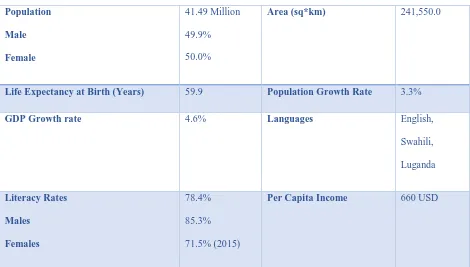
by President Yoweri K. Museveni, since 1986. Table 2.1 highlights the demographics of Uganda
21 Table 2.1: Demographics of Uganda
Population
Male
Female
41.49 Million 49.9% 50.0%
Area (sq*km) 241,550.0
Life Expectancy at Birth (Years) 59.9 Population Growth Rate 3.3%
GDP Growth rate 4.6% Languages English,
Swahili, Luganda
Literacy Rates
Males
Females
78.4% 85.3% 71.5% (2015)
Per Capita Income 660 USD
Source: (http://databank.worldbank.org/data/reports.aspx?source=2&country=UGA). Population Health Status
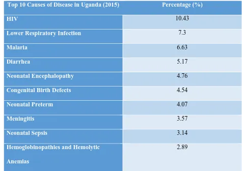
There are many health.related infections and diseases affecting the health of the Ugandan
population. From the Global Burden of Disease (GDB) Compare, a tree map of the world’s
health levels and trends from 1990-2015, the top 10 death related causes in Uganda are listed in
22 Table 2.2: Top 10 Causes of Death in Uganda (2015)
Top 10 Causes of Disease in Uganda (2015) Percentage (%)
HIV 10.43
Lower Respiratory Infection 7.3
Malaria 6.63
Diarrhea 5.17
Neonatal Encephalopathy 4.76
Congenital Birth Defects 4.54
Neonatal Preterm 4.07
Meningitis 3.57
Neonatal Sepsis 3.14
Hemoglobinopathies and Hemolytic
Anemias
2.89
Source:http://vizhub.healthdata.org/gbd.compare/
Agriculture Demographics
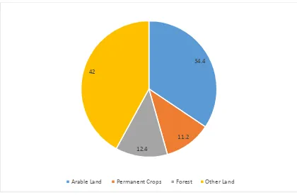
Uganda is approximately 19,997.38 hectares (ha). Figure 2.1 (FAO, 2015) shows the
distribution of land use throughout the country. As shown, arable land (land used for growing
23 Figure 2.1: Land Use in Uganda (2012)
Source:http://uganda.opendataforafrica.org/gallery/agriculture
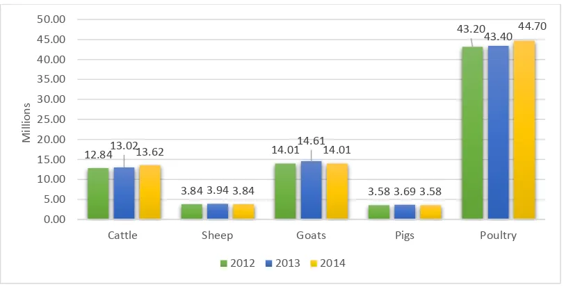
The primary food animal commodities in Uganda include cattle, sheep, goats, pigs, and
poultry. Figure 2.2 below shows the livestock numbers from 2012-2014. As a result of
exporting, sheep, goat, and pig numbers have decreased. Cattle and poultry demand in the
country has increased and seems to be continuing (Uganda Bureau of Statistics, 2015).
34.4
11.2 12.4
42
24 Figure 2.2: Livestock Numbers in Uganda (2012.2014)
Figure 2.3 displays the cattle production distributed throughout the country of Uganda
(Balikowa, 2011). Poultry production has not had a census since 1991, so there is a huge gap in
data to determine how many poultry producing farms are in Uganda (FAO, 2008).
Figure 2.3: Cattle Distribution in Uganda
12.84 3.84 14.01 3.58 43.20 13.02 3.94 14.61 3.69 43.40 13.62 3.84 14.01 3.58 44.70 0.00 5.00 10.00 15.00 20.00 25.00 30.00 35.00 40.00 45.00 50.00
Cattle Sheep Goats Pigs Poultry
M
ill
io
ns
25 Uganda Antimicrobial Use and Resistance
Antimicrobial Use: There are no available records in Uganda, regarding the quantities of imported
and exported antimicrobials. Existing reports indicate that many antimicrobials used in Uganda
were manufactured primarily in India. The antimicrobials used most often include ciprofloxacin
and amoxicillin (Foster, Sosa, Najjuka, & Mwenfa, 2011). Although the antimicrobials are being
imported, many believe that these drugs are of substandard quality (UNAS, CDDEP,
GARP-Uganda, Mpairwe, & Wamala, 2015). The most commonly used antimicrobials in veterinary
practice are tetracycline and penicillin (UNAS et al., 2015). As far as the quality of
antimicrobials, approximately 94% are acceptable but counterfeit drugs are encountered, and
companies from India have been banned from importing as a result. Due to limited resources,
The National Drug Administration (NDA) cannot control all the counterfeit drugs coming in
from Asia, Middle East, and Europe (UNAS et al., 2015).
The use of antimicrobials in agriculture, poultry, veterinary practice, milk and meat
products all have the same issues, which include inadequately trained staff, farm managers,
veterinary assistants, animal husbandry officers and veterinary practitioners. Use of
antimicrobials is often accomplished with little regard to proper evaluation and regulations for
treating animals. The Ministry of Agriculture (MOA) has the authority over antimicrobial use in
Uganda, but due to lack of resources and human power, there is limited control and enforcement
on how antimicrobials are being regulated. Because of the lack of regulation, farmers,
veterinarians, and managers take it upon themselves to diagnose and treat disease (UNAS et al.,
2015). Antimicrobials are accessible countrywide, often over-the-counter. Farm to table supervision that has not been implemented as recommended by the Food and Agriculture
26 are available to perform analysis, including antimicrobial susceptibility testing, on
microorganisms (UNAS et al., 2015).
Many modern testing platforms for conducting susceptibility testing are used by
economically advantaged countries. These include expensive, high throughput equipment such
as VITEK® (Biomeruix Inc) or Sensititre (Thermofisher Scientific). Inexpensive methods
including broth or agar dilution tests and disc diffusion are often used in resource-limited
countries like Uganda. Disk diffusion is routinely used and easy when performing quality
assurance. A set of standards for conducting disk diffusion as well guidelines provide
interpretive criteria for determining breakpoints of antimicrobials. North America uses the
Clinical Laboratory Standards Institute (CLSI) recommendation and guidelines for susceptibility
methodology; however, CLSI guidelines are not readily available to everyone because of costs
(Gelband, 2016). Other guidelines are available for free such as the European Committee on
Antimicrobial Susceptibility Testing (EUCAST) documents. However, both programs are not
identical, and CLSI is more widely used around the world (Gelband, 2016). To subside the costs
for countries with limited resources, WHONET was created as a free, user-friendly and
technically supported software program that includes all CLSI, EUCAST, and other interpretive
criteria needed to analyze susceptibility data (Gelband, 2016).
Uganda prescribers follow guidelines created by the Ministry of Health (MOH), Uganda
Clinical Guidelines. The MOH states “It is designed to provide updated, practical and
useful information for both upper and lower level health facilities on the diagnosis and
management of common conditions present in Uganda” (Ministry of Health, 2014). It was later
reported that these guidelines were never used because many questioned the source of
27 According to UNAS et al., three important factors are required for accelerating the
mitigation of antimicrobial resistance. These factors include economic power, knowledge, and
information. Uganda lacks economic power and consumer’s knowledge regarding many aspects
of antimicrobials including mode of action, misuse of over the counter drugs, and incorrect
self-medication (UNAS et al., 2015). Many healthcare workers do not have the knowledge or
information to provide proper health care to patients and animals. It was reported that almost all
(99%) of the healthcare workers did not know the guidelines related to prescription of effective
antimicrobials (Kamulegeya, William, & Rwenyonyi, 2011).
The MOH recognizes the need for surveillance for antimicrobial, antimalarial, and
antiretroviral resistance. They believe it will require minimizing importation of substandard
drugs, regulating use by prescribers, and self.medication controls (UNAS et al., 2015).
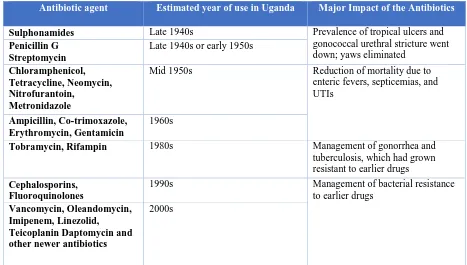
Antimicrobial Resistance:It is believed that the first antimicrobials introduced to Uganda were
sulphonimides. Table 2.3 was reported by UNAS et al. showing when antimicrobials were first
28 Table 2.3: Introduction of antimicrobials to Uganda
Antibiotic agent Estimated year of use in Uganda Major Impact of the Antibiotics
Sulphonamides Late 1940s Prevalence of tropical ulcers and
gonococcal urethral stricture went down; yaws eliminated
Penicillin G Streptomycin
Late 1940s or early 1950s
Chloramphenicol, Tetracycline, Neomycin, Nitrofurantoin,
Metronidazole
Mid 1950s Reduction of mortality due to enteric fevers, septicemias, and UTIs
Ampicillin, Co-trimoxazole, Erythromycin, Gentamicin
1960s
Tobramycin, Rifampin 1980s Management of gonorrhea and
tuberculosis, which had grown resistant to earlier drugs
Cephalosporins, Fluoroquinolones
1990s Management of bacterial resistance to earlier drugs
Vancomycin, Oleandomycin, Imipenem, Linezolid, Teicoplanin Daptomycin and other newer antibiotics
2000s
For Salmonella, several antimicrobial resistance studies utilizing human, cattle, and other
food samples were conducted from 1995 through 2014 (UNAS et al., 2015). It was reported that
resistance during that period was observed most often to co-trimoxazole, ampicillin, tetracycline,
and chloramphenicol. Resistance was observed in 50.85% of the isolates collected. About 20%
of the isolates were resistant to amikacin and ceftriaxone (UNAS et al., 2015). For E. coli,
studies show that there was a 40% resistance to the majority of the antimicrobials tested from
2007-2011(UNAS et al., 2015). Extended-beta lactamase (ESBL) testing has not been
conducted in Uganda. The prevalence from published studies ranged from 10.75% among
Gram-negative isolates. There is need surveillance on ESBL producing bacteria Uganda as it is
29 References
Acar, J. (1997). Broad. and narrow.spectrum antibiotics: an unhelpful categorization. Clin Microbiol Infect, 3(4), 395.396.
Arnott, A., Wang, Q., Bachmann, N., Sadsad, R., Biswas, C., Sotomayor, C., . . . Sintchenko, V. (2018). Multidrug.Resistant Salmonella enterica 4,[5],12:i:. Sequence Type 34, New South Wales, Australia, 2016.2017. Emerg Infect Dis, 24(4), 751.753. doi:
10.3201/eid2404.171619
Axelson, P. H. (2002). Essentials of Antimcrobial Pharmocology. Totowa, NJ: Humana Press. Balikowa, D. (2011). Dairy Development in Uganda: A Review of Uganda’s Dairy Industry. Bax, R., Bywater, R., Cornaglia, G., Goossens, H., Hunter, P., Isham, V., . . . White, A. (2001).
Surveillance of antimicrobial resistance..what, how and whither? Clin Microbiol Infect, 7(6), 316.325.
Berger.Bachi. (2002). Mechanisms of Resistance Against Different Antimicrobial Classes. Retrieved February 8, 2017, from
http://amrls.cvm.msu.edu/microbiology/bacterial.resistance.strategies/introduction/mecha nisms.of.resistance.against.different.antimicrobial.classes
Brenner, D. J., Krieg, N. R., & Staley, J. T. (2005). The Gammaproteobacteria (2 ed.). New York: Springer.
Bronzwaer, S. L., Cars, O., Buchholz, U., Molstad, S., Goettsch, W., Veldhuijzen, I. K., . . . Degener, J. E. (2002). A European study on the relationship between antimicrobial use and antimicrobial resistance. Emerg Infect Dis, 8(3), 278.282. doi:
10.3201/eid0803.010192
CDC. (1988). Guidelines for Evaluating Surveillance Systems. MMWR. Retrieved February 2, 2017, from https://www.cdc.gov/mmwr/preview/mmwrhtml/00001769.htm
CDC. (2012). A Public Health Action Plan to Combat Antimicrobial Resistance. Retrieved February 16, 2017, from https://www.cdc.gov/drugresistance/pdf/action.plan.2012.pdf Chen, L., Yang, J., Yu, J., Yao, Z., Sun, L., Shen, Y., & Jin, Q. (2005). VFDB: a reference
database for bacterial virulence factors. Nucleic Acids Res, 33(Database issue), D325.328. doi: 10.1093/nar/gki008
Cima G. (2013). Daniel E. Salmon helped reduce disease in animals and humans. JAVA news. Retrieved February 9, 2017, from
https://www.avma.org/News/JAVMANews/Pages/130301m.aspx
Clarke, R. C., & Gyles, C. L. (1986). Pathogenisis of Bacterial Infections in Animals. Ames, Iowa: Iowa State University Press.
Clinical and Laboratory Standards Institute. (2009). Performance standards for antimicrobial susceptibility testing Nineteenth informational supplement (pp. M100). PAClinical and Laboratory Standards Institute.
Corbett, D., Wise, A., Langley, T., Skinner, K., Trimby, E., Birchall, S., . . . Lister, T. (2017). Potentiation of Antibiotic Activity by a Novel Cationic Peptide: Potency and Spectrum of Activity of SPR741. Antimicrob Agents Chemother, 61(8). doi: 10.1128/aac.00200.17 Emborg, H., Ersboll, A. K., Heuer, O. E., & Wegener, H. C. (2001). The effect of discontinuing
the use of antimicrobial growth promoters on the productivity in the Danish broiler production. Prev Vet Med, 50(1.2), 53.70.
30 FAO. (2015). Land Use and Agriculture Inputs. Retrieved from:
http://uganda.opendataforafrica.org/dykiync/uganda.fao.stat.land.use.and.agricultural.inp uts
Fedorka.Cray, P. J., Bush, E., Thomas, L., Gray, J., & McKean, J. (1996). Salmonella Infection in Herds of Swine Swine Research Report.
Foster, S. D., Sosa, A., Najjuka, C., & Mwenfa, D. (2011). Drivers of antibiotic resistance in Uganda and Zambia. Paper presented at the Alliance for the Prudent Use of Antibiotics., Washington DC.
Gast, R. K., Porter, R. E., Jr., & Hold, P. S. (1997). Applying tests for specific yolk antibodies to predict contamination by Salmonella enteritidis in eggs from experimentally infected laying hens. Avian Dis, 41(1), 195.202.
Gelband, H. (2016). East Africa Public Health Laboratory Networking Project: CENTER FOR DISEASE DYNAMICS, ECONOMICS & POLICY.
Georgopapadakou, N. H. (1993). Penicillin.binding proteins and bacterial resistance to beta.lactams. Antimicrob Agents Chemother, 37(10), 2045.2053.
Giannella, R. A. (1996). Salmonella (4 ed.). Galveston, TX: University of Texas Medical Branch at Galveston.
Gleckman, R., Blagg, N., & Joubert, D. W. (1981). Trimethoprim: mechanisms of action, antimicrobial activity, bacterial resistance, pharmacokinetics, adverse reactions, and therapeutic indications. Pharmacotherapy, 1(1), 14.20.
Griffin, P. M., & Karmali, M. A. (2017). Emerging Public Health Challenges of Shiga Toxin– Producing Escherichia coli Related to Changes in the Pathogen, the Population, and the Environment. Clinical Infectious Diseases, 64(3), 371.376. doi: 10.1093/cid/ciw708 Groisman, E. A., & Ochman, H. (1996). Pathogenicity islands: bacterial evolution in quantum
leaps. Cell, 87(5), 791.794.
Guthrie, R. (1991). Salmonella (1 ed.): CRC Press. Hardy Diagnostics. (2017). XLT.4.
Hare, J. H. (1960). Antibiotic Potentiation . A Review. Can J Comp Med Vet Sci, 24(6), 171.176. Hebrard, M., Viala, J. P., Meresse, S., Barras, F., & Aussel, L. (2009). Redundant hydrogen
peroxide scavengers contribute to Salmonella virulence and oxidative stress resistance. J Bacteriol, 191(14), 4605.4614. doi: 10.1128/jb.00144.09
Hooper, D. C., & Jacoby, G. A. (2015). Mechanisms of drug resistance: quinolone resistance.
Ann N Y Acad Sci, 1354, 12.31. doi: 10.1111/nyas.12830
Ingledew, W. J., & Poole, R. K. (1984). The respiratory chains of Escherichia coli. Microbiol Rev, 48(3), 222.271.
Institute for Health Metrics and Evaluation. (2016). Uganda. Retrieved from: https://vizhub.healthdata.org/gbd.compare/
Jay, L. S., Davos, D., Dundas, M., Frankish, E., & Lightfoot, D. (2003). Salmonella (6 ed.). Sydney: Australian Institute of Food Science and Technology (NSW Branch).
Johnson, A. P. (2015). Surveillance of antibiotic resistance. Philos Trans R Soc Lond B Biol Sci, 370(1670), 20140080. doi: 10.1098/rstb.2014.0080
Jorgensen, J. H., & Ferraro, M. J. (2009). Antimicrobial susceptibility testing: a review of general principles and contemporary practices. Clin Infect Dis, 49(11), 1749.1755. doi: 10.1086/647952
31 Kamulegeya, A., William, B., & Rwenyonyi, C. M. (2011). Knowledge and Antibiotics
Prescription Pattern among Ugandan Oral Health Care Providers: A Cross.sectional Survey. J Dent Res Dent Clin Dent Prospects, 5(2), 61.66. doi: 10.5681/joddd.2011.013 Koser, C. U., Ellington, M. J., & Peacock, S. J. (2014). Whole.genome sequencing to control
antimicrobial resistance. Trends Genet, 30(9), 401.407. doi: 10.1016/j.tig.2014.07.003 Kuriyama, T., Karasawa, T., & Williams, D. W. (2014). Antimicrobial Chemotherapy:
Significance to Healthcare Biofilms in Infection Prevention and Control: Academic Press. Lessard, J. C. (2013). Growth media for E. coli. Methods Enzymol, 533, 181.189. doi:
10.1016/b978.0.12.420067.8.00011.8
Ling, L. L., Schneider, T., Peoples, A. J., Spoering, A. L., Engels, I., Conlon, B. P., . . . Lewis, K. (2015). A new antibiotic kills pathogens without detectable resistance. Nature, 517(7535), 455.459. doi: 10.1038/nature14098
Madigan, M. T., Martinko, J. M., & Parker, J. (1999). Brock's biology of microorganisms (9 ed.). Englewood Cliffs: Prentice Hall.
Mandell, L., & Tillotson, G. (2002). Safety of fluoroquinolones: An update. Can J Infect Dis, 13(1), 54.61.
Masterton, R. (2008). The importance and future of antimicrobial surveillance studies. Clin Infect Dis, 47 Suppl 1, S21.31. doi: 10.1086/590063
McDermott, P. F., Tyson, G. H., Kabera, C., Chen, Y., Li, C., Folster, J. P., . . . Zhao, S. (2016). Whole.Genome Sequencing for Detecting Antimicrobial Resistance in Nontyphoidal Salmonella. Antimicrob Agents Chemother, 60(9), 5515.5520. doi: 10.1128/aac.01030.16 Mckenna, M. (2014). The Coming Cost of Superbugs: 10 Million Deaths Per Year. Retrieved
April 2, 2017, from https://www.wired.com/2014/12/oneill.rpt.amr/ Ministry of Health. (2014). Uganda Clinical Guidelines. Kampala.
Nakajima, Y. (1999). Mechanisms of bacterial resistance to macrolide antibiotics. J Infect Chemother, 5(2), 61.74. doi: 10.1007/s101569900000
P.J. Fedorka.Cray, E. Bush, L. Thomas, J. Gray, & J. McKean. (1996). Salmonella Infection in Herds of Swine Swine Research Report.
Pootoolal, J., Neu, J., & Wright, G. D. (2002). Glycopeptide antibiotic resistance. Annu Rev Pharmacol Toxicol, 42, 381.408. doi: 10.1146/annurev.pharmtox.42.091601.142813 Popoff, M. Y., & Le Minor, L. (1997). Antigenic formulas of the Salmonella serovars. Paper
presented at the World Health Organization Collaborating Centre for Reference and Research on Salmonella, Paris, France.
Quinn, P., Markey, B., Carter, M., Donnelly, W., & Leonard, F. (2002). Antimicrobial Agents.
Veterinary Microbiology and Microbial Disease, 28.35.
Rempel, O. R., & Laupland, K. B. (2009). Surveillance for antimicrobial resistant organisms: potential sources and magnitude of bias. Epidemiol Infect, 137(12), 1665.1673. doi: 10.1017/s0950268809990100
Robinson, T. P., Bu, D. P., Carrique.Mas, J., Fevre, E. M., Gilbert, M., Grace, D., . . .
Woolhouse, M. E. (2016). Antibiotic resistance is the quintessential One Health issue.
Trans R Soc Trop Med Hyg, 110(7), 377.380. doi: 10.1093/trstmh/trw048 Rosenberger, C. M., Scott, M. G., Gold, M. R., Hancock, R. E., & Finlay, B. B. (2000).
Salmonella typhimurium infection and lipopolysaccharide stimulation induce similar changes in macrophage gene expression. J Immunol, 164(11), 5894.5904.
32 Russell, J. B., & Jarvis, G. N. (2001). Practical mechanisms for interrupting the oral.fecal
lifecycle of Escherichia coli. J Mol Microbiol Biotechnol, 3(2), 265.272.
Salyers, A. A., & Whitt, D. D. (1994). Bacterial Pathogenesis. Washington DC: ASM Press. Salyers and Whitte. (1994). Bacterial Pathogenisis. Washington, DC: ASM press.
Sarjit, A., & Dykes, G. A. (2015). Trisodium phosphate and sodium hypochlorite are more effective as antimicrobials against Campylobacter and Salmonella on duck as compared to chicken meat. Int J Food Microbiol, 203, 63.69. doi:
10.1016/j.ijfoodmicro.2015.02.026
Schoenknecht, F. D. (1973). The Kirby.Bauer technique in clinical medicine and its application to carbenicillin. J Infect Dis, 127, Suppl:111.115.
Scholar, E. (2007). Cephalosporins xPharm: The Comprehensive Pharmacology Reference. Shachar, D., & Yaron, S. (2006). Heat tolerance of Salmonella enterica serovars Agona,
Enteritidis, and Typhimurium in peanut butter. J Food Prot, 69(11), 2687.2691.
Shaikh, S., Fatima, J., Shakil, S., Rizvi, S. M., & Kamal, M. A. (2015). Antibiotic resistance and extended spectrum beta.lactamases: Types, epidemiology and treatment. Saudi J Biol Sci, 22(1), 90.101. doi: 10.1016/j.sjbs.2014.08.002
Short, L., Hennessy, M., & Campbell, J. (1996). Tracking the work. Family violence: Building a coordinated community response: A guide for communities.
Shulman, S. T., Friedmann, H. C., & Sims, R. H. (2007). Theodor Escherich: the first pediatric infectious diseases physician? Clin Infect Dis, 45(8), 1025.1029. doi: 10.1086/521946 Singleton, P. (1999). Bacteria in Biology, Biotechnology and Medicine (Wiley Ed. 5 ed.). Skold, O. (2000). Sulfonamide resistance: mechanisms and trends. Drug Resist Updat, 3(3),
155.160. doi: 10.1054/drup.2000.0146
Speer, B. S., Shoemaker, N. B., & Salyers, A. A. (1992). Bacterial resistance to tetracycline: mechanisms, transfer, and clinical significance. Clin Microbiol Rev, 5(4), 387.399. Stone, A. B. (1975). R factors: plasmids conferring resistance to antibacterial agents. Sci Prog,
62(245), 89.101.
Todar, K. (2008). Pathogenic E. coli. University of Wisconsin.Madison Department of Bacteriology.
Uganda Bureau of Statistics. (2015). 2015 Statistical Abstract. Retrieved February 20, 2017, from
http://www.ubos.org/onlinefiles/uploads/ubos/statistical_abstracts/Statistical%20Abstract %202015.pdf
UNAS, CDDEP, GARP.Uganda, Mpairwe, Y., & Wamala, S. (2015). Antibiotic Resistance in Uganda: Situation Analysis and Recommendations (pp. 107). Kampala, Uganda: Uganda National Academy of Sciences; Center for Disease Dynamics, Economics & Policy. Walsh, C. (2003). Antibiotics. Washington DC: ASM Press.
Wang, H., & Ryser, E. T. (2014). Efficacy of various sanitizers against Salmonella during simulated commercial packing of tomatoes. J Food Prot, 77(11), 1868.1875. doi: 10.4315/0362.028x.jfp.14.213
Wang, L., Rothemund, D., Curd, H., & Reeves, P. R. (2003). Species.wide variation in the Escherichia coli flagellin (H.antigen) gene. J Bacteriol, 185(9), 2936.2943.
Watts, J., & Lindeman, C. (2006). Antimicrobial susceptibility testing of bacteria of veterinary origin. Washington DC: ASM Press.
33 WHO. (2014). Antimicrobial resistance: global report on surveillance 2014 (pp. 257).
WHO. (2015). Global Action Plan for Antimicrobial Resistance: The World Health Organization.
WHO. (2016). Salmonella (non.typhoidal). Retrieved February 10, 2017, from http://www.who.int/mediacentre/factsheets/fs139/en/
WHO. (2017). Antimicrobial Resistance. Global Antimicrobial Resistance Surveillance System (GLASS). Retrieved February 16, 2017, from
http://www.who.int/antimicrobial.resistance/global.action.plan/surveillance/glass/en/ Worthington, R. J., & Melander, C. (2013). Combination approaches to combat
multidrug.resistant bacteria. Trends Biotechnol, 31(3), 177.184. doi: 10.1016/j.tibtech.2012.12.006
34
Chapter 3: Phenotypic Characterization of
Salmonella
and
E. coli
from
Cattle and Chicken Farms in the Wakiso District, Uganda
35
Phenotypic Characterization of
Salmonella
and
E. coli
from Cattle and
Chicken Farms in the Wakiso District, Uganda
Takiyah A. Ball, Paula J. Fedorka-Cray*, Maria Correa, Awa Aidara-Kane, Hongyu Ru, Siddhartha Thakur, Joy Horovitz, and Francis Ejobi
Abstract
Background: Antimicrobial resistance (AMR) is a global concern with over 10 million deaths and $10 trillion in costs estimated to occur annually by 2050. The development of AMR in food-borne pathogens is of particular concern as treatment options may be compromised. Surveillance programs are needed to monitor resistance trends over time and guide treatment options.
Objectives: Estimate the prevalence of Salmonella spp. and commensal E. coli on cattle and chicken farms in the Wakiso District of Uganda and determine AMR phenotype of recovered isolates. Determine if seasonal differences in prevalence are significant.
Methods: Forty chicken and dairy farms were visited each season (rainy and dry); ten fecal environmental samples were collected per farm. Samples were cultured using standard laboratory methods for Salmonella and E. coli. AMR profiles were determined using the SensitireTM system per manufacturer’s directions. Using a logistic regression model seasonal differences in prevalence were calculated.
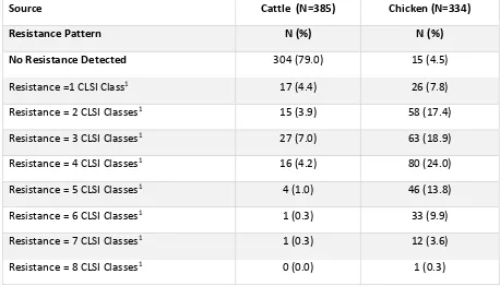
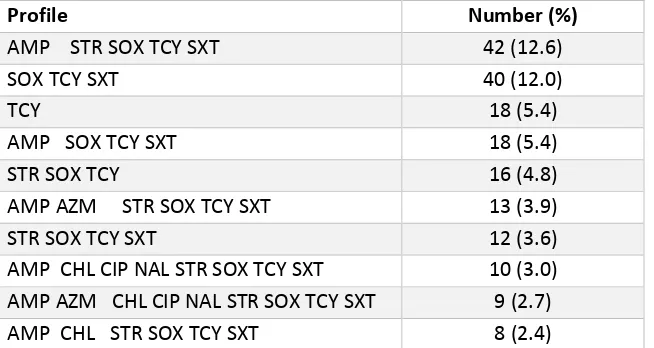
Results: Salmonella and E. coli were recovered from 379 and 400 chicken and cattle samples, respectively. From chicken, Salmonella and E. coli prevalence was 13.5% (N=51) and 88.1% (N= 334), respectively. From cattle, Salmonella and E. coli prevalence was 1.3% (N=5) and 96.3% (N=385), respectively. Salmonella Enteritidis (31.7%) and Kentucky (21.6%) were most often recovered on chicken farms. Collectively, Salmonella and E. coli were most often resistant totetracycline and sulfisoxazole. Eighty.nine percent and 23% of chicken and cattle isolates, respectively, were multi-drug resistant. All Salmonella Kentucky isolates were resistant to ciprofloxacin. ESBLs were detected in eight E. coli isolates. Resistance to the cephems, quinolones, and macrolides was observed among both organisms. A significant seasonal difference between chicken sampling periods was observed (p= 0.0017).
Conclusions: The emergence of MDR among both Salmonella and E. coli and the presentation of ESBLs among E. coli requires further investigation and monitoring over time. Further characterization of these isolates at the genotypic level is warranted.
36 Introduction
Salmonella is a food-borne pathogen and is one out of four leading causes of diarrheal
diseases worldwide. Salmonellosis has been linked to more than 33 million deaths annually.
Conversely, while most food-borne infections caused by Escherichia coli are less severe, some
strains are associated with severe disease (WHO, 2016).
In Uganda, there is no reporting of death related to food-borne diseases caused by
Salmonella spp. and commensal E. coli (UNAS et al., 2015). Unfortunately, most reports only
indicate numbers of diarrhea-related deaths of unknown etiology. Therefore, it is difficult to
estimate mortality linked to food-borne illnesses. Additionally, there is a lack of information on
antimicrobial resistant (AMR) food-borne pathogens and no coordinated AMR surveillance
system exists.
As a result of these information gaps, the World Health Organization (WHO) released a
call for research proposals in developing countries to collect data on the prevalence and AMR of
food-borne pathogens, including Salmonella and E. coli, in food animals, humans, and the
environment (WHO, 2015a). Antimicrobial resistance is a growing issue that can render current
treatment regimens ineffective resulting in higher health care costs for patients with resistant
infections. AMR is also associated with increased morbidity and mortality (WHO, 2017a). If no
changes or efforts are made to control AMR, by 2050, it is estimated that over 10 million deaths
per year will be related to AMR infections (Mckenna, 2014).
Antimicrobial resistance is common among many different bacterial genera that humans,
animals, and the environment harbor. Since it is difficult to separate the cause and effects of