Heterotopic accessory spleen with squamous epithelial cyst in
pancreas mimicking other pancreatic tumor: a case report
Jieli Luo1, Jianshe Chen1, Fengbo Huang2, Panpan Zhou3, Pintong Huang1
1Department of Ultrasound in Medicine, 2Department of Pathology, 3Department of Radiology, The Second Affiliated Hospital of Zhejiang University School of Medicine, Hangzhou 310000, China
Correspondence to: Pintong Huang, PhD. The Second Affiliated Hospital of Zhejiang University School of Medicine, No. 88 Jiefang Road, Hangzhou 310000, China. Email: huangpintong@zju.edu.cn.
Abstract: A 49-year-old female undergoing a periodic health examination at other hospital revealed a mass in the tail of pancreas. The patient denied any personal history of surgery except subtotal hysterectomy because of multiple myomas in uterus 7 years ago, family history of abdominal cancer and trauma. Physical examination and laboratory finding (including tumor marker) were unremarkable. Chest X-ray result was normal. Contrast enhanced ultrasound (CEUS) examination showed a well-defined hypoechoic pancreatic mass which was suggestive of solid pseudopapillary tumor. Contrast enhanced computed tomography (CE-CT) of the abdomen revealed a mass of hypodensity suggestive of intraductal papillary mucinous neoplasm. Because of the risk of bleeding and exclusion of surgical contraindications, patient underwent laparoscopic surgery. Intraoperatively, a solid mass was identified in the tail of pancreas, the intraoperative frozen pathological examination suggested a heterotopic accessary spleen (HAS) with squamous epithelial cyst. Partial pancreatectomy was performed. The uniqueness of this case is that the spleen can be ectopic to the pancreas, what is even more unexpected is that the HAS undergone cystic change. When encountering a pancreatic mass, we need to think about the possibility of HAS. In conclusion, it is important to diagnose HAS with squamous cyst in the pancreatic tail presenting as other pancreatic masses.
Keywords: Heterotopic accessory spleen (HAS); pancreas; contrast enhanced ultrasound (CEUS); contrast enhanced computed tomography (CE-CT); case report
Submitted Jan 16, 2020. Accepted for publication Feb 21, 2020. doi: 10.21037/atm.2020.03.79
View this article at: http://dx.doi.org/10.21037/atm.2020.03.79
Introduction
Heterotopic accessary spleen (HAS) is a separated ectopic splenic parenchyma due to an incomplete fusion of splenic masses during embryonic growth while arising from the midline to the left quadrant, which is often asymptomatic, however, its symptoms may be not-specific such as fatigue, abdominal pain, nausea. Pancreatic involvement is the most common presentation (1-3). However, HAS can appear in almost any other abdominal organ, including the stomach, kidney, greater omentum, mesenterium, and adnexal region
et al. (4,5). If HAS in the pancreas can be confirmed by radiographic imaging, no treatment is necessary unless the lesion is symptomatic (6). If abdominal symptoms are
actually related to pancreatic HAS, partial pancreatectomy is warranted. The pathologic examination revealed an encapsulated brownish mass, it was formed by red and white pulps.
There are no previous reports of HAS with squamous epithelial cyst in pancreas. Herein, we describe a case of HAS in the tail of pancreas mimic other pancreatic lesion, which was identified postoperatively in a 49-year-old female.
Case presentation
a mass in the tail of pancreas. Physical examination was normal, no obvious mass and anemic appearance. She had not significant past medical history. She had no surgery history except subtotal hysterectomy in 2012 because of multiple myomas in uterus. Her mother died of craniocerebral glioma in 2012, her father is suffering from hypertension and diabetes, and her sister is in good health. She denies history of similar disease, the family’s second-generation and third-second-generation infectious disease, genetic disease, psychosocial disease, familial disease and neoplastic disease. Laboratory data showed no abnormalities. The tumor marker such as CA 199, CA 125 and AFP values were within normal range. Chest X-ray was normal. Body mass index was 22.31 kg/m2.
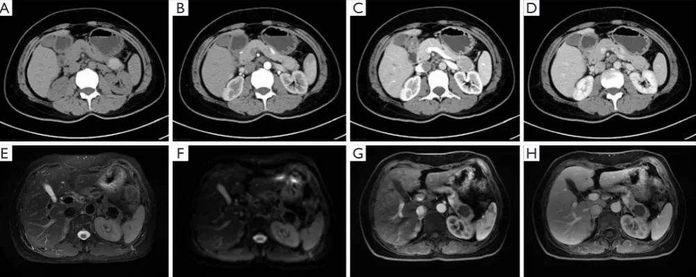
To further clarify the pancreatic mass, patient went to our hospital on Aug 5, 2019. Patient got reservation of triple phase helical contrast enhanced computed tomography (CE-CT) on Aug 8, 2019. Parameters were as follows: slice thickness 3 mm, tube voltage 120 kVp, tube current 400 mA. Iopamidol was used as a contrast medium and was administered at a dose of 2.5 mL/kg with a power injector. Precontrast, early arterial phase and late arterial phase were obtained. CE-CT revealed a hypo-intensity mass in the pancreas with heterogeneous wall enhancement in all phases, and the enhanced value of the mass was lower than that of the surrounding pancreatic parenchyma in all phases (Figure 1). No retroperitoneal lymph nodes showed
specific findings. Pancreatic mass, was considering solid pseudopapillary tumor with hemorrhage.
Contrast enhanced ultrasound examination (CEUS) using SonoVue was performed with a 1–5 MHz convex transducer (Mindray Reasona 7, China) on Aug 9, 2019. Sonography revealed a tail of pancreatic mass 26.7×18.1 mm2 in diameter showing a homogeneously hypoechoic pattern. The occupation was well-circumscribed, and pathological blood flow was not detected in color Doppler (Figure 2). The whole nodule was low-enhanced, ring-shaped high echoes can be seen in the periphery, and cord-like reinforcement can be seen in the nodule (Figure 3). Solid pseudopapillary tumor was first considered. Contrast enhanced magnetic resonance imaging (CE-MRI) on Aug 10, 2019 revealed slightly low signal in diffusion-weighted and low signal in T2-weighted measuring 22.7×23.8 mm2 in diameter. The capsule wall and separation were visible enhancement. CE-MRI indicated benign cystic mass in the tail of the pancreas combing bleeding, mucinous cystadenoma was first considered.
Considering the risk of major bleeding according to image, outpatient admitted to hospital with “pancreatic mass” on Aug 12, 2019. Owing to daily healthy body and exclusion of surgical contraindications (no asthma etc.), laparotomy was performed under general anesthesia on Aug 16, 2019, during which inspection of the pancreas revealed an oval mass with a smooth surface after ultrasound knife Figure 1 The CE-CT (A,B,C,D) and CE-MRI (E,F,G,H) images are demonstrating the enhanced pattern in all phases. A pancreatic mass was located at the tail. (A) Pre-contrast CT; (B) early arterial phase; (C) late arterial phase; (D) portal phase; (E) T2-weighted; (F) diffusion-weighted imaging; (G) arterial phase; (H) portal phase.
A
B
C
G
D
H
opened gastrointestinal ligament. Carefully released upward along the middle colonic vein and superior mesenteric vein to open the posterior channel of the pancreas, and suspended the pancreas with a cloth tape. The upper end of the pancreas had obvious adhesion to the arteriovenous of the spleen. Carefully isolated the arteriovenous of the spleen, cut the pancreatic body on the right side of cyst
with a cutting closure, and used electrocoagulate to stop pancreatic stump bleeding. The ultrasound knife separated the pancreas along the splenic arteriovenous, cut off the small branches between splenic arteriovenous and pancreas to prevent bleeding, and completely removed pancreatic body and tail within the lesion. 4-0 prolene interrupted sutured for hemostasis of pancreas. Intraoperative frozen Figure 2 The ultrasound images showing that a pancreatic mass was located at the tail. (A) B-mode; (B) color Doppler.
section histologically composed of a squamous epithelial cyst with abundant sinusoids and lymphoid tissue in the periphery, finding which was almost consistent with HAS. The pancreatic mass was diagnosed as HAS with squamous epithelial cyst form (Figure 4). Two flush drain tubes were indwelled in operation field. The operation was smooth, the anesthesia was stable during the operation, and the bleeding was about 300ml. The patient was sent to the resuscitation room after the operation. Cefmetazole was used to prevent infection, and symptomatic treatment was provided to protect the liver, stomach, analgesia, and enzyme inhibition. Postoperatively, the patient recovered well and was discharged on Aug 23, 2019, and kept a drainage tube for the pancreatic leakage. Discharged medicines were showing as follows: celecoxib capsule, compound lactobacillus acidophilus tablet, cefuroxime ester tablet, aspergillus oryzae trypsin tablet. Patient was advised to change gauze every three days and keep drainage tube unblocked after discharge, and sutures could be removed on Sep 6, 2019. She was followed up on Oct 6, 2019 with no complication which ultrasonography and lab test showed normal.
Discussion
During the spleen growth, it can develop anomalies such as complete agenesis, multiple spleens, HAS or persistent lobulation (7). HAS mostly appears in the hilum of the spleen followed by adjacent to the tail of the pancreas, left ovary, left testis, the greater omentum or mesentery of the small and large intestine (8,9). However, an atypical HAS with squamous epithelial cyst in the tail of pancreas has not been reported so far.
The HAS is an incidental finding of no clinical significance in most patients (10). The preoperative accurate diagnosis of an HAS is difficult, especially when HAS with squamous epithelial cyst, as it can be mistaken for other mass-forming lesions (11). Although ultrasound, CE-CT, CE-MRI and other imaging modalities have certain diagnostic value, lack specificity (12,13). On the ultrasound, typical HAS presents mostly as a solitary round or oval mass with a well-defined boundary. On CEUS, it can visualize vascular hilum and enhanced intensity of HAS dynamically. On CT scan, HAS is depicted as a clear margin mass. On the CECT, it shows as a homogeneously enhanced density mass lesion similar to that of spleen. On MRI, the signal intensity of HAS is consistent with the main spleen in both T1-weighted and T2-weighted images (14). For evaluating a functional accessory, scintigraphy with Tc-99m phytate may a useful method. However, it is difficult to make a clinically preoperative diagnosis despite availability of multiple diagnostic tools. HAS always mimics tumor or lymphadenopathy in atypical location, not always typical, like this case HAS with squamous epithelial cyst. In this case, HAS confused with other pancreatic mass based on CECT or enhanced ultrasound. The contrast pattern mimicked other pancreatic mass such as solid papillary neoplasm, serous cystadenoma neoplasm, mucinous cystadenoma.
In this case, no clinical symptom was apparent. However, it has been reported that some patient may present with abdominal pain, jaundice, malaise or other symptoms. Symptoms of HAS are atypical, requiring multiple imaging modalities for detection combined with clinical data. Routine laboratory tests are not usually helpful
A
B
some certain small blood vessels which representing splenic sinusoids. In the present case, HAS is seen squamous cyst with surrounding abundant sinusoids and lymphocytes. A HAS with squamous cyst in the tail of pancreas has not been reported so far and extremely rare. HAS, should be considered in the differential diagnosis of pancreatic masses. After laparoscopic surgery, the patient presented well with no complication during 1-month follow-up. In this case, the pancreatic body and tail was resected together with lesion. The limitation is that it destroys a large amount of normal pancreatic tissue and increases the risk of pancreatic leakage. Knowing the pathological result, the surgical method is reversed, and the local excision of the lesion is more suitable.
In conclusion, we reported a case of HAS with squamous cyst in the pancreatic tail presenting as other pancreatic masses. Although it is difficult to diagnose atypical HAS on multiple imaging modalities, it is important to note that HAS can present as other benign pancreatic masses, which are similar in appearance to HAS.
Acknowledgments
Funding: This study was funded by the National Natural Science Foundation of China (Grants No. 81420108018, 81527803), National Key R&D Program of China (Grants No. 2018YFC0115900), Zhejiang Science and Technology Project (Grants No. 2019C03077), and China ultrasound physician technology star program (Grants No. KJXX2018003).
Footnote
Conflicts of Interest:All authors have completed the ICMJE
uniform disclosure form (available at http://dx.doi.
org/10.21037/atm.2020.03.79). The authors have no
conflicts of interest to declare.
Ethical Statement: The authors are accountable for all aspects of the work in ensuring that questions related
License (CC BY-NC-ND 4.0), which permits the non-commercial replication and distribution of the article with the strict proviso that no changes or edits are made and the original work is properly cited (including links to both the formal publication through the relevant DOI and the license). See: https://creativecommons.org/licenses/by-nc-nd/4.0/.
References
1. Chauvet E, Spyropoulou V, Anooshiravani-Dumont M,
et al. Accessory Spleen Fracture: Report of a Pediatric
Case and Review of the Literature. Pediatr Emerg Care
2020;36:e10-e13.
2. Okabe Y, Ushijima T, Yasunaga M, et al. A rare case of sarcoidosis in accessory spleen. Gastrointest Endosc 2017;86:918-9.
3. Pandey A, Pandey P, Ghasabeh MA, et al. Accuracy of apparent diffusion coefficient in differentiating pancreatic
neuroendocrine tumour from intrapancreatic accessory
spleen. Eur Radiol 2018;28:1560-7.
4. Varga I, Babala J, Kachlik D. Anatomic variations of the spleen: current state of terminology, classification,
and embryological background. Surg Radiol Anat 2018;40:21-9.
5. Vikse J, Sanna B, Henry BM, et al. The prevalence and
morphometry of an accessory spleen: A meta-analysis
and systematic review of 22,487 patients. Int J Surg
2017;45:18-28.
6. Rodriguez Vargas D, Parada Blazquez MJ, Vargas
Serrano B. Diagnostic imaging of abnormalities in the number and location of the spleen. Radiologia
2019;61:26-34.
7. Palazzo M, Sauvanet A, Gincul R, et al. Impact of
needle-based confocal laser endomicroscopy on the therapeutic management of single pancreatic cystic lesions. Surg Endosc 2019. [Epub ahead of print].
8. Baugh KA, Villafane N, Farinas C, et al. Pancreatic
Incidentalomas: A Management Algorithm for Identifying
Splenic tissue in the ovary: Splenosis, accessory spleen or spleno-gonadal fusion? Pathol Res Pract
2019;215:152546.
10. Junus KL, Friedman JA, Rubin RR, et al. Accessory
spleen embolization: An option for refractory idiopathic thrombocytopenic purpura (ITP). Diagn Interv Imaging 2020;101:117-8.
11. Palumbo V, Mannino M, Teodoro M, et al. An extremely rare case of an oversized accessory spleen: case report and review of the literature. BMC Surg 2019;19:45.
12. Kim GE, Morris JD, Anand N, et al. Recognizing
intrapancreatic accessory spleen via EUS: Interobserver variability. Endosc Ultrasound 2019;8:392-7.
13. Matsumoto K, Kato H, Okada H. Epidermoid Cyst in an Intrapancreatic Accessory Spleen Diagnosed by Typical Radiographic Images and Endoscopic
Ultrasound Fine-Needle Aspiration Findings With Contrast Agent. Clin Gastroenterol Hepatol 2018;16:e13-4.
14. Morelli L, Guadagni S, Gianardi D, et al. Gray-scale,
Doppler and contrast-enhanced ultrasound in pancreatic allograft surveillance: A systematic literature review.
Transplant Rev (Orlando) 2019;33:166-72.
15. Feng Y, Shi Y, Wang B, et al. Multiple pelvic accessory
spleen: Rare case report with review of literature. Exp Ther Med 2018;15:4001-4.