Open Access
Research
A comprehensive analysis of the naturally occurring polymorphisms
in HIV-1 Vpr: Potential impact on CTL epitopes
Alagarsamy Srinivasan*
1, Velpandi Ayyavoo*
2, Sundarasamy Mahalingam
3,
Aarthi Kannan
1,4, Anne Boyd
1, Debduti Datta
3,
Vaniambadi S Kalyanaraman
5, Anthony Cristillo
5, Ronald G Collman
6,
Nelly Morellet
7, Bassel E Sawaya
8and Ramachandran Murali
9Address: 1Thomas Jefferson University, Department of Microbiology and Immunology, Jefferson Alumni Hall Rm 461, 1020 Locust Street,
Philadelphia, PA 19107, USA, 2University of Pittsburgh, Department of Infectious Diseases & Microbiology, Parran Hall Rm 439, 130 DeSoto
Street, Pittsburgh, PA 15261, USA, 3Department of Biotechnology, Indian Institute of Technology Madras, Chennai 600036, India, 4Wellesley
College, 21 Wellesley College Rd Unit 7430, Wellesley, MA 02481, USA, 5Advanced Bioscience Laboratories, Inc., 5510 Nicholson Lane,
Kensington, MD 20895, USA, 6University of Pennsylvania School of Medicine, 522 Johnson Pavilion, 36th and Hamilton Walk, Philadelphia PA
19104, USA, 7Unite de Pharmacologie Chimique et Genetique, INSERM, Avenue de l'Observatoire, Paris Cedex 06, France, 8Department of
Neuroscience, Center for Neurovirology, Temple University School of Medicine, Philadelphia, PA 19122, USA and 9University of Pennsylvania
School of Medicine, Dept of Pathology and Laboratory Medicine, 243 John Morgan, Philadelphia PA 19104, USA Email: Alagarsamy Srinivasan* - [email protected]; Velpandi Ayyavoo* - [email protected];
Sundarasamy Mahalingam - [email protected]; Aarthi Kannan - [email protected]; Anne Boyd - [email protected]; Debduti Datta - [email protected]; Vaniambadi S Kalyanaraman - [email protected];
Anthony Cristillo - [email protected]; Ronald G Collman - [email protected]; Nelly Morellet - [email protected]; Bassel E Sawaya - [email protected];
Ramachandran Murali - [email protected] * Corresponding authors
Abstract
The enormous genetic variability reported in HIV-1 has posed problems in the treatment of infected individuals. This is evident in the form of HIV-1 resistant to antiviral agents, neutralizing antibodies and cytotoxic T lymphocytes (CTLs) involving multiple viral gene products. Based on this, it has been suggested that a comprehensive analysis of the polymorphisms in HIV proteins is of value for understanding the virus transmission and pathogenesis as well as for the efforts towards developing anti-viral therapeutics and vaccines. This study, for the first time, describes an in-depth analysis of genetic variation in Vpr using information from global HIV-1 isolates involving a total of 976 Vpr sequences. The polymorphisms at the individual amino acid level were analyzed. The residues 9, 33, 39, and 47 showed a single variant amino acid compared to other residues. There are several amino acids which are highly polymorphic. The residues that show ten or more variant amino acids are 15, 16, 28, 36, 37, 48, 55, 58, 59, 77, 84, 86, 89, and 93. Further, the variant amino acids noted at residues 60, 61, 34, 71 and 72 are identical. Interestingly, the frequency of the variant amino acids was found to be low for most residues. Vpr is known to contain multiple CTL epitopes like protease, reverse transcriptase, Env, and Gag proteins of HIV-1. Based on this, we have also extended our analysis of the amino acid polymorphisms to the experimentally defined and predicted CTL epitopes. The results suggest that amino acid polymorphisms may contribute to the immune escape of the virus. The available data on naturally occurring polymorphisms will be useful to assess their potential effect on the structural and functional constraints of Vpr and also on the fitness of HIV-1 for replication.
Published: 23 August 2008
Virology Journal 2008, 5:99 doi:10.1186/1743-422X-5-99
Received: 7 July 2008 Accepted: 23 August 2008
This article is available from: http://www.virologyj.com/content/5/1/99
© 2008 Srinivasan et al; licensee BioMed Central Ltd.
Introduction
Humoral and cellular responses have been implicated in controlling viral and bacterial infections in addition to the host's innate immune responses. This is, indeed, demon-strated in the context of HIV-1 infection [1-3]. Specifically, CTL responses against the virus have been shown to limit the virus replication at a low level in the infected individ-uals. This is evident in the inverse correlation of CTL responses vs. virus load observed in acutely infected indi-viduals [4-6]. Utilizing the rhesus macaque/SIV infection model, a suppressive effect on virus replication was shown for CTLs [7]. However, the initial CTL responses are not able to contain the virus at a later stage, possibly due to the emergence of viral variants that evade the immune responses resulting in continued virus replica-tion [8,9]. Hence, an understanding of the CTL escape var-iants of HIV is important both in natural viral infections and also in the context of vaccine-induced immunity for developing effective CTL based polyvalent vaccines for containing diverse HIV-1 strains [10]. This is an area of research which is actively being pursued by several inves-tigators [11,12].
The genome of HIV-1 has been shown to code for two reg-ulatory proteins (Tat and Rev) and four auxiliary proteins (Vif, Vpr, Vpu and Nef) in addition to the Gag, Pol, and Env structural proteins [13]. The regulatory proteins Tat and Rev are essential for virus replication. Rev is involved in the transport of genomic and partially spliced subge-nomic mRNA from the nucleus to the cytoplasm [14]. Tat is known as an activator of transcription of viral and cel-lular RNA. Vif plays an important role in HIV-1 replica-tion in peripheral blood mononuclear cells (PBMC). Specifically, Vif prevents hypermutation in the newly made viral DNA through its interaction with APOBEC3G [15,16]. Vpr is known for its incorporation into the virus particles. The interaction of Vpr with the Gag enables its incorporation into the virus particle. Vpr is a multifunc-tional protein and is involved in the induction of apopto-sis, cell cycle arrest, and transcriptional activation [17]. Vpu plays a role in the particle release and degradation of CD4 [14,18,19]. The features of Nef include downregula-tion of cell surface receptors, interference with signal transduction pathways, enhancement of virion infectivity, induction of apoptosis in bystander cells, and protection of infected cells from apoptosis [20-24].
Based on the data reported so far, it is clear that HIV-1 employs multiple strategies to successfully replicate in the infected individuals [14,25,26]. The enormous genetic variation that is generated through errors of reverse tran-scriptase enzyme may provide a pool of variants to evade the host immune responses against the virus and also result in the emergence of drug resistant viruses during treatment. In addition, it is also likely that the
immuno-suppressive effects of HIV-1 encoded proteins may atten-uate the host immune responses in favor of the virus.
Upon infection of target cells by the virus, viral proteins are synthesized for carrying out the functions related to the virus replication and also exert effect on specific host cell functions. In addition, viral proteins are also targeted to the proteosomal degradation pathway. This process results in the generation of peptides, which are then trans-located to the ER through TAP and are presented on the cell surface in association with human leukocyte antigen (HLA) class I molecules. The genetic variability present in the coding sequences of the virus may result in viral pro-teins with alterations in the CTL epitopes, which may lead to defective processing, presentation or lack of recognition of the epitope by the reactive CTLs. This is the likely mech-anism of the CTL escape by HIV-1 and other viruses. The presence of multiple CTL epitopes has been demonstrated in HIV-1 proteins including Gag, Pol, Vif, Vpr, Tat, Rev, Vpu, Env and Nef. Though the characterization of the epitopes with respect to the viral proteins is achievable in individual cases, such an analysis at a population level is difficult to carry out for the following reasons: i) HIV-1 exhibits high genetic variation in different regions of the genome. The extent of heterogeneity among circulating HIV-1 strains is described to be in the range of 20% or more in relatively conserved proteins and up to 35% for Env protein [11]. In addition, there is also extensive diver-sity among HIV-1 within a subtype, ii) There are multiple subtypes of HIV-1, and iii) There are variables at the HLA loci. On the other hand, this limitation can be overcome to some extent by utilizing alternative approaches where information about CTL epitopes and their variants can be inferred from the sequences available for HIV-1 [27-29]. The HIV sequence database has information about the viral isolates from different parts of the world. This infor-mation can be used as a source to assess the extent of nat-urally occurring polymorphisms and their potential impact on CTL epitopes. We hypothesize that mutations or alterations in the residues which are part of the CTL epitope in the Vpr molecule are likely to affect the epitope at multiple levels (processing and recognition of the epitope). Recently, studies have addressed this issue using full length or partial HIV-1 genome sequences [30]. This has prompted us to carry out a comprehensive analysis of the extent of variation at the amino acid level in the aux-iliary gene product Vpr of HIV-1.
effect, vii) Vpr is present in the body fluids as an extracel-lular protein, viii) Vpr is highly immunogenic, ix) Vpr is a small protein comprising only 96 amino acids and x) Structural information for the whole Vpr molecule is available through NMR [17,31-34]. These features enable a detailed analysis of the polymorphisms in Vpr with respect to CTL epitopes, structure-function of the protein, and fitness of the virus for replication.
In this study, we have analyzed the predicted amino acid sequences of Vpr from global HIV-1 isolates available through the HIV database. Specifically, the extent of genetic variation in Vpr in the form of polymorphisms at the individual amino acid level was comprehensively ana-lyzed. Several of the amino acid polymorphisms were found to be part of the experimentally verified and pre-dicted CTL epitopes. The location and nature of the vari-ant amino acid were found to affect the CTL epitope considerably. Hence, our results provide a glimpse into the genetic footprints of immune evasion in Vpr.
Materials and methods
The goal of our studies is to assess the nature and extent of polymorphisms at the level of individual residues in the Vpr molecule. The sequences considered here comprise Vpr sequences derived from all the major subtypes of HIV-1. The details regarding the subtypes and the number of sequences from each subtype are presented in Table 1 and are taken from the HIV database http://www.hiv.lanl.gov [35-38]. In addition, we have included Vpr sequences derived from HIV-1 positive long term non-progressors (McKeithen et al., unpublished data). It should be noted
that we have also included Vpr from SIV isolated from chimpanzees, as this is likely the progenitor virus for HIV-1. Vpr sequences from the database were accessed in Jan-uary of 2007. The deletions in the Vpr molecule were excluded from our analysis. The alignment of Vpr sequences (which is available from the authors upon request) was analyzed manually for variant amino acids at the level of individual residue in Vpr from global and dis-tinct subtypes of HIV-1.
Results
Characteristics of Vpr sequences selected for this study
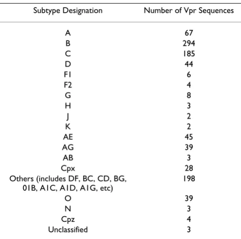
The alignment of Vpr sequences has enabled us to analyze the differences at the level of each residue from diverse HIV-1 isolates. A total of 976 Vpr sequences have been used for alignment. The polymorphisms, with respect to the length, have been noted in Vpr by several investigators [17,39]. As this may pose problem for our analysis, our alignment does not take into account both deletions and insertions. The Vpr alleles are from diverse subtypes and include 67, 294, 185 and 44 Vpr sequences representing subtype A, B, C, and D, respectively (Table 1). The O, AE, AG, and cpx groups represent 39, 45, 39 and 28 Vpr sequences, respectively. Since the Vpr sequences are derived from different sources such as viral RNA, cloned viral DNA and proviral DNA from tissues, we have not made attempts to classify them in our analysis.
Amino acid polymorphisms in the predicted Vpr sequences
Recently, the structure of full length Vpr has been resolved by NMR [40]. According to this study, Vpr consists of a flexible N-terminal domain (amino acids 1–16), helical domain I (HI) (residues 17–33), turn (residues 34–37), helical domain II (HII) (residues 38–50), turn (residues 51–54), helical domain III (HIII) (residues 55–77), and a flexible C-terminal domain (residues 78–96). Based on this structure, the polymorphisms observed in Vpr are pre-sented with respect to the individual domain.
N-terminus of Vpr (residues 1–16)
The results presented in Table 2 regarding the N-terminal domain of Vpr show that all the residues excluding the initiator methionine are susceptible for alterations. The altered amino acids or polymorphisms at each residue are indicated as variant amino acids or substitutions. For con-venience, we have used Vpr from NL4-3 proviral DNA as a reference sequence. The amino acid sequence of NL4-3 Vpr is similar to HIV-1 subtype B consensus Vpr except for residues 28(S), 77(Q) and 83(I). Interestingly, the residue 9, which is G, has only one variant amino acid. In an ear-lier study, it was noted that a change in residue 3 from Q to R was not associated with cytopathic effect [41]. In our analysis, variant amino acids H, L, M, and P were also noted for Q. Studies involving synthetic peptides corre-sponding to the N-terminus and also the full-length Vpr Table 1: Vpr sequences used for the analysis of amino acid
polymorphisms
Subtype Designation Number of Vpr Sequences
A 67
B 294
C 185
D 44
F1 6
F2 4
G 8
H 3
J 2
K 2
AE 45
AG 39
AB 3
Cpx 28
Others (includes DF, BC, CD, BG, 01B, A1C, A1D, A1G, etc)
198
O 39
N 3
Cpz 4
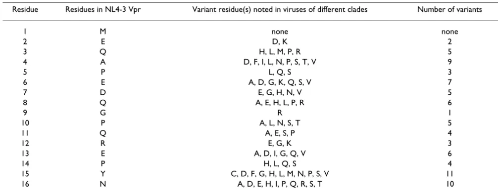
molecule have shown that the Vpr sequence (residues PHN) have the ability to form a γ-turn. The residue 15(H) exhibits eleven, residue 16 (N) shows ten and residue 14 (P) shows four variant amino acids. While residue 2 has two, residues 5 and 12 register three variant amino acids. Residues 3, 4, 6, 7, 8, 10, 11, and 13 contain multiple var-iant amino acids ranging from five to eleven. The N-termi-nal domain contains a total of 79 variant amino acids. Of these, non-conserved substitutions correspond to about 80% of the residues.
The impact of the majority of the polymorphisms on Vpr functions is not clear. Substitution of alanine for proline at residue 5 and 10 showed less or increased virion incor-poration of Vpr, respectively [42]. Similarly, substitution
of alanine for residue 12 reduced the cell cycle arrest func-tion of Vpr [43]. On the other hand, substitufunc-tion at resi-due 13 and 14 showed an increase in cell cycle arrest [42,44]. Hence, the naturally occurring polymorphisms are likely to affect the functions of Vpr.
Helical domain I (HI residues 17–33)
NMR studies of full length Vpr show that a region com-prising the residues 17–33 adapt a helical structure. This was also predicted by several algorithms. The polymor-phisms observed for the residues 17–33 are presented in Table 3. The characteristics of the residues with respect to the variant amino acids are the following: residues 18, 23 and 26 show two substitutions; residue 20 has three sub-stitutions; residues 25, and 29 show four subsub-stitutions; Table 2: The polymorphisms in the N-Terminus of Vpr (residues 1–16)
Residue Residues in NL4-3 Vpr Variant residue(s) noted in viruses of different clades Number of variants
1 M none none
2 E D, K 2
3 Q H, L, M, P, R 5
4 A D, F, I, L, N, P, S, T, V 9
5 P L, Q, S 3
6 E A, D, G, K, Q, S, V 7
7 D E, G, H, N, V 5
8 Q A, E, H, L, P, R 6
9 G R 1
10 P A, L, N, S, T 5
11 Q A, E, S, P 4
12 R E, G, K 3
13 E A, D, I, G, Q, V 6
14 P H, L, Q, S 4
15 Y C, D, F, G, H, L, M, N, P, S, V 11
16 N A, D, E, H, I, P, Q, R, S, T 10
Table 3: The polymorphisms in Helical Domain I of Vpr (residues 17–33)
Residue Residues in NL4-3 Vpr Variant residue(s) noted in viruses of different clades Number of variants
17 E A, D, G, Q, T, V 6
18 W G, R 2
19 T A, I, L, M, P, R, S, V 8
20 L I, M, V 3
21 E A, D, G, K, T 5
22 L F, I, M, P, T, V 6
23 L S, V 2
24 E D, G, K, Q, R 5
25 E A, D, G, K 4
26 L F, I 2
27 K I, M, N, Q, R 5
28 S A, D, E, G, H, I, K, N, Q, R, T, V 12
29 E D, G, Q, V 4
30 A D, P, S, T, V 5
31 V A, D, I, L, M, T 6
32 R G, K, Q, T, W, 5
residues 21, 24, 27, 30 and 32 show five substitutions; and residues 17, 22, and 31 register six substitutions and residue 19 has eight substitutions. Interestingly, residue 28 exhibits the highest number of substitutions and resi-due 33 has only one substitution. This domain exhibits a total of 80 variant amino acids and 61 of them are of non-conservative in nature.
Several laboratories including ours have reported on the importance of residues in the helical domain I for Vpr functions. Substitution of a proline residue for glutamic acid (residue 17, 21, 24, 25, and 29) has a drastic effect on the stability, subcellular localization, and virion incorpo-ration of Vpr [44-49]. The variant amino acids noted in this domain have the potential to destabilize and disrupt the function of Vpr. Similarly, substitution of alanine for leucine residue affected the stability and virion incorpora-tion of Vpr [45,48,50-53]. Based on the studies reported, varying amino acid arginine for histidine at residue 33 will affect the subcellular localization and virion incorpo-ration of Vpr [54].
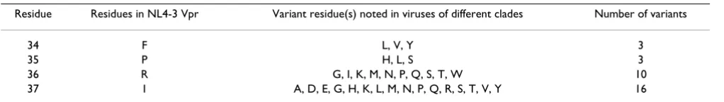
Interhelical domain I (residues 34–37)
This region is present between helical domains I and II and comprises only four residues. It has been shown that residues in this region have the ability to form a γ-turn. The naturally occurring polymorphisms in this region are presented in Table 4. Site-specific mutagenesis studies have shown an important role for residues in subcellular localization, cell cycle arrest, apoptosis and virion incor-poration of Vpr [42,44,51,55,56]. Residues 34 and 35 show only three substitutions. On the other hand, residue 36 and 37 register 10 and 16 substitutions, respectively. The variant amino acids reach a total of 31 and 21 of them are of non-conservative in nature.
Helical domain II (residues 38–50)
Studies with peptide (1–50 amino acids) and full-length Vpr have shown that residues 38–50 correspond to helical domain II of Vpr. The naturally occurring polymorphisms corresponding to the residues in this region are presented in Table 5. The characteristics of the substitution are the following: residues 39 and 47 exhibit a single substitu-tion; residues 43, 46 and 50 record two substitutions; res-idue 38 shows four substitutions; resres-idues 42, 45 and 49 show five substitutions; and residues 40 and 44 have eight
substitutions. Nine and thirteen substitutions were noted for residues 41 and 48, respectively. This domain contains 64 variant amino acids and non-conservative substitu-tions correspond to 41 residues. Several laboratories have carried out experiments addressing the role of residues in this region by utilizing site-specific mutagenesis. The alter-ation of hydrophobic residues severely affected the virion incorporation and transcriptional activation of Vpr [43,44,50,56].
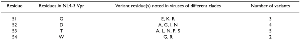
Interhelical domain II (residues 51–54)
This region is located between helical domains II and III. Of the four residues which are part of this domain, only the residue G51 has been shown to reduce G2/M cell cycle arrest through alanine substitution [44]. The naturally occurring polymorphisms corresponding to the residues in this region are presented in Table 6. The characteristics of the substitutions are the following: residue 54 shows two substitutions; residue 51 shows three substitutions; residue 52 shows four substitutions and residue 53 shows five substitutions. The variant amino acids reach a total of fourteen and the majority of them are non-conservative substitutions.
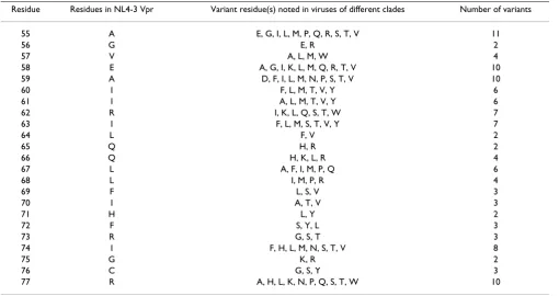
Helical domain III (residues 55–77)
The presence of helical domain III has been demonstrated by NMR [40]. Several laboratories including ours have shown the importance of this domain for the function of Vpr. The naturally occurring polymorphisms noted for the residues in this region are presented in Table 7. The char-acteristics of the substitutions are the following: residues 56, 64, 65, 71 and 75 exhibit two substitutions; residues 69, 70, 72, 73 and 76 register three substitutions; residues 57, 66 and 68 show four substitutions; residues 60, 61 and 67 show six substitutions; residues 62 and 63 have seven substitutions; residue 74 has eight substitutions; residues 58, 59, and 77 exhibit ten substitutions; and res-idue 55 shows eleven substitutions. While the variant amino acids reach a total of 108, 65 of them are of non-conservative nature. This region comprises LXXLL motif which is important for subcellular localization and also influences the virion incorporation of Vpr [44,57-62]. Additionally the LXXLL domain is also involved in Vpr-GR interaction and its subsequent role in virus replication [63,64].
Table 4: The polymorphisms in the Interhelical Domain 1 of Vpr (residues 34 – 37)
Residue Residues in NL4-3 Vpr Variant residue(s) noted in viruses of different clades Number of variants
34 F L, V, Y 3
35 P H, L, S 3
36 R G, I, K, M, N, P, Q, S, T, W 10
C-terminus of Vpr (residues 78–96)
The naturally occurring polymorphisms corresponding to the residues in the C-terminus of Vpr are presented in Table 8. The characteristics of the substitutions for the res-idues in this region are the following: residue 80 has only two substitutions; residues 78, 79, 82 and 92 have three substitutions; residues 81 and 90 have four substitutions; residues 91 and 96 have five substitutions. All of the other residues have substitutions ranging from six to thirteen. Of the 124 variant amino acids in this domain, 100 of them are of non-conservative nature.
This domain contains multiple arginine and serine resi-dues. It has been reported that the arginine residues are important for the cell cycle arrest and subcellular localiza-tion [65,66]. Vpr is known to undergo post-translalocaliza-tional modification and the serine residues located at 28, 79, 94, and 96 positions of the protein serve as substrates for the phosphorylation [67]. Vpr, devoid of phosphorylation through site-specific mutagenesis, severely affects replica-tion of HIV-1 in macrophages [68]. Residue 28 contains equivalent proportion of amino acids N (44%) and S (48%) and Vpr of SIV cpz contains N or T at this position. On the other hand, serine residues at 79, 94, and 96 are conserved in SIV cpz Vpr.
The naturally occurring polymorphisms for the whole Vpr molecule reach a total of 498 substitutions. The non-con-servative variant amino acids correspond to 72%. It is
important to note that all the residues in Vpr have the pro-pensity to accept variant amino acids. The data presented here also reveal that the variant amino acids noted with respect to some residues are identical. These include resi-dues 60(I), 61(I), 34(F), 71(H) and 72(F). We have car-ried out a detailed analysis of the variant amino acids noted in distinct subtypes (A, B, C, and D) of HIV-1. Such an analysis could not be carried out for several groups because of the limited information available regarding Vpr alleles. The data generated for subtype B Vpr alleles are presented in Tables 9, 10, 11, 12, 13, 14, 15. The anal-ysis of subtype B involves a total of 275 Vpr alleles. As expected, the extent of polymorphisms in subtype B is less in comparison to the total polymorphisms noted with all the Vpr alleles. Interestingly, there are several residues that did not have any variant amino acids. These include resi-dues 9, 18, 26, 34, 35, 38, 42, 46, 64, 66, and 79. On the other hand, the residues without variant amino acids in subtype C are different from that of subtype B except for 9, 26, and 64. In addition, the frequency of variant amino acids at the level of each residue was also determined for subtype B Vpr. The results indicate that the frequency of variant amino acids is low in most cases (0.4–1.1%) except for the residues 7, 19, 37, 41, 45, 55, 60, 63, 77, 80, 84, 85, 86, 89, and 93. Analysis involving a large number of Vpr alleles also showed frequency patterns consistent with the data presented in Tables 9, 10, 11, 12, 13, 14, 15. With respect to the N-terminus domain (Table 9), the res-idue 7 (D) has resres-idue N substitution with a frequency of Table 5: The polymorphisms in Helical Domain II of Vpr (residues 38 – 50)
Residue Residues in NL4-3 Vpr Variant residue(s) noted in viruses of different clades Number of variants
38 W C, F, T, Y 4
39 L F 1
40 H I, L, M, N, Q, R, T, Y 8
41 N A, D, E, G, H, Q, R, S, W 9
42 L C, I, F, M, V 5
43 G E, R 2
44 Q E, H, L, K, N, R, T, V 8
45 H F, L, Q, W, Y 5
46 I D, V 2
47 Y H 1
48 E A, D, G, H, I, K, N, Q, R, S, T, V, Y 13
49 T H, N, M, S, Y 5
50 Y H, S 2
Table 6: The polymorphisms in Interhelical Domain II of Vpr (residues 51 – 54)
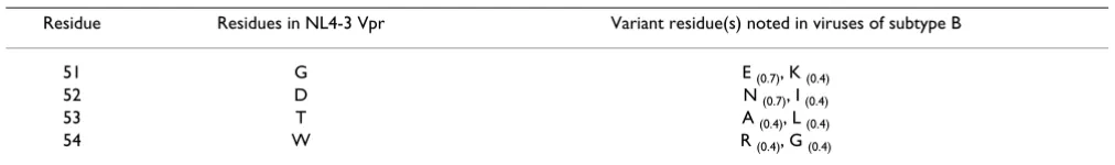
Residue Residues in NL4-3 Vpr Variant residue(s) noted in viruses of different clades Number of variants
51 G E, K, R 3
52 D A, G, I, N 4
53 T A, L, N, P, S 5
6.2%. Also, while the reference Vpr allele has Y at position 15, which is the predominant amino acid (85%), the var-iant amino acid F occurs to a limited extent (6.9%). Simi-lar scenario is also applicable to the residues 28, 77, and 83 (Tables 10 and 15). The residue R 80, which has been
implicated in cell cycle arrest function of Vpr, exhibits substitution of A with a frequency of 5.1%.
Table 7: The polymorphisms in Helical Domain III of Vpr (residues 54–77)
Residue Residues in NL4-3 Vpr Variant residue(s) noted in viruses of different clades Number of variants
55 A E, G, I, L, M, P, Q, R, S, T, V 11
56 G E, R 2
57 V A, L, M, W 4
58 E A, G, I, K, L, M, Q, R, T, V 10
59 A D, F, I, L, M, N, P, S, T, V 10
60 I F, L, M, T, V, Y 6
61 I A, L, M, T, V, Y 6
62 R I, K, L, Q, S, T, W 7
63 I F, L, M, S, T, V, Y 7
64 L F, V 2
65 Q H, R 2
66 Q H, K, L, R 4
67 L A, F, I, M, P, Q 6
68 L I, M, P, R 4
69 F L, S, V 3
70 I A, T, V 3
71 H L, Y 2
72 F S, Y, L 3
73 R G, S, T 3
74 I F, H, L, M, N, S, T, V 8
75 G K, R 2
76 C G, S, Y 3
77 R A, H, L, K, N, P, Q, S, T, W 10
Table 8: The polymorphisms in the Carboxy-Terminal Region of Vpr (residues 78–96)
Residue Residues in NL4-3 Vpr Variant residue(s) noted in viruses of different clades Number of variants
78 H L, R, Y 3
79 S N, R, T 3
80 R A, K 2
81 I G, M, R, V 4
82 G A, D, S 3
83 V H, I, L, M, N, P, T, V 8
84 T A, F, G, I, L, M, N, P, Q, S, V, W, Y 13
85 R A, H, I, L, P, Q, T, V, Y 9
86 Q E, G, H, M, P, R, S, T, V, Y 10
87 R A, E, G, K, M, N, P, Q, S, T 10
88 R A, E, G, I, S, T 6
89 A D, E, G, I, L, N, P, R, S, T, V 11
90 R G, I, N, S 4
91 N D, H, I, K, S 5
92 G A, E, R 3
93 A D, F, G, L, M, N, P, S, T, V 10
94 S D, E, F, G, H, N, R, V 8
95 R A, D, G, I, K, P, S, T 8
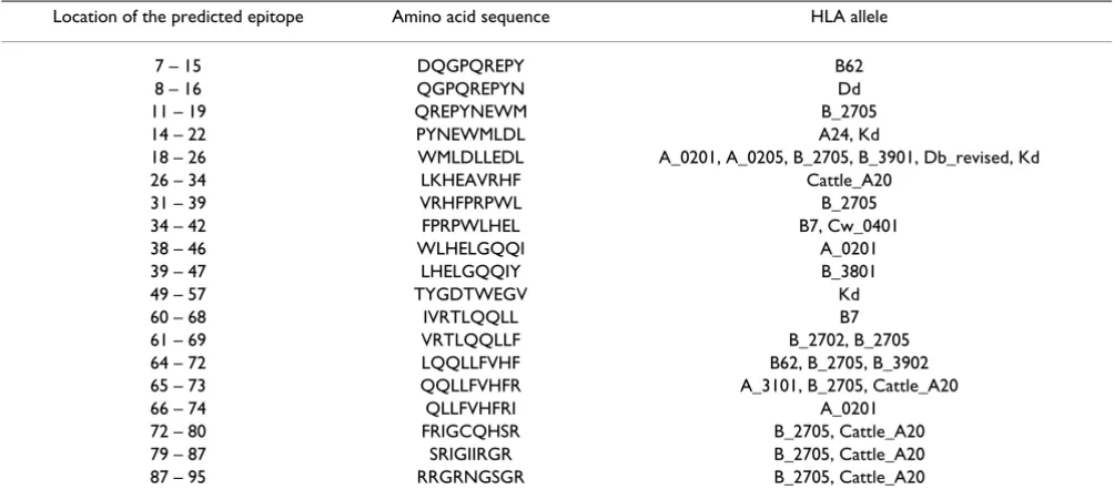
Impact of amino acid polymorphisms on defined and predicted CTL epitopes in Vpr
It has been shown that a single amino acid change in the epitope enables the virus to evade the T cell surveillance [9,69]. Hence, it is of interest to analyze the polymor-phisms in the context of both experimentally verified and predicted CTL epitopes. As Vpr is a highly immunogenic protein, several CTL epitopes have been already defined [12]. CD8+ epitopes are contiguous and nine amino acids long. The experimentally verified CTL epitopes in Vpr are presented in Table 16 with their location in the protein. We have presented the overall amino acid polymorphisms for each of the epitope. The experimentally verified CTL epitopes cluster in the region covering 1–70 residues of
Vpr. The total amino acid polymorphisms range from 36 to 107 for the individual epitopes. For example, the CTL epitope comprising the residues REPHNEWTL contains 53 variant amino acids. Residues at position 1 to 9 of the epitope show 3, 6, 4, 11, 10, 6, 2, 8, and 3 variant amino acids, respectively.
In addition, we have also utilized bioinformatics approach to assess the effect of polymorphisms on CTL epitope http://Bimas.dcrt.nih.gov/molbio/hla-bind. The predicted CTL epitopes with respect to several HLA class I alleles are presented in Table 17. The impact of polymor-phisms on the CTL epitope was assessed by determining the estimate of half-time of disassociation of the molecule Table 9: The frequency of variant amino acids in the N-Terminus of Vpr (Residues 1–16)
Residue Residues in NL4-3 Vpr Variant residue(s) noted in viruses of subtype B*
1 M no change
2 E D (0.4)
3 Q H (0.4), R (1.8)
4 A D (0.7), T (0.4), V (1.1)
5 P L (0.4), S (0.4)
6 E A (1.1), D (0.4), K (1.1), Q (0.7)
7 D N (6.2), V (0.4)
8 Q H (1.1)
9 G no change
10 P L (0.4), S (0.7)
11 Q A (0.7), E (0.4), P (1.8), S (1.8)
12 R K (0.4)
13 E I (0.4), Q (1.1), V (0.7)
14 P Q (0.4), S (0.7)
15 Y C (0.4), D (0.4), F (6.9), H (5.0), N (0.7), S (0.4), V (0.4) 16 N A (0.4), H (1.1), I (0.4), Q (0.7), P (0.4), R (0.4), S (0.4), T (0.7)
*275 Vpr alleles were used for analysis.
The numbers in the parentheses represent the percent frequency of the variant amino acid in the Vpr alleles analyzed.
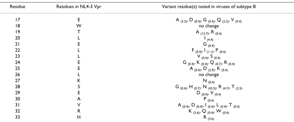
Table 10: The frequency of variant amino acids in Helical Domain I of Vpr (Residues 17–33)
Residue Residues in NL4-3 Vpr Variant residue(s) noted in viruses of subtype B
17 E A (3.3), D (0.4), G (0.4), Q (2.2), V (0.4)
18 W no change
19 T A (12.7), R (0.4)
20 L I (4.4)
21 E G (0.4)
22 L F (0.4), I (1.1), P (0.4)
23 L V (0.4), S (0.4)
24 E G (0.4), K (0.4), Q (0.7), R (0.4)
25 E A (0.4), D (2.9), K (0.4)
26 L no change
27 K N (0.4)
28 S G (0.4), H (0.7), N (43.5), R (4.7), T (2.5)
29 E D (0.4), V (0.4)
30 A P (0.4)
31 V A (0.4), D (0.4), I (0.4), L (0.4), T (0.4)
32 R K (3.6), Q (0.4), W (0.4)
containing the epitope. For this purpose, we have consid-ered 3, 1, 2, and 6 epitopes corresponding to HLA-A2, Cw-4, HLA B-7 and HLA B-2705, respectively. The influence of variant amino acids on the CTL epitope is presented in Table 18, 19, 20 with respect to HLA-A2 molecule. The epitopes considered for analysis correspond to residues 18–26, 38–46, and 66–74 of Vpr. While the reference pep-tide of the epitope located at residues 18–26 (Table 18) of Vpr shows the estimate of half time of disassociation value of 1213.356, the variant amino acid at position 1–9 in the epitope predicted a lower value. The substitution of vari-ant amino acids at residue position 2 of the epitope affected the half-time value considerably. Interestingly, substitution of R lowered the value to 0.233. Similarly, the substitution of F for L at position 9 of the epitope also lowered the value to 4.233. The analysis of the epitope corresponding to the residues 38–46 is shown in Table 19. The variant amino acids at residue 39 and 41 drastically lowered the value. The residue 46 showed contrasting val-ues based on the nature of the variant amino acid present. The impact of polymorphisms on the epitope correspond-ing to the residues 66–74 is shown in Table 20. The results show that both the location and nature of the amino acid have an effect on the half-time disassociation of the mol-ecule, which may lead to defective processing, presenta-tion, and recognition of the epitope.
Discussion
Viral infections in individuals generally lead to a scenario where the virus is confronted by the host immune system involving both innate and adaptive immune responses. Regarding the latter, cellular and humoral immune responses have been shown to play a role in the control of infections of viruses including HIV-1 [70,71]. It has been suggested that an understanding of the correlates of pro-tective immunity is an important requirement for the development of vaccines against HIV-1. Several studies have been published on this subject [71-73]. These studies point out a role for CD8+ and CD4+ T cell responses and neutralizing antibodies in the control of HIV-1 replica-tion. For example, it has been reported that CD8+ cells control HIV-1 in the acutely infected individuals [4-6]. The relevance of CD8+ T cells for the control of virus infec-tion was also shown in the case of SIV infected rhesus macaques [74,75]. Recently, the published data on CD8+ T cells in acute and chronic HIV-1 infection revealed that CTL epitopes are present in all of the proteins encoded by HIV-1. Virus replication, however, is not completely con-tained due to the emergence of CTL escape variant viruses. Based on this, it is suggested that vaccine efforts to control HIV-1 should take into account the high genetic variabil-ity noted among HIV-1.
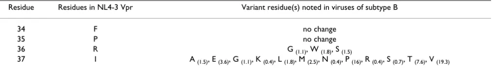
The continued emergence of genetic variants is a charac-teristic feature of RNA viruses. RNA dependent RNA polymerase and reverse transcriptase are error-prone Table 11: The frequency of variant amino acids in the Interhelical Domain 1 of Vpr (Residues 34–37)
Residue Residues in NL4-3 Vpr Variant residue(s) noted in viruses of subtype B
34 F no change
35 P no change
36 R G (1.1), W (1.8), S (1.5)
37 I A (1.5), E (3.6), G (1.1), K (0.4), L (1.8), M (2.5), N (0.4), P (16), R (0.4), S (0.7), T (7.6), V (19.3)
Table 12: The frequency of variant amino acids in Helical Domain II of Vpr (Residues 38–50)
Residue Residues in NL4-3 Vpr Variant residue(s) noted in viruses of subtype B
38 W no change
39 L F (0.4), I (0.4)
40 H L (2.2), N (0.4), Q (1.5), R (0.4), T (0.4), Y (0.4)
41 N A (0.7), D (0.7), E (0.4), G (52.0), S (30.5)
42 L no change
43 G E (0.4), R (0.4)
44 Q R (0.4)
45 H F (0.7), L (1.1), Q (0.4), Y (24.4)
46 I no change
47 Y H (0.4)
48 E A (0.4), D (2.5), G (1.1), K (0.4), Q (0.4), V (0.4)
49 T N (0.7)
enzymes and have been implicated as a cause for the gen-eration of variants [76,77]. The mutational changes in the protease and reverse transcriptase, depending on their location, may impact on their binding inhibitors targeting these enzymes. The viruses containing alterations may then be able to evade the inhibitory activities of the agents and are designated as drug-resistant variants. Similarly, the mutations in Env, Tat, and possibly other proteins can also evade the neutralizing antibody, CTL and T-helper cell responses [12,71]. The emergence of escape variants eventually repopulates the body in the face of immune responses against the virus. It has been suggested that immune escape may be a key step in the evolution of HIV-1 [30,78-80].
In an effort to understand the overall polymorphisms in a HIV-1 gene product, we undertook a comprehensive anal-ysis of the predicted amino acid sequences of Vpr from diverse HIV-1 subtypes. Considering the genetic variation noted in diverse HIV-1 [39], our hypothesis is that the dif-ferences in Vpr and other viral proteins may enable the
viruses to escape the host immunological pressures. To address this issue, we have initially compiled the poly-morphisms in Vpr at the level of individual amino acid. Vpr contains only 96 amino acids. Hence, the small size of the protein is an advantage for a comprehensive analy-sis. For this purpose, we have turned to the Vpr sequences which are available in the HIV database and also sequences from specific groups such as HIV-1 positive long-term non-progressors. A total of 976 predicted Vpr amino acid sequences were used for our studies. The anal-ysis revealed several characteristic features with respect to the individual amino acids in the Vpr. Of the 96 amino acids, all the amino acids except the initiator methionine have the propensity to change. This indicates that Vpr molecule is highly flexible in nature. The frequency of the variant amino acids, calculated for subtype B Vpr at the level of individual residue, revealed that substitution is very low for most of the residues. This suggests that many of the substitutions in Vpr may compromise the function and possibly the fitness of the virus. Interestingly, there are several amino acids that can accommodate ten or Table 13: The frequency of variant amino acids in Interhelical Domain II of Vpr (Residues 51 – 54)
Residue Residues in NL4-3 Vpr Variant residue(s) noted in viruses of subtype B
51 G E (0.7), K (0.4)
52 D N (0.7), I (0.4)
53 T A (0.4), L (0.4)
54 W R (0.4), G (0.4)
Table 14: The frequency of variant amino acids in Helical Domain III of Vpr (Residues 55–77)
Residue Residues in NL4-3 Vpr Variant residue(s) noted in viruses of subtype B
55 A E (2.2), P (1.1), Q (0.4), T (19.6), V (1.8)
56 G R (0.4), E (0.7)
57 V W (0.4)
58 E G (1.1), I (0.4), K (1.1), Q (0.7), V (0.4)
59 A L (0.4), P (0.4), S (0.4), V (0.4)
60 I L (16.4)
61 I L (0.4), M (1.1), T (3.3), V (1.5)
62 R K (0.7), L (0.4), S (0.4)
63 I M (5.8), S (1.8), T (11.3), V (1.8)
64 L no change
65 Q H (0.4)
66 Q no change
67 L M (1.5), P(0.7)
68 L M (1.5)
69 F L (1.8)
70 I T (2.9), V (1.1)
71 H L (0.4), Y (0.4)
72 F S (1.5), Y (0.4)
73 R T (0.7)
74 I L (0.4), M (0.4), V (0.4)
75 G R (1.1)
76 C G (1.1)
more alterations. We designate such amino acids as hot spots in Vpr which include residues 15, 16, 28, 36, 37, 48, 55, 58, 77, 84, 86 and 89. The underlying basis for the extensive genetic changes in specific regions of Vpr is not clear. It is likely that the error-prone reverse transcriptase, the secondary structure of RNA and other factors, either alone and/or in combination may play a role in the gen-eration of genetic variants. In this regard, Yusim et al. [28] have noted that Integrase (IN) exhibits the least variability and Vpu exhibits the highest variability. Boutwell and
Essex [27] also showed that the proportion of polymor-phic amino acids ranged from a low of 55% (RT, IN) to a high of 94% (Vpu). In our analysis, Vpr variability is high which may likely be due to the inclusion of diverse iso-lates including the HIV-1 progenitor virus SIVcpz.
Vpr is known as a highly immunogenic protein. The pres-ence of CTL epitopes verified through experimental approaches has been reported by several groups [12]. These include the region encompassing residues 9–70 of Table 15: The frequency of variant amino acids in the Carboxy-Terminal Region of Vpr (Residues 78–96)
Residue Residues in NL4-3 Vpr Variant residue(s) noted in viruses of subtype B
78 H L (0.7)
79 S no change
80 R A (5.1)
81 I G (0.4), M (0.7), V (0.4)
82 G D (0.7), S (0.7)
83 V I (86.9), L (0.4), T (0.7)
84 T A (0.4), F (0.4), G (0.7), I (30.9), L (2.5), M (0.4), N (0.7), S (0.4), V (0.4) 85 R H (0.4), I (0.4), L (2.5), P (15.6), Q (28.4), T (0.4), Y (1.1)
86 Q M (0.4), P (1.1), R (21.1), S (1.5), V (1.1)
87 R A (0.4), G (1.5), K (0.4), M (0.4), N (0.4), S (3.3), T (3.6)
88 R A (2.2), G (1.5), I (0.4), S (0.4), T (0.7)
89 A E (0.7), G (0.4), P (0.7), R (2.2), S (2.2), T (10.2), V (0.4)
90 R G (0.4), N (0.4), S (0.4)
91 N D (1.5), H (0.4), I (0.4), K (0.4)
92 G A (0.4), E (0.4), R (0.7)
93 A D (0.4), L (0.4), P (0.7), S (6.5), T (2.2), V (0.4)
94 S F (0.4), G (2.5), N (1.1), R (3.3), V (0.4)
95 R A (0.4), D (0.4), I (0.4), K (0.4), T (1.1), S (0.4)
96 S P (4.0)
Table 16: The extent of amino acid polymorphisms in experimentally defined CTL epitopes
Location of the epitope in Vpr Amino acid sequence Total number of variant amino acids in the CTL epitope
Reference
1 – 18 MEQAPENQGLQREPYNEW 87 [86]
9 – 26 GPQREPYNEWTLELLEEL 87 [87]
12 – 20 REPHNEWTL 53 [28,88,89]
19 – 28 TLEILEELKN 51 [86]
25 – 40 ELKNEAVRHFPRIWLH 87 [90]
29 – 37 EAVRHFPRI 52 [91-94]
30 – 38 AVRHFPRIW 52 [28,88,95]
31 – 50 VRHFPRWLHSLGQYIYETY 107 [96]
31 – 39 VRHFPRIWL 48 [97]
34 – 42 FPRIWLHGL 58 [28,87,89,97-103]
41 – 49 SLGQHIYET 49 [99]
41 – 57 GLGQYIYETYGDTWTGV 82 [87]
46 – 54 IYETYGDTW 36 [104]
48 – 57 ETYGDTWTGV 50 [87,97]
52 – 62 DTWAGVEAIIR 66 [97]
53 – 63 TWAVEAIIRI 69 [92]
55 – 70 AGVEAIIRILQQLLFI 86 [28]
59 – 67 AIIRILQQL 49 [10,28,89,96,98,105,106]
Vpr. Of the 96 residues, 62 (65%) have been shown to be associated with experimentally defined CTL epitopes. The data presented in Table 16 show that there are polymor-phisms with respect to the experimentally verified CTL epitopes. The presence of variant amino acids at distinct locations within the epitope is likely to impact the CTL epitope. Further, we have also evaluated the effect of Vpr polymorphisms on CTL epitopes using the bioinformatics approach by calculating the estimate of half time of disas-sociation of the molecule containing the epitope. Such an analysis predicted several CTL epitopes all over Vpr including the C-terminus with respect to specific HLA class 1 molecules. The detailed analysis was carried out for different HLA alleles (A2, Cw-4, B7 and HLA-B2705) involving a total of 12 epitopes. The polymor-phisms have also been analyzed for three predicted epitopes corresponding to residues 18–26, 38–46, and 66–74. The substitution of the variant amino acids for the residues comprising the epitope resulted in a drastic reduction in the value corresponding to the half time of the disassociation of the molecule containing the epitope. It should, however be noted that additional in vitro bind-ing studies are necessary to confirm the predicted values.
Based on the data presented here, the amino acid poly-morphisms noted in Vpr have the potential to contribute to the escape of the virus along with the epitopes present in other HIV-1 proteins [30]. It is also likely that the infor-mation regarding the polymorphisms at the CTL epitope will provide an opportunity to create an epitope-based vaccine that will exert control over viral isolates from dif-ferent parts of the world. It is important to mention that the extensive HLA-associated amino acid polymorphisms
noted here may also impact on the structure/function of Vpr and fitness of the virus [10,81-85]. The biological sources used for generating the sequence information of
vpr include tissues from infected individuals, plasma viral RNA, and cloned viral DNA. For this reason, the Vpr sequences considered here for the analysis may be derived from both infectious and non-infectious viral genomes. Hence, there is a possibility that the amino acid polymor-phisms noted here may or may not have a chance to be acted upon by CTL and T-helper cell pressures. It is known that amino acids in the proximal region of the epitope can also influence their immunogenic potential. The amino acid polymorphisms noted in the putative CTL epitopes can have an effect at a single and/or multiple levels in the generation of immune response: i) The mutations may eliminate the binding of the peptide to the appropriate HLA molecule, which will be presented on the cell surface. ii) Mutations may also disrupt the interaction with the T-cell receptors. iii) Mutations may disrupt the intraT-cellular processing of the peptides. This results in the escape of the cells expressing the viral proteins from the surveillance of CD8+ T cells. The variant amino acids present in the prox-imal or far away from the epitope could influence through interference with the processing of the peptide from the protein. With regard to the latter, the variant amino acids may be either independent or compensatory in relation to changes in specific residues of Vpr. In addition, variant amino acids, which are part of overlapping epitopes pre-sented by different HLA molecule, can also exert an influ-ence on the epitope [30].
HIV variability is an important factor that should be taken into account in the efforts directed towards the develop-Table 17: The predicted HLA Class 1 CTL epitopes in HIV-1 Vpr
Location of the predicted epitope Amino acid sequence HLA allele
7 – 15 DQGPQREPY B62
8 – 16 QGPQREPYN Dd
11 – 19 QREPYNEWM B_2705
14 – 22 PYNEWMLDL A24, Kd
18 – 26 WMLDLLEDL A_0201, A_0205, B_2705, B_3901, Db_revised, Kd
26 – 34 LKHEAVRHF Cattle_A20
31 – 39 VRHFPRPWL B_2705
34 – 42 FPRPWLHEL B7, Cw_0401
38 – 46 WLHELGQQI A_0201
39 – 47 LHELGQQIY B_3801
49 – 57 TYGDTWEGV Kd
60 – 68 IVRTLQQLL B7
61 – 69 VRTLQQLLF B_2702, B_2705
64 – 72 LQQLLFVHF B62, B_2705, B_3902
65 – 73 QQLLFVHFR A_3101, B_2705, Cattle_A20
66 – 74 QLLFVHFRI A_0201
72 – 80 FRIGCQHSR B_2705, Cattle_A20
79 – 87 SRIGIIRGR B_2705, Cattle_A20
ment of vaccines against HIV-1. In order for the vaccines to be effective against diverse HIV-1, strategies that are being considered include consensus sequence approaches and polyvalent vaccines in the form of a mixture of genes/ proteins from different subtypes of HIV-1. Despite the extensive variability reported for HIV-1, the nature and extent of variation has not been systematically investi-gated. Such an analysis is difficult to carry out for HIV-1 Gag, Pol or Env protein due to its size. It is for this reason that we have selected Vpr, a small protein. The results pre-sented for Vpr here are interesting and novel as they describe genetic variation involving global HIV-1. Surpris-ingly, the frequency of the variant amino acids for most of the residues is low. This suggests that majority of the resi-Table 18: Effect of variant amino acids on CTL epitope
corresponding to residues 18–26 of Vpr
Amino Acid Sequence of Predicted Epitope Scoreβ
Prototype sequence (start position 18)α
WMLDLLEDL 1,213.356
Natural variations observed at this epitope
GMLDLLEDL 263.773
RMLDLLEDL 263.773
WALDLLEDL 23.334
WILDLLEDL 231.004
WLLDLLEDL 1,680.031
WPLDLLEDL 10.967
WRLDLLEDL 0.233
WSLDLLEDL 10.967
WVLDLLEDL 147.003
WTLDLLEDL 23.334
WMIDLLEDL 327.934
WMMDLLEDL 1,213.356
WMVDLLEDL 327.934
WMLALLEDL 295.940
WMLELLEDL 1,213.356
WMLGLLEDL 295.940
WMLKLLEDL 295.940
WMLTLLEDL 295.940
WMLDLSEDL 527.546
WMLDLVEDL 1,213.356
WMLDLLDDL 1,213.356
WMLDLLGDL 321.911
WMLDLLKDL 2,476.237
WMLDLLQDL 2,476.237
WMLDLLRDL 495.247
WMLDLLEDF 4.233
WMLDLLEDI 592.569
αAccession No.: A1.TZ.01.A341_AY253314
βEstimate of Half Time of Disassociation of a Molecule Containing
This Epitope
Table 19: Effect of variant amino acids on CTL Epitope corresponding to residues 38–46 of Vpr
Amino Acid Sequence of Predicted Epitope Scoreβ
Prototype sequence (start position 38)α
WLHELGQQI 196.763
Natural variations observed at this epitope
CLHELGQQI 42.774
FLHELGQQI 196.763
TLHELGQQI 42.774
YLHELGQQI 196.763
WFHELGQQI 0.137
WLIELGQQI 196.763
WLLELGQQI 728.022
WLMELGQQI 728.022
WLNELGQQI 196.763
WLQELGQQI 196.763
WLRELGQQI 14.954
WLTELGQQI 196.763
WLYELGQQI 629.64
WLHALGQQI 47.991
WLHDLGQQI 196.763
WLHGLGQQI 47.991
WLHHLGQQI 47.991
WLHNLGQQI 47.991
WLHQLGQQI 47.991
WLHRLGQQI 47.991
WLHSLGQQI 47.991
WLHWLGQQI 47.991
WLHECGQQI 196.763
WLHEFGQQI 747.698
WLHEIGQQI 196.763
WLHEMGQQI 196.763
WLHEVGQQI 196.763
WLHELGEQI 96.414
WLHELGHQI 196.763
WLHELGKQI 196.763
WLHELGLQI 196.763
WLHELGNQI 196.763
WLHELGRQI 39.353
WLHELGTQI 196.763
WLHELGVQI 196.763
WLHELGQFI 1082.194
WLHELGQHI 196.763
WLHELGQLI 196.763
WLHELGQWI 1082.194
WLHELGQYI 1082.194
WLHELGQQD 0.281
WLHELGQQV 1311.751
αAccession No.: A1.TZ.01.A341_AY253314
βEstimate of Half Time of Disassociation of a Molecule Containing
dues cluster around a sequence shared by HIV-1 isolates of different subtypes. It is likely that the influence of the res-idues on the fitness of the virus counters the variability, thus limiting the genetic variation. The information on Vpr polymorphisms will be of value for the development of vaccines based on the auxiliary genes of HIV-1.
Authors' contributions
AS, VA, AK, AB, VS, RC and AC participated in the analysis of the predicted amino acid sequences of Vpr. SM, DD and BS provided information regarding the structure-function of Vpr. NM and RM contributed to the analysis of poly-morphisms in Vpr from the structural angle. AS, VA, SM, VS, AC, and RC were involved in the preparation of the manuscript. All authors read and approved the final man-uscript.
Acknowledgements
This work was supported in part by grant R56-AI50463 from NIAID, National Institute of Health to VA.
References
1. Alter G, Altfeld M: NK cell function in HIV-1 infection. Curr Mol Med 2006, 6:621-629.
2. Benito JM, Lopez M, Soriano V: The role of CD8+ T-cell response in HIV infection. AIDS Rev 2004, 6:79-88.
3. Maecker HT, Maino VC: T cell immunity to HIV: defining parameters of protection. Curr HIV Res 2003, 1:249-259. 4. Koup RA: Virus escape from CTL recognition. J Exp Med 1994,
180:779-782.
5. Ogg GS, Jin X, Bonhoeffer S, Dunbar PR, Nowak MA, Monard S, Segal JP, Cao Y, Rowland-Jones SL, Cerundolo V, et al.: Quantitation of HIV-1-specific cytotoxic T lymphocytes and plasma load of viral RNA. Science 1998, 279:2103-2106.
6. Borrow P, Lewicki H, Hahn BH, Shaw GM, Oldstone MB: Virus-spe-cific CD8+ cytotoxic T-lymphocyte activity associated with control of viremia in primary human immunodeficiency virus type 1 infection. J Virol 1994, 68:6103-6110.
7. Matano T, Kobayashi M, Igarashi H, Takeda A, Nakamura H, Kano M, Sugimoto C, Mori K, Iida A, Hirata T, et al.: Cytotoxic T lym-phocyte-based control of simian immunodeficiency virus replication in a preclinical AIDS vaccine trial. J Exp Med 2004, 199:1709-1718.
8. O'Connor D, Friedrich T, Hughes A, Allen TM, Watkins D: Under-standing cytotoxic T-lymphocyte escape during simian immunodeficiency virus infection. Immunol Rev 2001, 183:115-126.
9. Allen TM, O'Connor DH, Jing P, Dzuris JL, Mothe BR, Vogel TU, Dun-phy E, Liebl ME, Emerson C, Wilson N, et al.: Tat-specific cytotoxic T lymphocytes select for SIV escape variants during resolu-tion of primary viraemia. Nature 2000, 407:386-390.
10. Goulder PJ, Watkins DI: HIV and SIV CTL escape: implications for vaccine design. Nat Rev Immunol 2004, 4:630-640.
11. Fischer W, Perkins S, Theiler J, Bhattacharya T, Yusim K, Funkhouser R, Kuiken C, Haynes B, Letvin NL, Walker BD, et al.: Polyvalent vaccines for optimal coverage of potential T-cell epitopes in global HIV-1 variants. Nat Med 2007, 13:100-106.
12. Korber BTM, Brander C, Hayens BF, Koup R, Moore JP, Walker BD, Watkins DI: HIV Molecular Immunology Los Alamos National Labora-tory, Los Alamos, NM; 2007.
13. Freed EO: HIV-1 replication. Somat Cell Mol Genet 2001, 26:13-33. 14. Freed EO, Martin MA: HIVs and their replication. In "Fields' Virol-ogy" Edited by: Knipe DM, Howley PM. Lippincott Williams & Wilkins, Philadelphia; 2001:1971-2041.
15. Sheehy AM, Gaddis NC, Choi JD, Malim MH: Isolation of a human gene that inhibits HIV-1 infection and is suppressed by the viral Vif protein. Nature 2002, 418:646-650.
16. Zhang H, Yang B, Pomerantz RJ, Zhang C, Arunachalam SC, Gao L: The cytidine deaminase CEM15 induces hypermutation in newly synthesized HIV-1 DNA. Nature 2003, 424:94-98. 17. Tungaturthi PK, Sawaya BE, Ayyavoo V, Murali R, Srinivasan A:
HIV-1 Vpr: genetic diversity and functional features from the per-spective of structure. DNA Cell Biol 2004, 23:207-222.
18. Varthakavi V, Smith RM, Bour SP, Strebel K, Spearman P: Viral pro-tein U counteracts a human host cell restriction that inhibits HIV-1 particle production. Proc Natl Acad Sci USA 2003, 100:15154-15159.
19. Willey RL, Maldarelli F, Martin MA, Strebel K: Human immunode-ficiency virus type 1 Vpu protein induces rapid degradation of CD4. J Virol 1992, 66:7193-7200.
20. James CO, Huang MB, Khan M, Garcia-Barrio M, Powell MD, Bond VC: Extracellular Nef protein targets CD4+ T cells for apop-tosis by interacting with CXCR4 surface receptors. J Virol
2004, 78:3099-3109.
21. Arold ST, Baur AS: Dynamic Nef and Nef dynamics: how struc-ture could explain the complex activities of this small HIV protein. Trends Biochem Sci 2001, 26:356-363.
22. Geleziunas R, Xu W, Takeda K, Ichijo H, Greene WC: HIV-1 Nef inhibits ASK1-dependent death signalling providing a poten-tial mechanism for protecting the infected host cell. Nature
2001, 410:834-838. Table 20: Effect of variant amino acids on CTL Epitope
corresponding to residues 66–74 of Vpr
Amino Acid Sequence of Predicted Epitope Scoreβ
Prototype sequence (start position 66)α
QLLFVHFRI 223.888
Natural variations observed at this epitope
HLLFVHFRI 7.612
βEstimate of Half Time of Disassociation of a Molecule Containing
23. Okada H, Takei R, Tashiro M: HIV-1 Nef protein-induced apop-totic cytolysis of a broad spectrum of uninfected human blood cells independently of CD95(Fas). FEBS Lett 1997, 414:603-606.
24. Das SR, Jameel S: Biology of the HIV Nef protein. Indian J Med Res 2005, 121:315-332.
25. Cohen OJ, Fauci AS: Pathogenesis and medical aspects of HIV-1 infection. In "Fields' Virology" Lippincott Williams & Wilkins, Phila-delphia; 2001:2043-2094.
26. Levy JA: "HIV and the Pathogenesis of AIDS". ASM Press, Washington, DC; 1998.
27. Boutwell CL, Essex M: Identification of HLA class I-associated amino acid polymorphisms in the HIV-1C proteome. AIDS Res Hum Retroviruses 2007, 23:165-174.
28. Yusim K, Kesmir C, Gaschen B, Addo MM, Altfeld M, Brunak S, Chi-gaev A, Detours V, Korber BT: Clustering patterns of cytotoxic T-lymphocyte epitopes in human immunodeficiency virus type 1 (HIV-1) proteins reveal imprints of immune evasion on HIV-1 global variation. J Virol 2002, 76:8757-8768.
29. Rousseau CM, Birditt BA, McKay AR, Stoddard JN, Lee TC, McLaugh-lin S, Moore SW, Shindo N, Learn GH, Korber BT, et al.: Large-scale amplification, cloning and sequencing of near full-length HIV-1 subtype C genomes. J Virol Methods 2006, 136:118-125. 30. Rousseau CM, Daniels MG, Carlson JM, Kadie C, Crawford H,
Pren-dergast A, Matthews P, Payne R, Rolland M, Raugi DN, et al.: HLA Class-I Driven Evolution of Human Immunodeficiency Virus Type 1 Subtype C Proteome: Immune Escape and Viral Load. J Virol 2008, 82:6434-46.
31. Azad AA: Could Nef and Vpr proteins contribute to disease progression by promoting depletion of bystander cells and prolonged survival of HIV-infected cells? Biochem Biophys Res Commun 2000, 267:677-685.
32. Emerman M: HIV-1, Vpr and the cell cycle. Curr Biol 1996, 6:1096-1103.
33. Bukrinsky M, Adzhubei A: Viral protein R of HIV-1. Rev Med Virol
1999, 9:39-49.
34. Emerman M, Malim MH: HIV-1 regulatory/accessory genes: keys to unraveling viral and host cell biology. Science 1998, 280:1880-1884.
35. Luk KC, Holzmayer V, Yamaguchi J, Swanson P, Brennan CA, Ngan-sop C, Mbanya D, Gayum H, Djuidje MN, Ndembi N, et al.: Near full-length genome characterization of three additional HIV type 1 CRF13_cpx strains from Cameroon. AIDS Res Hum Retrovi-ruses 2007, 23:297-302.
36. Reinis M, Weiser B, Kuiken C, Dong T, Lang D, Nachman S, Zhang Y, Rowland-Jones S, Burger H: Genomic analysis of HIV type 1 strains derived from a mother and child pair of long-term nonprogressors. AIDS Res Hum Retroviruses 2007, 23:309-315. 37. Bell CM, Connell BJ, Capovilla A, Venter WD, Stevens WS,
Papatha-nasopoulos MA: Molecular characterization of the HIV type 1 subtype C accessory genes vif, vpr, and vpu. AIDS Res Hum Ret-roviruses 2007, 23:322-330.
38. Shen C, Gupta P, Wu H, Chen X, Huang X, Zhou Y, Chen Y: Molec-ular Characterization of the HIV Type 1 vpr Gene in Infected Chinese Former Blood/Plasma Donors at Different Stages of Diseases. AIDS Res Hum Retroviruses 2008, 24:661-666.
39. Kuiken CL, Cornelissen MT, Zorgdrager F, Hartman S, Gibbs AJ, Goudsmit J: Consistent risk group-associated differences in human immunodeficiency virus type 1 vpr, vpu and V3 sequences despite independent evolution. J Gen Virol 1996, 77(Pt 4):783-792.
40. Morellet N, Bouaziz S, Petitjean P, Roques BP: NMR structure of the HIV-1 regulatory protein VPR. J Mol Biol 2003, 327:215-227. 41. Somasundaran M, Sharkey M, Brichacek B, Luzuriaga K, Emerman M, Sullivan JL, Stevenson M: Evidence for a cytopathogenicity determinant in HIV-1 Vpr. Proc Natl Acad Sci USA 2002, 99:9503-9508.
42. Votteler J, Studtrucker N, Sorgel S, Munch J, Rucker E, Kirchhoff F, Schick B, Henklein P, Fossen T, Bruns K, et al.: Proline 35 of human immunodeficiency virus type 1 (HIV-1) Vpr regulates the integrity of the N-terminal helix and the incorporation of Vpr into virus particles and supports the replication of R5-tropic HIV-1 in human lymphoid tissue ex vivo. J Virol 2007, 81:9572-9576.
43. Iijima S, Nitahara-Kasahara Y, Kimata K, Zhong Zhuang W, Kamata M, Isogai M, Miwa M, Tsunetsugu-Yokota Y, Aida Y: Nuclear
localiza-tion of Vpr is crucial for the efficient replicalocaliza-tion of HIV-1 in primary CD4+ T cells. Virology 2004, 327:249-261.
44. Di Marzio P, Choe S, Ebright M, Knoblauch R, Landau NR: Muta-tional analysis of cell cycle arrest, nuclear localization and virion packaging of human immunodeficiency virus type 1 Vpr. J Virol 1995, 69:7909-7916.
45. Mahalingam S, Khan SA, Jabbar MA, Monken CE, Collman RG, Srini-vasan A: Identification of residues in the N-terminal acidic domain of HIV-1 Vpr essential for virion incorporation. Virol-ogy 1995, 207:297-302.
46. Mahalingam S, Khan SA, Murali R, Jabbar MA, Monken CE, Collman RG, Srinivasan A: Mutagenesis of the putative alpha-helical domain of the Vpr protein of human immunodeficiency virus type 1: effect on stability and virion incorporation. Proc Natl Acad Sci USA 1995, 92:3794-3798.
47. Paxton W, Connor RI, Landau NR: Incorporation of Vpr into human immunodeficiency virus type 1 virions: requirement for the p6 region of gag and mutational analysis. J Virol 1993, 67:7229-7237.
48. Mueller SM, Lang SM: The first HxRxG motif in simian immun-odeficiency virus mac239 Vpr is crucial for G(2)/M cell cycle arrest. J Virol 2002, 76:11704-11709.
49. Zhao Y, Chen M, Wang B, Yang J, Elder RT, Song XQ, Yu M, Saksena NK: Functional conservation of HIV-1 Vpr and variability in a mother-child pair of long-term non-progressors. Virus Res
2002, 89:103-121.
50. Thotala D, Schafer EA, Tungaturthi PK, Majumder B, Janket ML, Wag-ner M, Srinivasan A, Watkins S, Ayyavoo V: Structure-functional analysis of human immunodeficiency virus type 1 (HIV-1) Vpr: role of leucine residues on Vpr-mediated transactiva-tion and virus replicatransactiva-tion. Virology 2004, 328:89-100.
51. Jacquot G, Le Rouzic E, David A, Mazzolini J, Bouchet J, Bouaziz S, Niedergang F, Pancino G, Benichou S: Localization of HIV-1 Vpr to the nuclear envelope: impact on Vpr functions and virus replication in macrophages. Retrovirology 2007, 4:84.
52. Forget J, Yao XJ, Mercier J, Cohen EA: Human immunodeficiency virus type 1 vpr protein transactivation function: mechanism and identification of domains involved. J Mol Biol 1998, 284:915-923.
53. Yao XJ, Subbramanian RA, Rougeau N, Boisvert F, Bergeron D, Cohen EA: Mutagenic analysis of human immunodeficiency virus type 1 Vpr: role of a predicted N-terminal alpha-helical structure in Vpr nuclear localization and virion incorpora-tion. J Virol 1995, 69:7032-7044.
54. Chen M, Elder RT, Yu M, O'Gorman MG, Selig L, Benarous R, Yamamoto A, Zhao Y: Mutational analysis of Vpr-induced G2 arrest, nuclear localization, and cell death in fission yeast. J Virol 1999, 73:3236-3245.
55. Vodicka MA, Koepp DM, Silver PA, Emerman M: HIV-1 Vpr inter-acts with the nuclear transport pathway to promote macro-phage infection. Genes Dev 1998, 12:175-185.
56. Singh SP, Tomkowicz B, Lai D, Cartas M, Mahalingam S, Kalyanaraman VS, Murali R, Srinivasan A: Functional role of residues corre-sponding to helical domain II (amino acids 35 to 46) of human immunodeficiency virus type 1 Vpr. J Virol 2000, 74:10650-10657.
57. Kamata M, Aida Y: Two putative alpha-helical domains of human immunodeficiency virus type 1 Vpr mediate nuclear localization by at least two mechanisms. J Virol 2000, 74:7179-7186.
58. Bolton DL, Lenardo MJ: Vpr cytopathicity independent of G2/M cell cycle arrest in human immunodeficiency virus type 1-infected CD4+ T cells. J Virol 2007, 81:8878-8890.
59. Sherman MP, de Noronha CM, Eckstein LA, Hataye J, Mundt P, Wil-liams SA, Neidleman JA, Goldsmith MA, Greene WC: Nuclear export of Vpr is required for efficient replication of human immunodeficiency virus type 1 in tissue macrophages. J Virol
2003, 77:7582-7589.
60. Mahalingam S, Ayyavoo V, Patel M, Kieber-Emmons T, Weiner DB: Nuclear import, virion incorporation, and cell cycle arrest/ differentiation are mediated by distinct functional domains of human immunodeficiency virus type 1 Vpr. J Virol 1997, 71:6339-6347.