1
Unfolded Protein Response-Dependent Communication and Contact
among Endoplasmic Reticulum, Mitochondria and Plasma Membrane
Atsushi Saito1* and Kazunori Imaizumi2
1 Department of Stress Protein Processing, Institute of Biomedical & Health Sciences,
Hiroshima University, 1-2-3 Kasumi, Minami-ku, Hiroshima 734-8553, Japan.
2 Department of Biochemistry, Institute of Biomedical & Health Sciences, Hiroshima
University, 1-2-3 Kasumi, Minami-ku, Hiroshima 734-8553, Japan.
*Corresponding author: Atsushi Saito
Department of Stress Protein Processing, Institute of Biomedical & Health Sciences,
Hiroshima University, 1-2-3 Kasumi, Minami-ku, Hiroshima 734-8553, Japan.
Tel: +81-82-257-5131; Fax: +81-82-257-5134; E-mail: saitoa@hiroshima-u.ac.jp
2 Abstract
The function of the endoplasmic reticulum (ER) can be impaired by the alternation of
the extra- and intracellular environment such as disruption of calcium homeostasis,
expression of mutated proteins and oxidative stress. In response to disruptions to ER
homeostasis, eukaryotic cells activate canonical branches of signal transduction cascades,
collectively termed the unfolded protein response (UPR). The UPR attempts to recover
the protein folding capacity and avoid irreversible cellular damage. Additionally, the UPR
has been shown to play unique physiological roles in the regulation of diverse cellular
events, including cell differentiation and development and lipid biosynthesis. Recent
studies have indicated that these important cellular events are also regulated by contact
and communication among organelles. These reports suggest strong involvement among
the UPR, organelle communication and regulation of cellular homeostasis. However, the
precise mechanisms for the formation of contact sites and the regulation of its dynamics
by UPR remain unresolved. In this review, we summarized the current understanding of
how the UPR regulates morphological changes to the ER and the formation of contact
sites between the ER and other organelles. We also review how UPR-dependent
connections between the ER and other organelles affect cellular and physiological
functions.
3 1. Introduction
The endoplasmic reticulum (ER) is the intracellular organelle responsible for the
synthesis, folding, modification and assembly of secretory proteins. This organelle has a
unique system, collectively known as the unfolded protein response (UPR), to maintain
an optimal environment for protein quality control 1-2. Accumulation of unfolded and/or
misfolded proteins in the ER lumen leads to ER dysfunction and apoptotic cell death 3.
The UPR is activated to resolve protein misfolding events and thus ameliorate the ER
environment and maintain homeostasis 4. Additionally, the function of the UPR has been
extended from maintenance of protein quality control to fine-tuning of cellular
homeostasis and biological functions such as cell development, differentiation,
glycogenesis and lipid metabolism 5-10. Previous reports have indicated that regulation of
these physiological processes is controlled by signal transduction events derived from the
ER including the UPR 11-15. Moreover, morphological changes and dynamics of the
developed ER also manipulate cellular homeostasis through communication with other
organelles 16-19. Here, we discuss the current knowledge of the diverse functions and
prospects of the UPR for regulating ER morphology and the formation of contact sites
with mitochondria and the plasma membrane (PM).
2. ER stress transducers and key molecules of the UPR
The ER is a critical organelle for lipid synthesis, calcium storage, protein synthesis
and post-translational modifications of many secretory and membrane proteins. An
altered environment to the ER and/or cellular malfunction such as calcium depletion in
the ER lumen, expression of mutated proteins, oxidative stress and ischemia cause
4
functions. These abnormal conditions are collectively known as ER stress 1-3. Three major
canonical ER stress transducers, inositol-requiring kinase 1 (IRE1) 20, protein kinase
R-like ER kinase (PERK) 21 and activating transcription factor 6 (ATF6) 22, are activated in
response to ER stress. These three proteins initiate signaling events that induce the
expression of chaperone molecules, attenuate protein translation and degrade unfolded
proteins, and collectively these are referred to as the UPR 1-2, 4. These ER stress
transducers localize at the ER membrane. ER stress triggers IRE1 dimerization and
trans-autophosphorylation 20. Activated IRE1 processes a 26-nucleotide intron of the x-box
binding protein 1 (Xbp1u) mRNA (unspliced form of Xbp1) via its RNase activity. The
splicing produces the mature Xbp1s mRNA (spliced form of Xbp1) 23-25, generating the
transcription factor XBP1s. The target genes of XBP1s include molecular chaperones and
ER-associated degradation (ERAD)-related genes 25. PERK also oligomerizes and
auto-phosphorylates in response to ER stress. Phosphorylated PERK directly auto-phosphorylates
the subunit of eukaryotic initiation factor 2 (eIF2α). The phosphorylated eIF2α
accelerates the disassembly of the 80S ribosome, and eventually this process suppresses
global protein synthesis 21, 26-27. Anomalistically, ATF4 escapes translational attenuation
by phosphorylated eIF2α. The gene coding for ATF4 has open reading frames (ORFs) in
its 5′-untranslated region. These upstream ORFs prevent translation of native ATF4 under
normal conditions. The phosphorylation of eIF2α and the disassembly of the 80S
ribosome circumvent these pseudo ORFs and promote the translation of native ATF4 26, 28. ATF4 drives the expression of several genes involved in amino acid metabolism 26, 29-30. In turn, ATF4 induces the transcription of the transcription factor, CCAAT enhancer
binding protein homologous protein (CHOP) 31-33. Prolonged expression of CHOP
5
stress. This protein undergoes subsequent processing by site-1 and site-2 proteases,
respectively 35-36. The cleaved N-terminal fragments move into the nucleus. The ATF6
N-terminal fragments act as transcription factors and induce the expression of ER
molecular chaperones such as binding immunoglobulin protein (BiP) 22, 37. Interestingly,
recent studies have revealed that the UPR regulates biological functions and cellular
homeostasis, as well as the removal of accumulated proteins 5. These beneficial outcomes
also regulate the ER morphology and the communication of the ER network with other
organelles 16-18. Thus, the UPR may orchestrate overall cellular homeostasis through ER
dynamics and cooperative attachment among organelles. The perturbation of these
systems can lead to the pathogenesis of various diseases including neurodegenerative
diseases.
3. Morphological changes to the ER by the UPR
The regulation of ER biogenesis and expansion is regulated by the UPR. ER expansion
is necessary to increase protein folding capacity to handle unfolded proteins that
accumulate in the ER lumen 38-39. XBP1, the downstream transcription factor of IRE1, is
mainly responsible for ensuring sufficient regulation of ER biogenesis, including an
increase in biosynthesis of ER proteins and lipid biogenesis 40-41. Phophatidylcholine
(PtdCho) is the most abundant cellular phospholipid and a major component of ER
membranes 42. PtdCho is primarily produced from cytidine diphosphocholine
(CDP-choline) 42. Choline cytidylyltransferase (CCT) converts phosphocholine to CDP-choline
in the presence of CTP 43. The residual phosphocholine is transferred to diacylglycerol
(DAG), yielding PtdCho 42. Cholinephosphotransferase (CPT1) 44 or
6
and synthesis of CCT is upregulated in fibroblasts overexpressing a spliced form of XBP1
46. The increase in activity of CCT accelerates the production of PtdCho. Increasing the
synthesis of PtdCho by transduction of CCT is sufficient for only a small expansion of
rough ER. In contrast, the transduction of the spliced form of XBP1 yields a clear increase
in PtdCho synthesis and promotes expression of abundant ER proteins, which leads to
robust expansion of the ER. Therefore, XBP1 may orchestrate ER biogenesis by
coordinating phospholipid biosynthesis and the expression of ER proteins. These ER
expansions and morphological changes by UPR components may facilitate
communication and contact between the ER and other organelles.
4. The mitochondria-associated ER membrane (MAM) and the UPR
The ER is physically and biologically connected to mitochondria. The specialized
subdomain of the ER is called the mitochondria-associated ER membrane (MAM) 19.
MAM is an intracellular lipid raft-like structure intimately involved in calcium
homeostasis, lipid metabolism, apoptosis and mitochondrial functions 19, 47-48. In
mammalian cells, several types of connectors for MAM have been identified. Mitofusin
2 (MFN2) is a dynamin-related GTPase localized at the ER surface and mitochondria 49.
MFN2 contributes to the tethering between the ER and mitochondria by homologous
interaction of ER-associated MFN2 with mitochondrial MFN2. MFN2 also forms a
heterologous interaction with MFN1, a homologue protein only localized at the outer
mitochondrial membrane 49. Vesicle-associated membrane protein-associated protein B
(VAPB), an ER protein, also forms a connection between the ER and mitochondria by
interacting with protein tyrosine phosphatase-interacting protein 51 (PTPIP51) that is
7
mitochondrial voltage-dependent anion channel 1 (VDAC1) form a complex with
subtype 3 of the 1, 4, 5-triphosphate receptor (IP3R3). This complex serves as a
calcium exchange platform at MAM 51. B-cell receptor-associated protein 31 (BAP31)
localized at the ER interacts with mitochondrial fission 1 homolog (FIS1) and
phosphofurin acidic cluster sorting protein-2 (PACS-2) as MAM connectors related to
the induction of apoptosis 52-53. Multiple molecules involved in protein quality control,
autophagy, mitochondrial dynamics, lipid synthesis and the inflammasome, are recruited
to MAM, suggesting that diverse signaling derived from MAM orchestrates these
physiological events 54-57.
The induction of various MAM proteins correlates with ER stress. Of those proteins,
Rab32 is a GTPase and localizes to the ER and mitochondria 58-59. This protein regulates
ER-mitochondria interactions and mitochondrial dynamics 60. The expression of Rab32
is upregulated upon brain inflammation in a mouse model and lesions of multiple sclerosis
(MS) brain tissues 61-62. The treatment of human neuroblastoma SH-SY5Y cells with ER
stress inducers results in the transcriptional activation of Rab32, indicating that the
expression of Rab32 may be under the control of ER stress-induced UPR. The induction
of Rab32 in vivo parallels those of ER stress-related genes in active lesions of MS brain
62. Rab32 is also known as a modulator for MAM properties 59. The expressions of the
other MAM regulatory proteins including MFN2, GRP75 and PACS-2 are upregulated in
active lesions of MS brain, which is consistent with that observed for Rab32 expression
62. Overexpression of Rab32 or expression of the dominant-active form of Rab32 in
SH-SY5Y cells results in impaired mitochondrial dynamics and distribution, and the
inhibition of neurite outgrowth. UPR-dependent Rab32 may manipulate neurite
8
Sigma 1 receptor (Sig1R) is another MAM protein that is a chaperone protein highly
expressed in spinal motor neurons and several peripheral organs including lung, kidney,
liver pancreas, spleen, adrenal gland and heart 63-65. This protein specifically localizes at
MAM and forms a complex with ER chaperones under normal conditions. Calcium
depletion in the ER lumen accelerates the dissociation of Sig1R from ER chaperones,
followed by regulation of a variety of cellular functions such as the transfer of calcium
signaling between the ER and mitochondria 64, 66. A recent study has shown that
Sig1R-deficiency attenuates ER-mitochondria crosstalk and triggers the degradation of moderate
motor neurons in Sig1R-deficient mice, suggesting that Sig1R is a key factor for the
integrity of MAM 67. Sig1R-deficiency disrupts ER-mitochondria contacts, which impairs
intracellular calcium signaling, and mitochondrial dynamics and transport. Interestingly,
the expression of Sig1R is upregulated in response to ER stress 68. Sig1R is
transcriptionally upregulated by treatment with ER stress inducers. Many transcription
factors including XBP1 (IRE1 pathway), ATF4 (PERK pathway) and ATF6 (ATF6
pathway) are activated downstream of the UPR. Knockdown experiments indicate that
suppressing the expression of ATF4 decreases the level of Sig1R. Furthermore, strong
binding of ATF4 to a 5′ upstream region of Sig1R was observed by a chromatin
immunoprecipitation assay, suggesting that the expression of Sig1R is regulated by the
PERK pathway of a UPR branch. These previous observations indicate that the UPR may
fine-tune the functions of MAM through PERK pathway-dependent expression of Sig1R.
Whereas, Sig1R can regulate UPR through the direct interaction with ER stress
transducers. Sig1R binds to monomeric form of IRE1 at MAM under ER stress condition
69. The interaction of Sig1R with IRE1 leads to correct the conformation of IRE1,
9
the dimerization and the autophosphorylation of IRE1, the stabilized IRE1 enables to
exert a long-lasting activation. The knockdown of Sig1R causes the upset of IRE1-XBP1
signaling, resulting in the induction of apoptosis by ER stress. The report suggests that
the stabilization of IRE1 by Sig1R at MAM serves the resistance against ER stress by
ensuring the long-lasting activation of IRE1-XBP1 signaling.
Recent studies have reported that Sig1R may be involved in the etiology of
neurodegenerative diseases. Alzheimer’s disease (AD) is now accepted as being caused
by amyloid β (Aβ) plaques and tau neurofibrillary tangles 70-71. Aβ is generated at MAM
and may affect the functions of the ER, mitochondria and MAM 72. Knockdown of Sig1R
in hippocampal neurons has been shown to cause neuronal degeneration. The
uncontrolled expression of Sig1R leads to abnormal calcium shuttling from the ER to
mitochondria 72. Additionally, impaired expression of Sig1R is observed in the brain of
APPSwe/Lon mice, the AD mouse model [Swedish (K670/M671) and London (V717I)
mutations] 73, and postmortem cortical brain tissue of AD patients. The downregulation
of Sig1R is also detected in putamen of Parkinson’s disease (PD) and in the lumbar spinal
cord of amyotrophic lateral sclerosis (ALS) patients 74-75. Sig1R-knockdown cells exhibit
vulnerability to dopamine toxicity, which is involved in the etiology of PD, resulting in
the induction of apoptosis 76. Sig1R-deficient mice show muscle weakness and loss of
motor neurons 67. The pathogenesis is similar to those of ALS. Sig1R-deficiency triggers
impaired mitochondrial fission and transport in axons, leading to axonal degeneration.
The pathogenesis of these neurodegenerative diseases involves the induction of ER stress
and the UPR 77. Thus, disturbance of the UPR-Sig1R axis may cause aberrant functions
10
In contrast to the regulation of MAM by UPR-related molecules, several MAM
connectors can modulate the UPR. The three branches (IRE1, PERK and ATF6 pathways)
of the UPR induced by ER stress show excessive activation in MFN2-deficient mouse
embryonic fibroblasts 78. The over-activation of IRE1 and PERK pathways triggers the
attenuation of ER stress-dependent apoptosis and autophagy, respectively, in these
deficient cells. MFN2 interacts with PERK to suppress its activation under normal
conditions. Loss of function of MFN2 causes an increase in reactive oxygen species
(ROS) production, mitochondrial calcium overload and impaired mitochondrial
morphology through the sustained activation of PERK. These data suggest that MAM
connector MFN2 has unique roles in cooperatively orchestrating mitochondrial dynamics
and the UPR. Additionally, PERK localized at MAM is also known as a MAM connector
79. This connector promotes apoptosis following insults, requiring the transfer of
ROS-mediated signals between the ER and mitochondria. Accelerated ROS production and
the over-activation of PERK because of MFN2 deficiency, and an increase in ROS
damage by PERK localized at MAM may synergistically accelerate ROS-based apoptosis.
Consequently, the UPR and MAM may have a bidirectional communication that enables
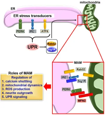
regulation of the ER and mitochondrial dynamics and cellular homeostasis (Figure 1).
5. ER-PM contact sites and the UPR
Regions of the ER closely apposed to the PM (the distance is typically within 10 to 30
nm) were first revealed by electron microscopic analysis 80. ER-PM contact sites have
emerged as key regulators of intracellular calcium dynamics 81-84. Previous studies have
shown that calcium depletion in the ER lumen triggers extracellular calcium influx
11
Figure 1. Schematic describing the formation of MAM and the UPR. The UPR induces the expression of MAM connectors Rab32 and Sig1R, followed by fine-tuning
of calcium signaling, calcium shuttling, mitochondrial dynamics, ROS production and
neurite outgrowth through the formation of MAM. Sig1R can bind to IRE1 to regulate its
stabilization. The stabilized IRE1 causes the long-lasting activation, and promotes the
cellular survival under ER stress condition. A second MAM connector, MFN2, interacts
with PERK to inhibit its activity for regulating ROS production, calcium shuttling and
12
lumen 85-86. Stromal-interacting molecule 1 (STIM1) is an integral ER protein that
regulates the formation of ER-PM contact sites in response to calcium depletion in the
luminal ER 87. STIM1 senses a decrease in the intraluminal ER calcium level and
undergoes conformational changes. The exposed domains following the conformational
change to STIM1 preferentially target phosphatidylinositol 4,5-biphosphate (PI(4, 5)P2)
enriched ER-PM contact sites 85, 88-89. STIM1 directly recruits and forms a complex with
Orai1, calcium channels localized at the PM, followed by replenishing calcium levels in
the ER lumen 85, 90.
ER dynamics is regulated by microtubule- and actin-binding proteins 91-92. Of those
proteins, filamin A (FLNA) is known as a connector between the ER and actin
cytoskeleton 91. PERK has been identified as an interacting partner of FLNA at the ER
membrane by a proximity-dependent biotin identification (BioID) assay 93. PERK acts as
a scaffold molecule for FLNA, enabling interlocking between F-actin and ER dynamics.
The dimerization of PERK induced by calcium depletion in the ER lumen is necessary
for the interaction between PERK and FLNA. Simultaneously, the decrease in calcium
concentration in the ER lumen leads to activation of STIM1. The PERK-FLNA axis
accelerates the remodeling and alters the polymerization dynamics of F-actin, followed
by the relocalization of cortical ER containing STIM1 to the PM. These morphological
changes to the ER are regulated by the PERK-FLNA connection and allow for the
efficient formation of ER-PM contact sites and calcium influx to replenish the luminal
calcium level. Thus, PERK manipulates morphological changes to the ER and the
formation of ER-PM contact sites by its dimerization, but not via signal transduction.
The other ER stress transducer, IRE1, is also involved in cytoskeleton remodeling via
13
cytosolic domain of IRE1 94. The dimerization of IRE1 is an essential step for the
physiological interaction with FLNA. The IRE1 dimer acts as a scaffold molecule, and
recruits FLNA and protein kinase C type (PKC). FLNA is phosphorylated by PKC,
followed by an increase in the remodeling of the actin cytoskeleton and cell migration.
The interaction between IRE1 and FLNA implicates significant roles for ER functions
and ER-derived signaling including the UPR at lamellipodia and filopodia, where active
actin dynamics are observed. Additionally, the connection between IRE1 and FLNA
implies the involvement of IRE1 in the formation of ER-PM contact sites, like those of
PERK. As mentioned in Section 3, the IRE1 pathway is responsible for the regulation of
ER biogenesis through manipulation of lipid biosynthesis. ER-PM contact sites are
known to play roles in the supply of membrane lipids from the ER membrane to the PM
81. Lipid transfer at ER-PM contact sites may facilitate extension of the PM and alter
cellular morphology. The IRE1 pathway may bidirectionally regulate the formation of
ER-PM contact sites via modulation of actin dynamics and the supply of membrane lipids
at contact sites via fine-tuning of lipid biosynthesis. Collectively, these reports on PERK
and IRE1 serve as reminders to the importance of defining the interactome of ER stress
transducers, which may reveal unexpected mechanisms for cooperative regulation among
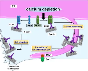
the UPR, actin dynamics and ER-PM contact sites (Figure 2).
6. Concluding remarks
Understanding how ER morphology is regulated is essential for understanding how
the ER communicates with other organelles. Moreover, characterizing this regulation is
important for comprehending cellular homeostasis because a well-developed ER that
14
Figure 2. Schematic describing the formation of ER-PM contact sites and the UPR.
PERK dimerizes in response to calcium depletion in the ER lumen. The PERK dimer
binds to FLNA, which accelerates formation of ER-PM contact sites containing STIM1
and Orai1. The contact sites promote calcium influx, which returns calcium levels to
normal values in the ER lumen. The IRE1 dimer also interacts with FLNA. Although the
effect of the IRE1-FLNA interaction on the formation of ER-PM contact sites remains
15
signal transmission through the formation of contact sites. These organelle contacts
regulate the dynamics of each organelle and signal transduction, and ultimately
orchestrate cellular homeostasis and biological functions. The UPR is a major signaling
system found in the ER and is involved in regulating morphological changes to the ER.
The mechanisms that regulate contact sites between the ER and other organelles are not
fully understood. However, further studies will uncover new signaling cascades and
concepts for organelle communication that is regulated by the UPR. Although not
discussed in this review, research has also focused on characterizing contact sites between
the ER and cellular components other than mitochondria and the PM (e.g., endosome and
Golgi apparatus). Such studies may provide novel information that further elucidates the
comprehensive regulation of cellular functions by the ER and UPR. These types of
approaches may provide insight into new therapeutic targets for a variety of diseases
including neurodegenerative diseases associated with the UPR and the organelle contact
sites. The expansion of a broad spectrum of UPR signaling to regulation of morphological
changes and contact sites should assist with deciphering the mechanisms for manipulating
an intricate organelle network, which may lead to breakthroughs in therapeutic strategies
that target various diseases.
Acknowledgments
We thank the members of our laboratory, M. Kaneko, R. Asada, K. Matsuhisa and T.
Okamoto for valuable discussions related to this work. This work was partly supported
by grants from the Japan Society for the Promotion of Science KAKENHI (JP17H06416,
16
and The Sumitomo Electric Industries Group Corporate Social Responsibility Foundation.
We thank Edanz Group (www.edanzediting.com/ac) for editing a draft of this manuscript.
Conflicts of interest
17 References
1. Kaufman, R. J., Orchestrating the unfolded protein response in health and
disease. The Journal of clinical investigation 2002,110 (10), 1389-98.
2. Ron, D., Translational control in the endoplasmic reticulum stress response. The
Journal of clinical investigation 2002,110 (10), 1383-8.
3. Rutkowski, D. T.; Kaufman, R. J., A trip to the ER: coping with stress. Trends
in cell biology 2004,14 (1), 20-8.
4. Ron, D.; Walter, P., Signal integration in the endoplasmic reticulum unfolded
protein response. Nature reviews. Molecular cell biology 2007,8 (7), 519-29.
5. Wu, J.; Kaufman, R. J., From acute ER stress to physiological roles of the
Unfolded Protein Response. Cell death and differentiation 2006,13 (3), 374-84.
6. Reimold, A. M.; Iwakoshi, N. N.; Manis, J.; Vallabhajosyula, P.;
Szomolanyi-Tsuda, E.; Gravallese, E. M.; Friend, D.; Grusby, M. J.; Alt, F.; Glimcher, L. H., Plasma
cell differentiation requires the transcription factor XBP-1. Nature 2001,412 (6844),
300-7.
7. Zhang, K.; Shen, X.; Wu, J.; Sakaki, K.; Saunders, T.; Rutkowski, D. T.; Back,
S. H.; Kaufman, R. J., Endoplasmic reticulum stress activates cleavage of CREBH to
induce a systemic inflammatory response. Cell 2006,124 (3), 587-99.
8. Vecchi, C.; Montosi, G.; Zhang, K.; Lamberti, I.; Duncan, S. A.; Kaufman, R.
J.; Pietrangelo, A., ER stress controls iron metabolism through induction of hepcidin.
Science (New York, N.Y.) 2009,325 (5942), 877-80.
9. Lee, A. H.; Scapa, E. F.; Cohen, D. E.; Glimcher, L. H., Regulation of hepatic
lipogenesis by the transcription factor XBP1. Science (New York, N.Y.) 2008,320 (5882),
18
10. Mao, T.; Shao, M.; Qiu, Y.; Huang, J.; Zhang, Y.; Song, B.; Wang, Q.; Jiang, L.;
Liu, Y.; Han, J. D.; Cao, P.; Li, J.; Gao, X.; Rui, L.; Qi, L.; Li, W.; Liu, Y., PKA
phosphorylation couples hepatic inositol-requiring enzyme 1alpha to glucagon signaling
in glucose metabolism. Proceedings of the National Academy of Sciences of the United
States of America 2011,108 (38), 15852-7.
11. Iwakoshi, N. N.; Lee, A. H.; Glimcher, L. H., The X-box binding protein-1
transcription factor is required for plasma cell differentiation and the unfolded protein
response. Immunological reviews 2003,194, 29-38.
12. Gass, J. N.; Gifford, N. M.; Brewer, J. W., Activation of an unfolded protein
response during differentiation of antibody-secreting B cells. The Journal of biological
chemistry 2002,277 (50), 49047-54.
13. Saito, A.; Hino, S.; Murakami, T.; Kanemoto, S.; Kondo, S.; Saitoh, M.;
Nishimura, R.; Yoneda, T.; Furuichi, T.; Ikegawa, S.; Ikawa, M.; Okabe, M.; Imaizumi,
K., Regulation of endoplasmic reticulum stress response by a BBF2H7-mediated Sec23a
pathway is essential for chondrogenesis. Nature cell biology 2009,11 (10), 1197-204.
14. Murakami, T.; Saito, A.; Hino, S.; Kondo, S.; Kanemoto, S.; Chihara, K.; Sekiya,
H.; Tsumagari, K.; Ochiai, K.; Yoshinaga, K.; Saitoh, M.; Nishimura, R.; Yoneda, T.;
Kou, I.; Furuichi, T.; Ikegawa, S.; Ikawa, M.; Okabe, M.; Wanaka, A.; Imaizumi, K.,
Signalling mediated by the endoplasmic reticulum stress transducer OASIS is involved
in bone formation. Nature cell biology 2009,11 (10), 1205-11.
15. Sha, H.; He, Y.; Chen, H.; Wang, C.; Zenno, A.; Shi, H.; Yang, X.; Zhang, X.;
Qi, L., The IRE1alpha-XBP1 pathway of the unfolded protein response is required for
19
16. West, M.; Zurek, N.; Hoenger, A.; Voeltz, G. K., A 3D analysis of yeast ER
structure reveals how ER domains are organized by membrane curvature. The Journal of
cell biology 2011,193 (2), 333-46.
17. Allison, R.; Edgar, J. R.; Pearson, G.; Rizo, T.; Newton, T.; Gunther, S.; Berner,
F.; Hague, J.; Connell, J. W.; Winkler, J.; Lippincott-Schwartz, J.; Beetz, C.; Winner, B.;
Reid, E., Defects in ER-endosome contacts impact lysosome function in hereditary
spastic paraplegia. The Journal of cell biology 2017,216 (5), 1337-1355.
18. Grimm, S., The ER-mitochondria interface: the social network of cell death.
Biochimica et biophysica acta 2012,1823 (2), 327-34.
19. Csordas, G.; Renken, C.; Varnai, P.; Walter, L.; Weaver, D.; Buttle, K. F.; Balla,
T.; Mannella, C. A.; Hajnoczky, G., Structural and functional features and significance
of the physical linkage between ER and mitochondria. The Journal of cell biology 2006,
174 (7), 915-21.
20. Tirasophon, W.; Welihinda, A. A.; Kaufman, R. J., A stress response pathway
from the endoplasmic reticulum to the nucleus requires a novel bifunctional protein
kinase/endoribonuclease (Ire1p) in mammalian cells. Genes & development 1998,12 (12),
1812-24.
21. Harding, H. P.; Zhang, Y.; Ron, D., Protein translation and folding are coupled
by an endoplasmic-reticulum-resident kinase. Nature 1999,397 (6716), 271-4.
22. Yoshida, H.; Okada, T.; Haze, K.; Yanagi, H.; Yura, T.; Negishi, M.; Mori, K.,
ATF6 activated by proteolysis binds in the presence of NF-Y (CBF) directly to the
cis-acting element responsible for the mammalian unfolded protein response. Molecular and
20
23. Calfon, M.; Zeng, H.; Urano, F.; Till, J. H.; Hubbard, S. R.; Harding, H. P.; Clark,
S. G.; Ron, D., IRE1 couples endoplasmic reticulum load to secretory capacity by
processing the XBP-1 mRNA. Nature 2002,415 (6867), 92-6.
24. Yoshida, H.; Matsui, T.; Yamamoto, A.; Okada, T.; Mori, K., XBP1 mRNA is
induced by ATF6 and spliced by IRE1 in response to ER stress to produce a highly active
transcription factor. Cell 2001,107 (7), 881-91.
25. Yoshida, H.; Matsui, T.; Hosokawa, N.; Kaufman, R. J.; Nagata, K.; Mori, K.,
A time-dependent phase shift in the mammalian unfolded protein response.
Developmental cell 2003,4 (2), 265-71.
26. Harding, H. P.; Novoa, I.; Zhang, Y.; Zeng, H.; Wek, R.; Schapira, M.; Ron, D.,
Regulated translation initiation controls stress-induced gene expression in mammalian
cells. Molecular cell 2000,6 (5), 1099-108.
27. Shi, Y.; Vattem, K. M.; Sood, R.; An, J.; Liang, J.; Stramm, L.; Wek, R. C.,
Identification and characterization of pancreatic eukaryotic initiation factor 2
alpha-subunit kinase, PEK, involved in translational control. Molecular and cellular biology
1998,18 (12), 7499-509.
28. Vattem, K. M.; Wek, R. C., Reinitiation involving upstream ORFs regulates
ATF4 mRNA translation in mammalian cells. Proceedings of the National Academy of
Sciences of the United States of America 2004,101 (31), 11269-74.
29. Barbosa-Tessmann, I. P.; Chen, C.; Zhong, C.; Siu, F.; Schuster, S. M.; Nick, H.
S.; Kilberg, M. S., Activation of the human asparagine synthetase gene by the amino acid
response and the endoplasmic reticulum stress response pathways occurs by common
21
30. Roybal, C. N.; Hunsaker, L. A.; Barbash, O.; Vander Jagt, D. L.; Abcouwer, S.
F., The oxidative stressor arsenite activates vascular endothelial growth factor mRNA
transcription by an ATF4-dependent mechanism. The Journal of biological chemistry
2005,280 (21), 20331-9.
31. Wang, X. Z.; Lawson, B.; Brewer, J. W.; Zinszner, H.; Sanjay, A.; Mi, L. J.;
Boorstein, R.; Kreibich, G.; Hendershot, L. M.; Ron, D., Signals from the stressed
endoplasmic reticulum induce C/EBP-homologous protein (CHOP/GADD153).
Molecular and cellular biology 1996,16 (8), 4273-80.
32. Ma, Y.; Brewer, J. W.; Diehl, J. A.; Hendershot, L. M., Two distinct stress
signaling pathways converge upon the CHOP promoter during the mammalian unfolded
protein response. Journal of molecular biology 2002,318 (5), 1351-65.
33. Harding, H. P.; Zhang, Y.; Zeng, H.; Novoa, I.; Lu, P. D.; Calfon, M.; Sadri, N.;
Yun, C.; Popko, B.; Paules, R.; Stojdl, D. F.; Bell, J. C.; Hettmann, T.; Leiden, J. M.; Ron,
D., An integrated stress response regulates amino acid metabolism and resistance to
oxidative stress. Molecular cell 2003,11 (3), 619-33.
34. Marciniak, S. J.; Yun, C. Y.; Oyadomari, S.; Novoa, I.; Zhang, Y.; Jungreis, R.;
Nagata, K.; Harding, H. P.; Ron, D., CHOP induces death by promoting protein synthesis
and oxidation in the stressed endoplasmic reticulum. Genes & development 2004,18 (24),
3066-77.
35. Ye, J.; Rawson, R. B.; Komuro, R.; Chen, X.; Dave, U. P.; Prywes, R.; Brown,
M. S.; Goldstein, J. L., ER stress induces cleavage of membrane-bound ATF6 by the same
22
36. Chen, X.; Shen, J.; Prywes, R., The luminal domain of ATF6 senses endoplasmic
reticulum (ER) stress and causes translocation of ATF6 from the ER to the Golgi. The
Journal of biological chemistry 2002,277 (15), 13045-52.
37. Yamamoto, K.; Sato, T.; Matsui, T.; Sato, M.; Okada, T.; Yoshida, H.; Harada,
A.; Mori, K., Transcriptional induction of mammalian ER quality control proteins is
mediated by single or combined action of ATF6alpha and XBP1. Developmental cell
2007,13 (3), 365-76.
38. Schroder, M.; Kaufman, R. J., The mammalian unfolded protein response.
Annual review of biochemistry 2005,74, 739-89.
39. Sriburi, R.; Jackowski, S.; Mori, K.; Brewer, J. W., XBP1: a link between the
unfolded protein response, lipid biosynthesis, and biogenesis of the endoplasmic
reticulum. The Journal of cell biology 2004,167 (1), 35-41.
40. Shaffer, A. L.; Shapiro-Shelef, M.; Iwakoshi, N. N.; Lee, A. H.; Qian, S. B.;
Zhao, H.; Yu, X.; Yang, L.; Tan, B. K.; Rosenwald, A.; Hurt, E. M.; Petroulakis, E.;
Sonenberg, N.; Yewdell, J. W.; Calame, K.; Glimcher, L. H.; Staudt, L. M., XBP1,
downstream of Blimp-1, expands the secretory apparatus and other organelles, and
increases protein synthesis in plasma cell differentiation. Immunity 2004,21 (1), 81-93.
41. Lee, A. H.; Iwakoshi, N. N.; Glimcher, L. H., XBP-1 regulates a subset of
endoplasmic reticulum resident chaperone genes in the unfolded protein response.
Molecular and cellular biology 2003,23 (21), 7448-59.
42. Lykidis, A.; Jackowski, S., Regulation of mammalian cell membrane
biosynthesis. Progress in nucleic acid research and molecular biology 2001,65, 361-93.
43. Kent, C., CTP:phosphocholine cytidylyltransferase. Biochimica et biophysica
23
44. Henneberry, A. L.; Wistow, G.; McMaster, C. R., Cloning, genomic organization,
and characterization of a human cholinephosphotransferase. The Journal of biological
chemistry 2000,275 (38), 29808-15.
45. Henneberry, A. L.; McMaster, C. R., Cloning and expression of a human
choline/ethanolaminephosphotransferase: synthesis of phosphatidylcholine and
phosphatidylethanolamine. The Biochemical journal 1999,339 ( Pt 2), 291-8.
46. Sriburi, R.; Bommiasamy, H.; Buldak, G. L.; Robbins, G. R.; Frank, M.;
Jackowski, S.; Brewer, J. W., Coordinate regulation of phospholipid biosynthesis and
secretory pathway gene expression in XBP-1(S)-induced endoplasmic reticulum
biogenesis. The Journal of biological chemistry 2007,282 (10), 7024-34.
47. Fujimoto, M.; Hayashi, T.; Su, T. P., The role of cholesterol in the association
of endoplasmic reticulum membranes with mitochondria. Biochemical and biophysical
research communications 2012,417 (1), 635-9.
48. Hayashi, T.; Rizzuto, R.; Hajnoczky, G.; Su, T. P., MAM: more than just a
housekeeper. Trends in cell biology 2009,19 (2), 81-8.
49. de Brito, O. M.; Scorrano, L., Mitofusin 2 tethers endoplasmic reticulum to
mitochondria. Nature 2008,456 (7222), 605-10.
50. De Vos, K. J.; Morotz, G. M.; Stoica, R.; Tudor, E. L.; Lau, K. F.; Ackerley, S.;
Warley, A.; Shaw, C. E.; Miller, C. C., VAPB interacts with the mitochondrial protein
PTPIP51 to regulate calcium homeostasis. Human molecular genetics 2012,21 (6),
1299-311.
51. Mendes, C. C.; Gomes, D. A.; Thompson, M.; Souto, N. C.; Goes, T. S.; Goes,
24
inositol 1,4,5-trisphosphate receptor preferentially transmits apoptotic Ca2+ signals into
mitochondria. The Journal of biological chemistry 2005,280 (49), 40892-900.
52. Iwasawa, R.; Mahul-Mellier, A. L.; Datler, C.; Pazarentzos, E.; Grimm, S., Fis1
and Bap31 bridge the mitochondria-ER interface to establish a platform for apoptosis
induction. The EMBO journal 2011,30 (3), 556-68.
53. Simmen, T.; Aslan, J. E.; Blagoveshchenskaya, A. D.; Thomas, L.; Wan, L.;
Xiang, Y.; Feliciangeli, S. F.; Hung, C. H.; Crump, C. M.; Thomas, G., PACS-2 controls
endoplasmic reticulum-mitochondria communication and Bid-mediated apoptosis. The
EMBO journal 2005,24 (4), 717-29.
54. van Vliet, A. R.; Verfaillie, T.; Agostinis, P., New functions of mitochondria
associated membranes in cellular signaling. Biochimica et biophysica acta 2014, 1843
(10), 2253-62.
55. Bononi, A.; Bonora, M.; Marchi, S.; Missiroli, S.; Poletti, F.; Giorgi, C.; Pandolfi,
P. P.; Pinton, P., Identification of PTEN at the ER and MAMs and its regulation of Ca(2+)
signaling and apoptosis in a protein phosphatase-dependent manner. Cell death and
differentiation 2013,20 (12), 1631-43.
56. Zhou, R.; Yazdi, A. S.; Menu, P.; Tschopp, J., A role for mitochondria in NLRP3
inflammasome activation. Nature 2011,469 (7329), 221-5.
57. Vance, J. E., MAM (mitochondria-associated membranes) in mammalian cells:
lipids and beyond. Biochimica et biophysica acta 2014,1841 (4), 595-609.
58. Alto, N. M.; Soderling, J.; Scott, J. D., Rab32 is an A-kinase anchoring protein
25
59. Bui, M.; Gilady, S. Y.; Fitzsimmons, R. E.; Benson, M. D.; Lynes, E. M.; Gesson,
K.; Alto, N. M.; Strack, S.; Scott, J. D.; Simmen, T., Rab32 modulates apoptosis onset
and mitochondria-associated membrane (MAM) properties. The Journal of biological
chemistry 2010,285 (41), 31590-602.
60. Ortiz-Sandoval, C. G.; Hughes, S. C.; Dacks, J. B.; Simmen, T., Interaction with
the effector dynamin-related protein 1 (Drp1) is an ancient function of Rab32 subfamily
proteins. Cellular logistics 2014,4 (4), e986399.
61. Liang, Y.; Lin, S.; Zou, L.; Zhou, H.; Zhang, J.; Su, B.; Wan, Y., Expression
profiling of Rab GTPases reveals the involvement of Rab20 and Rab32 in acute brain
inflammation in mice. Neuroscience letters 2012,527 (2), 110-4.
62. Haile, Y.; Deng, X.; Ortiz-Sandoval, C.; Tahbaz, N.; Janowicz, A.; Lu, J. Q.;
Kerr, B. J.; Gutowski, N. J.; Holley, J. E.; Eggleton, P.; Giuliani, F.; Simmen, T., Rab32
connects ER stress to mitochondrial defects in multiple sclerosis. Journal of
neuroinflammation 2017,14 (1), 19.
63. Mavlyutov, T. A.; Epstein, M. L.; Andersen, K. A.; Ziskind-Conhaim, L.; Ruoho,
A. E., The sigma-1 receptor is enriched in postsynaptic sites of C-terminals in mouse
motoneurons. An anatomical and behavioral study. Neuroscience 2010,167 (2), 247-55.
64. Hayashi, T.; Su, T. P., Sigma-1 receptor chaperones at the ER-mitochondrion
interface regulate Ca(2+) signaling and cell survival. Cell 2007,131 (3), 596-610.
65. Bhuiyan, M. S.; Fukunaga, K., Targeting sigma-1 receptor signaling by
endogenous ligands for cardioprotection. Expert opinion on therapeutic targets 2011,15
26
66. Kourrich, S.; Hayashi, T.; Chuang, J. Y.; Tsai, S. Y.; Su, T. P.; Bonci, A.,
Dynamic interaction between sigma-1 receptor and Kv1.2 shapes neuronal and behavioral
responses to cocaine. Cell 2013,152 (1-2), 236-47.
67. Bernard-Marissal, N.; Medard, J. J.; Azzedine, H.; Chrast, R., Dysfunction in
endoplasmic reticulum-mitochondria crosstalk underlies SIGMAR1 loss of function
mediated motor neuron degeneration. Brain : a journal of neurology 2015, 138 (Pt 4), 875-90.
68. Mitsuda, T.; Omi, T.; Tanimukai, H.; Sakagami, Y.; Tagami, S.; Okochi, M.;
Kudo, T.; Takeda, M., Sigma-1Rs are upregulated via PERK/eIF2alpha/ATF4 pathway
and execute protective function in ER stress. Biochemical and biophysical research
communications 2011,415 (3), 519-25.
69. Mori, T.; Hayashi, T.; Hayashi, E.; Su, T. P., Sigma-1 receptor chaperone at the
ER-mitochondrion interface mediates the mitochondrion-ER-nucleus signaling for
cellular survival. PloS one 2013,8 (10), e76941.
70. Selkoe, D. J., Translating cell biology into therapeutic advances in Alzheimer's
disease. Nature 1999,399 (6738 Suppl), A23-31.
71. Price, D. L.; Sisodia, S. S., Mutant genes in familial Alzheimer's disease and
transgenic models. Annual review of neuroscience 1998,21, 479-505.
72. Hedskog, L.; Pinho, C. M.; Filadi, R.; Ronnback, A.; Hertwig, L.; Wiehager, B.;
Larssen, P.; Gellhaar, S.; Sandebring, A.; Westerlund, M.; Graff, C.; Winblad, B.; Galter,
D.; Behbahani, H.; Pizzo, P.; Glaser, E.; Ankarcrona, M., Modulation of the endoplasmic
reticulum-mitochondria interface in Alzheimer's disease and related models. Proceedings
27
73. Hutter-Paier, B.; Huttunen, H. J.; Puglielli, L.; Eckman, C. B.; Kim, D. Y.;
Hofmeister, A.; Moir, R. D.; Domnitz, S. B.; Frosch, M. P.; Windisch, M.; Kovacs, D.
M., The ACAT inhibitor CP-113,818 markedly reduces amyloid pathology in a mouse
model of Alzheimer's disease. Neuron 2004,44 (2), 227-38.
74. Toyohara, J.; Sakata, M.; Ishiwata, K., Imaging of sigma1 receptors in the human
brain using PET and [11C]SA4503. Central nervous system agents in medicinal
chemistry 2009,9 (3), 190-6.
75. Prause, J.; Goswami, A.; Katona, I.; Roos, A.; Schnizler, M.; Bushuven, E.;
Dreier, A.; Buchkremer, S.; Johann, S.; Beyer, C.; Deschauer, M.; Troost, D.; Weis, J.,
Altered localization, abnormal modification and loss of function of Sigma receptor-1 in
amyotrophic lateral sclerosis. Human molecular genetics 2013,22 (8), 1581-600.
76. Mori, T.; Hayashi, T.; Su, T. P., Compromising sigma-1 receptors at the
endoplasmic reticulum render cytotoxicity to physiologically relevant concentrations of
dopamine in a nuclear factor-kappaB/Bcl-2-dependent mechanism: potential relevance to
Parkinson's disease. The Journal of pharmacology and experimental therapeutics 2012,
341 (3), 663-71.
77. Lindholm, D.; Wootz, H.; Korhonen, L., ER stress and neurodegenerative
diseases. Cell death and differentiation 2006,13 (3), 385-92.
78. Munoz, J. P.; Ivanova, S.; Sanchez-Wandelmer, J.; Martinez-Cristobal, P.;
Noguera, E.; Sancho, A.; Diaz-Ramos, A.; Hernandez-Alvarez, M. I.; Sebastian, D.;
Mauvezin, C.; Palacin, M.; Zorzano, A., Mfn2 modulates the UPR and mitochondrial
function via repression of PERK. The EMBO journal 2013,32 (17), 2348-61.
79. Verfaillie, T.; Rubio, N.; Garg, A. D.; Bultynck, G.; Rizzuto, R.; Decuypere, J.
28
ER-mitochondrial contact sites to convey apoptosis after ROS-based ER stress. Cell death
and differentiation 2012,19 (11), 1880-91.
80. Porter, K. R.; Palade, G. E., Studies on the endoplasmic reticulum. III. Its form
and distribution in striated muscle cells. The Journal of biophysical and biochemical
cytology 1957,3 (2), 269-300.
81. Saheki, Y.; De Camilli, P., Endoplasmic Reticulum-Plasma Membrane Contact
Sites. Annual review of biochemistry 2017,86, 659-684.
82. Henne, W. M.; Liou, J.; Emr, S. D., Molecular mechanisms of inter-organelle
ER-PM contact sites. Current opinion in cell biology 2015,35, 123-30.
83. Dickson, E. J.; Jensen, J. B.; Hille, B., Regulation of calcium and
phosphoinositides at endoplasmic reticulum-membrane junctions. Biochemical Society
transactions 2016,44 (2), 467-73.
84. Stefan, C. J.; Manford, A. G.; Emr, S. D., ER-PM connections: sites of
information transfer and inter-organelle communication. Current opinion in cell biology
2013,25 (4), 434-42.
85. Carrasco, S.; Meyer, T., STIM proteins and the endoplasmic reticulum-plasma
membrane junctions. Annual review of biochemistry 2011,80, 973-1000.
86. Lewis, R. S., Store-operated calcium channels: new perspectives on mechanism
and function. Cold Spring Harbor perspectives in biology 2011,3 (12).
87. Zhang, S. L.; Yu, Y.; Roos, J.; Kozak, J. A.; Deerinck, T. J.; Ellisman, M. H.;
Stauderman, K. A.; Cahalan, M. D., STIM1 is a Ca2+ sensor that activates CRAC
channels and migrates from the Ca2+ store to the plasma membrane. Nature 2005,437
29
88. Varnai, P.; Toth, B.; Toth, D. J.; Hunyady, L.; Balla, T., Visualization and
manipulation of plasma membrane-endoplasmic reticulum contact sites indicates the
presence of additional molecular components within the STIM1-Orai1 Complex. The
Journal of biological chemistry 2007,282 (40), 29678-90.
89. Maleth, J.; Choi, S.; Muallem, S.; Ahuja, M., Translocation between
PI(4,5)P2-poor and PI(4,5)P2-rich microdomains during store depletion determines STIM1
conformation and Orai1 gating. Nature communications 2014,5, 5843.
90. Sharma, S.; Quintana, A.; Findlay, G. M.; Mettlen, M.; Baust, B.; Jain, M.;
Nilsson, R.; Rao, A.; Hogan, P. G., An siRNA screen for NFAT activation identifies
septins as coordinators of store-operated Ca2+ entry. Nature 2013,499 (7457), 238-42.
91. Lynch, C. D.; Gauthier, N. C.; Biais, N.; Lazar, A. M.; Roca-Cusachs, P.; Yu, C.
H.; Sheetz, M. P., Filamin depletion blocks endoplasmic spreading and destabilizes
force-bearing adhesions. Molecular biology of the cell 2011,22 (8), 1263-73.
92. Park, S. H.; Zhu, P. P.; Parker, R. L.; Blackstone, C., Hereditary spastic
paraplegia proteins REEP1, spastin, and atlastin-1 coordinate microtubule interactions
with the tubular ER network. The Journal of clinical investigation 2010,120 (4),
1097-110.
93. van Vliet, A. R.; Giordano, F.; Gerlo, S.; Segura, I.; Van Eygen, S.; Molenberghs,
G.; Rocha, S.; Houcine, A.; Derua, R.; Verfaillie, T.; Vangindertael, J.; De Keersmaecker,
H.; Waelkens, E.; Tavernier, J.; Hofkens, J.; Annaert, W.; Carmeliet, P.; Samali, A.;
Mizuno, H.; Agostinis, P., The ER Stress Sensor PERK Coordinates ER-Plasma
Membrane Contact Site Formation through Interaction with Filamin-A and F-Actin
30
94. Urra, H.; Henriquez, D. R.; Canovas, J.; Villarroel-Campos, D.; Carreras-Sureda,
A.; Pulgar, E.; Molina, E.; Hazari, Y. M.; Limia, C. M.; Alvarez-Rojas, S.; Figueroa, R.;
Vidal, R. L.; Rodriguez, D. A.; Rivera, C. A.; Court, F. A.; Couve, A.; Qi, L.; Chevet, E.;
Akai, R.; Iwawaki, T.; Concha, M. L.; Glavic, A.; Gonzalez-Billault, C.; Hetz, C.,
IRE1alpha governs cytoskeleton remodelling and cell migration through a direct