Parasitic Infections in Pregnancy
Decrease Placental Transfer of
Antipneumococcus Antibodies
Noah D. McKittrick,aDavid M. Vu,bIndu Malhotra,cCharles H. King,c Francis Mutuku,dA. Desiree LaBeaudb
Division of Infectious Diseases, Department of Medicine, Stanford University School of Medicine, Stanford, California, USAa; Division of Infectious Diseases, Department of Pediatrics, Lucille Packard Children's Hospital at Stanford School of Medicine, Stanford, California, USAb; Center for Global Health and Diseases, Case Western Reserve University School of Medicine, Cleveland, Ohio, USAc; Department of Environment and Health Sciences, Technical University of Mombasa, Mombasa, Kenyad
ABSTRACT Many factors can influence maternal placental antibody transfer to the fetus, which confers important immune protection to the newborn infant. However, little is known about the effect of maternal parasitic infection on placental antibody transfer. To investigate this, we selected from a parent study of 576 pregnant Ke-nyan women four groups of women with term deliveries (ⱖ37 weeks), including un-infected women (n ⫽ 30) and women with solo infections with malaria (n ⫽ 30), hookworm (n⫽30), or schistosomiasis (n⫽10). Maternal plasma at delivery and in-fant cord blood were tested via multiplex fluorescent bead assay for IgG against 10 pneumococcal serotypes (PnPs 1, 4, 5, 6B, 7F, 9V, 14, 18C, 19F, and 23F), diphtheria toxoid, and Haemophilus influenzae type B. Infants born to mothers with prenatal malaria, hookworm, or Schistosoma haematobium infections were associated with a significantly reduced ratio of maternal to infant cord blood antibody concentration
forStreptococcus pneumoniaeserotypes 1, 4, 5, 6B, 7F, 9V, and 18C compared to
in-fants of uninfected mothers. Anti-diphtheria toxoid and anti-H. influenzaetype B IgG ratios were not significantly different among infection groups. Prenatal parasitic in-fections decrease the transfer of maternal IgG antibodies to infants for several sero-types ofS. pneumoniae.
KEYWORDS humoral immunity, parasites, placental antibody transfer, pregnancy
T
he newborn infant is immunologically disadvantaged. Its naive adaptive immune system places it at risk for infection from many pathogens early in life. Morbidity and mortality due to infectious diseases often are greatest during the first few months of life, leading to a high global burden of infectious diseases in young infants. Much of the infant’s early protection from infectious diseases results from passive immunity acquired from its mother, both from the placental transfer of IgG antibodiesin uteroand in the acquisition of (primarily) mucosal IgA protection via breastfeeding (1). Transplacental antibody transfer, in particular, is the underpinning of many prenatal vaccination strategies (2).
Maternal-fetal antibody transfer is an active process, whereby IgG molecules are transported across the placenta from maternal to fetal circulation (3). This is accom-plished via Fc␥receptors [FcRn] on the syncytiotrophoblast. This process favors certain IgG subtypes over others, IgG1 being the most preferentially transferred, followed in order by IgG4, IgG3, and finally IgG2 (4). Transfer of maternal antibodies begins as early as the second trimester, but most activity occurs in the second half of the third trimester (5). Various factors have been shown to affect the magnitude of this transfer, from gestational age and low birth weight to maternal hypergammaglobulinemia (6, 7).
Received9 February 2017Returned for
modification24 March 2017Accepted7
April 2017
Accepted manuscript posted online12
April 2017
CitationMcKittrick ND, Vu DM, Malhotra I, King
CH, Mutuku F, Labeaud AD. 2017. Parasitic infections in pregnancy decrease placental transfer of antipneumococcus antibodies. Clin Vaccine Immunol 24:e00039-17.https://doi .org/10.1128/CVI.00039-17.
EditorPatricia P. Wilkins, CDC
Copyright© 2017 American Society for
Microbiology.All Rights Reserved.
Address correspondence to Noah D. McKittrick, [email protected].
crossm
on August 17, 2020 by guest
http://cvi.asm.org/
Prenatal infections can dynamically alter this process of antibody transfer. HIV infection has been shown to decrease transplacental antibody transfer for various pathogen-specific antibodies, including antibodies against Haemophilus influenzae, pertussis, pneumococcus, measles, and tetanus (8, 9, 10). In addition, placental malaria has been associated with decreased transfer of antibodies to measles, pneumococcus, tetanus, and respiratory syncytial virus (RSV) (11, 12). The effect of other maternal infections on antibody transfer is not as well understood, however. In the developing world, other parasitic infections such as soil-transmitted helminths and schistosomiasis are common and represent a significant public health challenge (13, 14). Prenatal screening and treatment for these infections have been a part of standard WHO guidelines for many years, but maternal infection with these parasites continues to occur at significant rates. In this study, we investigated the effects of prenatal parasitic infections with malaria, hookworm, and schistosomiasis on transplacental antibody transfer of maternal IgG antibody against 10Streptococcus pneumoniaeserotype poly-saccharides (PnPs 1, 4, 5, 6B, 7F, 9V, 14, 18C, 19F, and 23F), diphtheria toxoid, and
Haemophilus influenzaetype B [Hib] polysaccharide. As these three bacterial diseases
can be particularly fatal to young infants, it is especially important to better understand the maternal-fetal interface for acquisition of immunity and how this may be perturbed by common parasitic infections.
RESULTS
This research was completed as part of an ongoing cohort study in Kenya, with enrollment beginning in July in 2013 and follow-up ending in July 2016, investigating the effect of maternal parasitic infections on infant immunity. In that parent study, 576 pregnant women were enrolled in the Msambweni District Hospital Antenatal Clinic (ANC) and monitored for the course of their pregnancy. Infants born to these mothers were subsequently monitored after birth until up to 3 years of age. The study was performed in a high-risk area for parasitic disease in a predominantly rural location in the southern coastal region of Kenya.
Four stratified maternal subgroups were selected for analysis, along with their newborns. These groups were (i) uninfected women (n⫽ 30), (ii) women with only malaria infection (n⫽30), (iii) women with only hookworm infection (n⫽30), and (iv) women with only Schistosoma haematobium infection (n ⫽ 10). “Uninfected” was defined as having no evidence of parasitic infections based on testing either in the prenatal clinic or at delivery. The infection groups were limited to women with single infections to prevent confounding by polyparasitism. To account for previously ob-served effects of gestational age on transplacental antibody transfer, only mothers who delivered at term (ⱖ37 weeks) were included in the study. Since trained ultrasound technicians and equipment were not available, this was estimated by the revised Dubowitz clinical measurement, which includes 34 physical and neurologic assess-ments to predict the gestational age at birth (15) and has been validated in populations similar to our cohort (16, 17).
Baseline characteristics of the four groups are shown in Table 1. There were no significant differences among the groups for most individual features; however, two baseline characteristics did show significant differences among the groups. First, mean maternal body mass index (BMI [measured at first prenatal visit]) was significantly higher in the uninfected group (26.8) compared to the infected groups (23.5 for malaria, 23.7 for hookworm, and 22.4 for schistosomiasis; P⫽ 0.023). In addition, on a self-reported survey regarding estimated household expenditures in thousands of Kenyan shillings (Ksh) per month, there was a significantly larger amount of mean monthly expenditures in the uninfected group (4.87 thousand Ksh/month) versus the other groups, with 4.53 thousand Ksh/month for malaria, 4.40 thousand Ksh/month for hookworm, and 4.10 thousand Ksh/month for schistosomiasis (P⫽0.007).
Evaluation of transplacental antibody transfer.Serum antibody levels to the 10
S. pneumoniaeserotype polysaccharide antigens, Hib polyribosylribitol phosphate (PRP)
polysaccharide, and diphtheria toxoid (Dpt) were measured using a fluorescent
on August 17, 2020 by guest
http://cvi.asm.org/
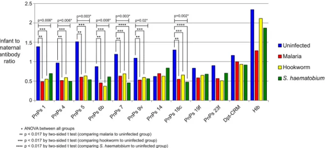
plexed bead-based immunoassay. The ratio of the geometric means of infant and maternal plasma antibody concentrations (cord/maternal ratio [CMR]) was used to measure placental antibody transfer. The observed CMR values are shown in Fig. 1. There was a significant difference for seven of the pneumococcal serotypes when comparing uninfected mothers to the infected parasite groups (Table 2). All three infections showed a reduction in the rate of antibody transfer for PnPs 1 (malaria, 65%; hookworm, 60%; and schistosomiasis, 50%), with similar patterns in the other pneu-mococcal serotypes. Significant reductions in antibody transfer were observed among the study groups via analysis of variance (ANOVA) for PnPS 4 (P⫽0.006), 5 (P⫽0.003), 6B (P ⫽ 0.008), 7F (P ⫽ 0.003), 9V (P ⫽ 0.02), and 18C (P ⫽ 0.002). In pairwise comparisons of infected versus uninfected mothers, for malaria and hookworm the differences in CMR were significant (Pⱕ0.017,ttest) for all 7 antibodies (PnPs 1, 4, 5, 6B, 7F, 9V, and 18C). For the schistosomiasis group, significance was only reached for antibodies against PnPs 7F and 18C. For PnPs 14, 19F, and 23F, there was no significant difference among the 4 groups in CMR. Rates of placental transfer of diphtheria toxoid (Dpt-CRM)- and Haemophilus influenzae type B (Hib)-specific antibodies were not significantly different among the study subgroups.
Given the differences in BMIs and monthly expenditures between the uninfected and infected groups, we performed linear regression analyses to test the hypothesis that these baseline variables could be associated with the antibody transfer process by comparing them to the CMR for each antigen. There were no significant associations found between the CMRs and BMIs or monthly expenditures for any of the tested antigens. Our cohort had a relatively low prevalence of HIV, with a total of 6 mothers (6%) infected and an equal distribution among the groups (P⫽0.876). In unpairedt
TABLE 1Baseline characteristics
Characteristic
Result for:
Pvalue by ANOVA Uninfected
(nⴝ30)
Malaria (nⴝ30)
Hookworm (nⴝ30)
Schistosomiasis (nⴝ10)
Maternal age, mean yr 26.1 26.5 27.2 22.5 0.248
Previous pregnancies, mean no. 1.67 2.63 2.6 1.7 0.168
Household expenditures, mean 1,000 Ksh/mo 4.87 4.53 4.4 4.1 0.007
Maternal mean BMI 26.8 23.5 23.7 22.4 0.023
Maternal Hgb at delivery, mean g/dl 9.62 9.97 10.15 10.3 0.067
Maternal HIV infection, total no. (%) 2 (6.7) 1 (3.3) 2 (6.7) 1 (10) 0.876
Newborn wt, mean g 3,061.3 3,035.7 3,044.7 3,087 0.986
Newborn head circumference, mean cm 33.3 33.5 33.7 33.4 0.504
Gestational age by Dubowitz score, mean wk 40 41.3 41.3 38.5 0.132
FIG 1Ratios of infant to maternal antibody concentrations (geometric means) by infection status. PnPs,S. pneumoniaepolysaccharide; Dpt-CRM, diphtheria-CRM; Hib,Haemophilus influenzaetype b. ANOVA com-parisons among all groups are indicated forPvalues of 0.006, 0.003, 0.008, 0.02, and 0.002.
on August 17, 2020 by guest
http://cvi.asm.org/
tests comparing the geometric mean antibody concentration ratios of the 12 antibod-ies in HIV⫹versus HIV⫺groups, no significant differences were detected.
DISCUSSION
In this study, we observed that prenatal malaria, hookworm, andS. haematobium
infections are associated with a decrease in the transplacental transfer of several anti-S.
pneumoniaeserotype antigen-specific antibodies compared to the levels observed for
uninfected mothers and their infants. Previous studies have reported the effect of infections such as HIV and malaria on placental antibody transfer. Data on the effect of helminth infections on this process are scarce (18), and this is the first time that prenatal hookworm and schistosomiasis have been shown to affect the maternal transfer of antipneumococcal antibodies. In addition, we have also observed that there can be differences in antipneumococcal antibody transfer among the different tested sero-types.
Although there were no differences in most baseline characteristics among our different study subgroups, we did find significant differences in terms of maternal BMIs and average monthly expenditures. With a higher average BMI in the uninfected group, there may have been an association between nutritional status and the efficiency of placental antibody transfer; malnourishment during pregnancy has previously been associated with a 14% reduction in placental antibody transfer (19). The uninfected group also reported a larger monthly expenditure than the other groups, a variable that serves as a proxy for socioeconomic status in our study, suggesting a correlation of monthly income and parasite burden.
Unlike previous studies, our results did not find a significant difference in
Haemo-philus influenzaetype B placental antibody transfer in malaria-infected women (9). This
difference could be related to the fact that, in our study, the malaria cohort was chosen for inclusion if they had any evidence of infection throughout their pregnancy, whereas other studies have only looked at the role of placental malaria at the time of delivery (8, 20).
At present, there is no routine prenatal vaccination forS. pneumoniaein Kenya, and participants in our maternal cohort were born before the 2011 introduction of child-hood pneumococcal conjugate vaccine (PCV) vaccination there. Data on pneumococcal seroprevalence rates in Africa are limited. One group from Burkina Faso surveyed the serotype-specific antibody concentrations of pneumococcal IgG during a meningococ-cal outbreak in 2006 and showed that antibody concentrations increased with age in the absence of any prior vaccination programs, which suggested that natural immunity develops over time as a result of exposure to circulating pneumococcal serotypes or TABLE 2Placental transfer of antigen-specific antibodies by infection status
Antigena
CMR (95% CI) forb:
Pvalue by ANOVA Uninfected Malaria Hookworm Schistosomiasis
PnPs 1 1.39 (0.67–2.86) 0.49 (0.37–0.64) 0.55 (0.45–0.67) 0.69 (0.33–1.43) 0.006c
PnPs 4 0.96 (0.63–1.47) 0.52 (0.43–0.62) 0.59 (0.48–0.72) 0.49 (0.38–0.65) 0.006c
PnPs 5 1.51 (0.80–2.85) 0.60 (0.52–0.69) 0.63 (0.45–0.89) 0.53 (0.32–0.88) 0.003c
PnPs 6b 0.87 (0.60–1.27) 0.46 (0.35–0.59) 0.37 (0.23–0.58) 0.61 (0.31–1.18) 0.008c
PnPs 7 1.19 (0.89–1.60) 0.63 (0.48–0.82) 0.69 (0.51–0.93) 0.45 (0.20–1.04) 0.003d
PnPs 9v 1.09 (0.67–1.77) 0.53 (0.41–0.68) 0.59 (0.43–0.83) 0.56 (0.37–0.85) 0.02c
PnPs 14 0.62 (0.39–0.98) 0.69 (0.54–0.89) 0.62 (0.43–0.90) 0.83 (0.55–1.25) 0.827 PnPs 18c 1.30 (0.74–2.30) 0.55 (0.47–0.64) 0.65 (0.50–0.86) 0.47 (0.37–0.61) 0.002d
PnPs 19f 0.83 (0.50–1.39) 0.58 (0.48–0.71) 0.65 (0.47–0.89) 0.68 (0.43–1.06) 0.547 PnPs 23f 0.90 (0.54–1.49) 0.57 (0.49–0.66) 0.51 (0.32–0.81) 0.70 (0.44–1.12) 0.185 Dpt-CRM 1.16 (0.82–1.65) 1.00 (0.87–1.15) 0.93 (0.71–1.23) 0.91 (0.66–1.26) 0.612 Hib 2.34 (1.17–4.69) 1.28 (0.71–2.32) 2.11 (1.16–3.85) 1.87 (0.62–5.65) 0.53
aPnPs,S. pneumoniaepolysaccharide; Dpt-CRM, diphtheria toxoid; Hib,Haemophilus influenzaetype B. bCMR, geometric mean antibody concentration ratio of infant cord plasma to maternal delivery plasma; CI,
confidence interval.
cPⱕ0.017 by unpairedttest compared to uninfected for malaria and hookworm groups. dPⱕ0.017 by unpairedttest compared to uninfected for all infection groups.
on August 17, 2020 by guest
http://cvi.asm.org/
from other bacteria with cross-reacting polysaccharides (21). By age 10, depending on the serotype, 50 to 80% of those individuals had a level of antibody presumed to be protective against invasive disease (ⱖ0.35g/ml, based on prior studies [22]). The same researchers looked at pneumococcal carriage prevalence in Burkina Faso and found that most serotypes had a 0.5 to 5% prevalence of asymptomatic carriage in the population (23). In our group, the percentage of mothers with protective antibody levels varied from 5 to 83% among the different pneumococcal serotypes and between 5 to 79% in the infants. Diphtheria protection (defined asⱖ0.1 IU/ml [24]) was seen in 73% of mothers and 74% of infants, while Hib protective levels (ⱖ1g/ml (25) were only 3% in mothers and 11% in the infants.
It has been observed that the majority of antipneumococcal antibody produced is of the IgG2 subclass, similar to other polysaccharide antigens (26), although antipneu-mococcal antibodies of all IgG subclasses can be detected (27). While IgG2 has the least transplacental transfer of all the subclasses, there is on average a 50% transfer of this class (4). One study documented total IgG transfer rates between 77 and 116% in serotypes 1, 3, 6B, 9V, and 14, with no correlation found between the concentrations of serotype-specific IgG subclasses and the transplacental transfer of these antibodies (28). Our study did not look at IgG subclass-specific data, as it was not possible to selectively measure these data with the multiplex platform we utilized.
Our reports of differences between antibody transfer between pneumococcal sero-types and the lack of an effect onH. influenzaeand diphtheria antibody transfer may be explained by several possible mechanisms. It has been observed that parasitic infections can raise levels of total IgG in the host, and since Fc␥receptors can be saturated (5), there could be a decrease in serotype-specific transfer in mothers having higher levels of IgG, as a consequence of subclass competition for limited Fc binding sites. There may also be a role for antigen specificity in transport, where higher levels of maternal IgG against herpes simplex virus, tetanus toxoid, streptolysin O, and S.
pneumoniaehave been shown to have an inverse relationship with transfer of these
antibodies to babies (8). It may be possible that IgGs against certain serotypes of pneumococcus are more easily transferred than others (supported by the variable transfer rates of serotypes [28]) and that this could be further altered by the inflam-matory changes induced by different parasitic infections. Ultimately, more research is warranted to better understand the specifics of this process.
This study was observational, with the potential for experimental bias and con-founding. A more robust randomized clinical trial, however, would have been difficult given the current standard of care for antiparasitic treatment in pregnancy. We could not control for the timing of the infection with this study, as infection status was determined not by proximity to birth but instead by the presence of parasites at any point throughout the pregnancy. Because the majority of antibody transfer occurs later in the third trimester, it remains unclear how differences in the timing of exposure might ultimately affect placental antibody transfer by the time of birth. All of the women in the study received antiparasitic therapy for malaria and intestinal helminths during their pregnancies, which involved intermittent preventive treatment (IPTp) and mebendazole, so it is not possible to analyze the effect of treatment on the antibody transfer process in this group. Despite this consistent antiparasitic antenatal treatment, many mothers in the parent study from which this group was selected were found to be infected at delivery with malaria (8.5%), hookworm (5.7%), and/orS. haematobium
(29%). These diseases are continuously circulating in the community, and none of the mothers were on continuous prophylaxis— only intermittent treatment. Furthermore, mebendazole does not effectively treat all intestinal helminths, particularly Trichuris; whileS. haematobiuminfections, which require praziquantel therapy, went untreated for the duration of the pregnancy based on Kenya Ministry of Health (MOH) guidelines. HIV infections have been shown to have a significant effect on placental antibody transfer in previous studies (8, 9, 10), a potential source for confounding in these results. Our cohort had an HIV prevalence of 6%, comparable to recent prevalence data for this region (5.9% in Kwale County as of 2016 [29]), and infected participants were evenly
on August 17, 2020 by guest
http://cvi.asm.org/
distributed within the study groups. When comparing the CMRs of the 12 antibodies in HIV⫹versus HIV⫺groups, there was no association found between infection status and
antibody transfer.
This study was limited by a smaller sample size in the schistosomiasis group. Despite over 40% of women in the parent study being infected with this parasite, the majority of participants withSchistosomaexposure had other parasitic infections. Because the presence of two or more concurrent infections may have led to confounding, the infected groups in this study were selected from mothers with single-parasite infections only. With that exclusion, there remained only a limited number of women infected
withSchistosomaonly.
Vaccine-preventable infectious diseases remain a serious health issue for very young infants in developing countries. Where there is a potential for prenatal parasitic infections to decrease the transfer of protective maternal antibodies to infants via transplacental transport, it will be important to develop prenatal care and vaccination strategies to mitigate this effect. The present study adds to the evidence in favor of antiparasite control among expectant mothers, so that their infants may have the best possible protection before they receive their standard vaccinations. Because this ob-servational study was limited in its ability to provide a detailed analysis of the effects of treatment on the transfer of antibodies from mother to fetus, future prospective studies are needed to further define this important aspect of early immunity.
MATERIALS AND METHODS
Ethical oversight.Ethical approval of this study was obtained from the Kenyatta National Hospital Ethical Review Committee (protocol no. P85/03/2013), from the Institutional Review Board for Human Studies at Case Western Reserve University (IRB no. 01-13-13), and from the Stanford University School of Medicine Institutional Review Board (protocol no. IRB-31468). Mothers provided written informed consent for their own participation and that of their infants.
Inclusion and exclusion criteria.To be included as a study participant, mothers must have (i) been permanent residents of Msambweni, (ii) been willing to participate in prenatal and postnatal care at ANC, (iii) exclusively used study health facilities for primary health care, (iv) delivered at Msambweni District Hospital (now Msambweni County Referral Hospital), (v) been willing to provide blood, urine, and stool samples, and (vi) been willing to allow examination, blood, urine, and stool testing of their infants. Pregnant mothers were excluded if they (i) presented with a complicated delivery resulting in significant infant morbidity at birth, (ii) delivered an infant atⱕ27 weeks gestation and/or (iii) had known chronic illness (e.g., tuberculosis [TB], diabetes, or renal failure), (iv) severe anemia requiring hospitalization (hemoglobin [Hgb] level of ⬍6 g/dl accompanied by symptoms requiring urgent treatment), (v) permanent disability that impeded study participation and/or comprehension, (vi) known multiple pregnancy and/or (vii) plans to relocate after delivery.
Clinical testing.Maternal participants were screened at each prenatal visit and at delivery for malaria (blood smear by light microscopy and DNA PCR from red blood cell [RBC] pellet [30]), schistosomiasis (urine filtration egg counts [31] and plasma anti-soluble worm adult protein [anti-SWAP] IgG4 [32]), and intestinal helminths (stool microscopy following the Ritchie method [33]). Mothers found positive for malaria and/or intestinal helminths received treatment within 72 h per Kenya Ministry of Health [MOH] guidelines. The current standard of care includes a single dose of mebendazole for helminths and 4 monthly doses of intermittent preventive treatment (IPTp) for malaria with sulfadoxine/pyrimethamine. This prophylaxis and treatment for malaria followed MOH guidelines for antenatal care. Any pregnant woman developing symptoms of infection between study visits was asked to return for further evalu-ation and care. For the mother-infant pairs included in this study, both maternal venous blood and umbilical cord blood were collected at delivery, as previously described (34). Plasma was stored at⫺80°C until antibody assays were performed.
Determination of antibody levels.Serum antibody levels to the 10S. pneumoniaepolysaccharide serotypes, Hib PRP polysaccharide, and diphtheria toxoid were measured using a fluorescent multiplexed bead-based immunoassay employing Luminex multiple analyte profiling technology (Luminex, Austin, TX) (35, 36). Briefly, the 10 pneumococcal polysaccharide (PnPs) antigens (serotypes 1, 4, 5, 6B, 7F, 9V, 14, 18C, 19F, and 23F) were obtained from the American Type Culture Collection (Manassas, VA) and conjugated to carboxylated microspheres (Luminex, Austin, TX) using 4-(4,6-dimethoxy[1,3,5]triazin-2-yl)-4-methyl-morpholinium [DMTMM] (37). Hib PRP capsular polysaccharide conjugated to human albu-min and diphtheria toxoid were obtained from the National Institute for Biological Standards and Control (NIBSC [Potters Bar, United Kingdom]). These were coupled to carboxylated microspheres (Luminex, Austin, TX) using a two-step carbodiimide reaction, as previously described (38).
For the assay standard, a 1:1 mixture of 007SP (anti-PnPs human serum; NIBSC) and 09/222 (anti-Hib human serum; NIBSC) reference sera was used. Dilutions of the mixture were tested against each reference serum, as well as the reference serum for diphtheria toxoid (10/26; NIBSC), to define the concentrations of all 12 antibodies in the 007SP-09/222 mixture. A series of seven 3-fold dilutions of the standard serum starting at 1:50 was prepared using a standard diluent buffer consisting of
on August 17, 2020 by guest
http://cvi.asm.org/
buffered saline (PBS), 1% bovine serum albumin (BSA), and 0.05% Tween 20, with 5g/ml of pneumo-coccal cell wall polysaccharide (CWPS [Statens Serum Institute, Copenhagen, Denmark]) added to quench nonspecific binding ofS. pneumoniaeantibodies (39). Mother and infant test serum samples were diluted 1:50 in PBS–1% BSA– 0.05% Tween 20 containing 5g/ml CWPS at 50l/well and tested in duplicate. Antigen-coupled beads were added to the samples in a mixture containing 1,000 beads/antigen target in the same diluent serum at 50l/well of a 96-well plate and incubated on a rotator plate at 4°C overnight. After incubation, the beads were washed with PBS– 0.05% Tween 20 and stained with goat anti-human IgG Fc␥-specific R-phycoerythrin (R-PE) conjugate for an additional 30 min at room temper-ature. After a final wash, the beads were resuspended in 100l PBS– 0.05% Tween 20 and data were acquired using a BioPlex Magpix multiplex reader (Bio-Rad, Hercules, CA).
Luminex data analysis was performed using Bio-plex Manager 6.1 software (Bio-Rad). Antibody concentrations were derived by interpolating the measured median fluorescence intensity (MFI) values of samples against a 5-parameter logistic curve fit from MFI values of the standard curve. We used the geometric means of the infant (cord blood) antibody concentrations and maternal (delivery blood) antibody concentrations due to the variability and skewed distribution of IgG concentrations between individuals.
Statistical analysis.Results were analyzed in Excel (Microsoft, 2011) and on STATA (Statacorp, 2015). The study groups for this research were chosen as a pilot study, with cost and sample availability limiting the group selection. Assuming a mean antibody transfer ratio in healthy pregnancies of 120%⫾40% (40), a sample size of 28 per group would give 80% power to detect a 25% decrease in placental antibody transfer. Significance among groups was first confirmed by analysis of variance (ANOVA). Subsequent two-sided t tests were performed for comparisons between each infection group and the uninfected group, with Bonferroni correction for multiple comparisons settingPⱕ0.017 as significant.
ACKNOWLEDGMENTS
This work was supported by the Bill and Melinda Gates Foundation Healthy Growth Award (OPP1066865). Fellowship support for Noah McKittrick was obtained from the Child Health Research Institute at Stanford.
We acknowledge the women and their children who participated in this study, as well as the team of nurses, technicians, and staff in the Msambweni County Referral Hospital and their Antenatal Clinic.
REFERENCES
1. Simon AK, Hollander GA, McMichael A. 2015. Evolution of the immune system in humans from infancy to old age. Proc Biol Sci 282:20143085. https://doi.org/10.1098/rspb.2014.3085.
2. Swamy GK, Heine RP. 2015. Vaccinations for pregnant women. Obstet Gynecol 125:212–226.https://doi.org/10.1097/AOG.0000000000000581. 3. Kristoffersen EK. 1996. Human placental Fc gamma-binding proteins in the maternofetal transfer of IgG. APMIS Suppl 64:5–36.https://doi.org/ 10.1111/j.1600-0463.1996.tb05583.x.
4. Costa-Carvalho BT, Vieria HM, Dimantas RB, Arslanian C, Naspitz CK, Solé D, Carneiro-Sampaio MM. 1996. Transfer of IgG subclasses across pla-centa in term and preterm newborns. Braz J Med Biol Res 29:201–204. 5. Saji F, Samejima Y, Kamiura S, Koyama M. 1999. Dynamics of immuno-globulins at the feto-maternal interface. Rev Reprod 4:81– 89.https://doi .org/10.1530/ror.0.0040081.
6. Munoz FM, Englund JA. 2000. A step ahead. Infant protection through maternal immunization. Pediatr Clin North Am 47:449 – 463.https://doi .org/10.1016/S0031-3955(05)70217-0.
7. Munoz FM, Englund JA. 2001. Vaccines in pregnancy. Infect Dis Clin North Am 15:253–271.https://doi.org/10.1016/S0891-5520(05)70278-6. 8. de Moraes-Pinto MI, Almeida AC, Kenj G, Filgueiras TE, Tobias W, Santos
AM, Carneiro-Sampaio MM, Farhat CK, Milligan PJ, Johnson PM, Hart CA. 1996. Placental transfer and maternally acquired neonatal IgG immunity in human immunodeficiency virus infection. J Infect Dis 173:1077–1084. https://doi.org/10.1093/infdis/173.5.1077.
9. Mulholland K, Suara RO, Siber G, Roberton D, Jaffar S, N=Jie J, Baden L, Thompson C, Anwaruddin R, Dinan L, Glezen WP, Francis N, Fritzell B, Greenwood BM. 1996. Maternal immunization withHaemophilus influ-enzaetype b polysaccharide-tetanus protein conjugate vaccine in The Gambia. JAMA 275:1182–1188.
10. Hood N, Chan MC, Maxwell SM, Familusi JB, Hart CA. 1994. Placental transfer of tetanus toxoid antibodies in Nigerian mothers. Ann Trop Paediatr 14:179 –182. https://doi.org/10.1080/02724936.1994 .11747714.
11. Brair ME, Brabin BJ, Milligan P, Maxwell S, Hart CA. 1994. Reduced
transfer of tetanus antibodies with placental malaria. Lancet 343: 208 –209.https://doi.org/10.1016/S0140-6736(94)90991-1.
12. Cumberland P, Shulman CE, Maple PA, Bulmer JN, Dorman EK, Kawuondo K, Marsh K, Cutts FT. 2007. Maternal HIV infection and placental malaria reduce transplacental antibody transfer and tetanus antibody levels in newborns in Kenya. J Infect Dis 196:550 –557.https:// doi.org/10.1086/519845.
13. Pullan RL, Smith JL, Jasrasaria R, Brooker SJ. 2014. Global numbers of infection and disease burden of soil transmitted helminth infections in 2010. Parasit Vectors 7:37.https://doi.org/10.1186/1756-3305-7-37. 14. King CH, Dickman K, Tisch DJ. 2005. Reassessment of the cost of chronic
helminthic infection: a meta-analysis of disability-related outcomes in endemic schistosomiasis. Lancet 365:1561–1569. https://doi.org/10 .1016/S0140-6736(05)66457-4.
15. Dubowitz LM, Dubowitz V, Goldberg C. 1970. Clinical assessment of gestational age in the newborn infant. J Pediatr 77:1.https://doi.org/10 .1016/S0022-3476(70)80038-5.
16. Raghu MB, Patel YS, Gupta K. 1981. Estimation of gestational age in Zambian newborn infants. Ann Trop Paediatr 1:245–247.https://doi.org/ 10.1080/02724936.1981.11748097.
17. Sunjoh F, Njamnshi AK, Tietche F, Kago I. 2004. Assessment of gesta-tional age in the Cameroonian newborn infant: a comparison of four scoring methods. J Trop Pediatr 50:285–291.https://doi.org/10.1093/ tropej/50.5.285.
18. Gebreegziabiher D, Desta K, Desalegn G, Howe R, Abebe M. 2014. The effect of maternal helminth infection on maternal and neonatal immune function and immunity to tuberculosis. PLoS One 9:e93429.https://doi .org/10.1371/journal.pone.0093429.
19. Cavalcante RS, Kopelman BI, Costa-Carvalho BT. 2008. Placental transfer ofHaemophilus influenzaetype b antibodies in malnourished pregnant women. Braz J Infect Dis 12:47–51.https://doi.org/10.1590/S1413-8670 2008000100011.
20. Okoko BJ, Wesumperuma LH, Ota MO, Pinder M, Banya W, Gomez SF, McAdam KP, Hart AC. 2001. The influence of placental malaria infection
on August 17, 2020 by guest
http://cvi.asm.org/
and maternal hypergammaglobulinemia on transplacental transfer of antibodies and IgG subclasses in a rural West African population. J Infect Dis 184:627– 632.https://doi.org/10.1086/322808.
21. Yaro S, Njanpop-Lafourcade BM, Drabo A, Idohou RS, Kroman SS, Sanou O, Traoré Y, Sangaré L, Diagbouga SP, Koeck JL, Borrow R, Gessner BD, Mueller JE. 2014. Antipneumococcal seroprevalence and pneumococcal carriage during a meningococcal epidemic in Burkina Faso. J Infect Dis 209:1241–1250.https://doi.org/10.1093/infdis/jit641.
22. Plotkin SA. 2010. Correlates of protection induced by vaccination. Clin Vaccine Immunol 17:1055–1065.https://doi.org/10.1128/CVI.00131-10. 23. Mueller JE, Yaro S, Ouédraogo MS, Levina N, Njanpop-Lafourcade BM,
Tall H, Idohou RS, Sanou O, Kroman SS, Drabo A, Nacro B, Millogo A, van der Linden M, Gessner BD. 2012. Pneumococci in the African meningitis belt: meningitis incidence and carriage prevalence in children and adults. PLoS One 7:e52464. https://doi.org/10.1371/journal.pone .0052464.
24. Hasselhorn HM, Nübling M, Tiller FW, Hofmann F. 1998. Factors influ-encing immunity against diphtheria in adults. Vaccine 16:70 –75.https:// doi.org/10.1016/S0264-410X(97)00148-5.
25. Käyhty H, Peltola H, Karanko V, Mäkelä PH. 1983. The protective level of serum antibodies to the capsular polysaccharide of Haemophilus influ-enzae type b. J Infect Dis 147:1100.https://doi.org/10.1093/infdis/147.6 .1100.
26. Barrett DJ, Ayoub EM. 1986. IgG2 subclass restriction of antibody to pneumococcal polysaccharides. Clin Exp Immunol 63:127–134. 27. Bardardottir E, Jonsson S, Jonsdottir I, Sigfusson A, Valdimarsson H. 1990.
IgG subclass response and opsonization ofStreptococcus pneumoniae
after vaccination of healthy adults. J Infect Dis 162:482– 488.https://doi .org/10.1093/infdis/162.2.482.
28. Carvalho BT, Carneiro-Sampaio MM, Solé D, Naspitz C, Leiva LE, Sorensen RU. 1999. Transplacental transmission of serotype-specific pneumococcal antibodies in a Brazilian population. Clin Diagn Lab Immunol 6:50 –54.
29. National AIDS Control Council. 2016. Kenya HIV county profiles, p 93–97. Kenya Ministry of Health, Nairobi, Kenya.
30. McNamara DT, Thomson JM, Kasehagen LJ, Zimmerman PA. 2004. De-velopment of a multiplex PCR-ligase detection reaction assay for diag-nosis of infection by the four parasite species causing malaria in humans. J Clin Microbiol 42:2403–2410. https://doi.org/10.1128/JCM.42.6.2403 -2410.2004.
31. Peters PAS, Mahmoud AAF, Warren KS, Ouma JH, Siongok TKA. 1976. Field studies of a rapid, accurate means of quantifyingSchistosoma haematobium eggs in urine samples. Bull World Health Organ 54: 159 –162.
32. DuVall AS, Fairley JK, Sutherland L, Bustinduy AL, Mungai PL, Muchiri EM, Malhotra I, Kitron U, King CH. 2014. Development of a specimen-sparing multichannel bead assay to detect antiparasite IgG4 for the diagnosis of
SchistosomaandWuchereriainfections on the coast of Kenya. Am J Trop Med Hyg 90(4):638 – 645.https://doi.org/10.4269/ajtmh.13-0292. 33. Ritchie LS. 1948. An ether sedimentation technique for routine stool
examinations. Bull US Army Med Dep 8:326.
34. Malhotra I, Mungai P, Muchiri E, Ouma J, Sharma S, Kazura JW, King CL. 2005. Distinct Th1- and Th2-type prenatal cytokine responses to Plas-modium falciparum erythrocyte invasion ligands. Infect Immun 73: 3462–3470.https://doi.org/10.1128/IAI.73.6.3462-3470.2005.
35. Pickering JW, Martins TB, Schroder MC, Hill HR. 2002. Comparison of a multiplex flow cytometric assay with enzyme-linked immunosorbent assay for quantitation of antibodies to tetanus, diphtheria, and Haemo-philus influenzaetype b. Clin Diagn Lab Immunol 9:872– 876. 36. Malhotra I, McKibben M, Mungai P, McKibben E, Wang X, Sutherland LJ,
Muchiri EM, King CH, King CL, LaBeaud AD. 2015. Effect of antenatal parasitic infections on anti-vaccine IgG levels in children: a prospective birth cohort study in Kenya. PLoS Negl Trop Dis 9:e0003466.https://doi .org/10.1371/journal.pntd.0003466.
37. Schlottmann SA, Jain N, Chirmule N, Esser MT. 2006. A novel chemistry for conjugating pneumococcal polysaccharides to Luminex micro-spheres. J Immunol Methods 309:75– 85.https://doi.org/10.1016/j.jim.2005 .11.019.
38. Lal G, Balmer P, Stanford E, Martin S, Warrington R, Borrow R. 2005. Development and validation of a nonaplex assay for the simultaneous quantitation of antibodies to nineStreptococcus pneumoniaeserotypes. J Immunol Methods 296:135–147.https://doi.org/10.1016/j.jim.2004.11 .006.
39. Siber GR, Priehs C, Madore DA. 1989. Standardization of antibody assays for measuring the response to pneumococcal infection and immuniza-tion. Pediatr Infect Dis J 8(Suppl 1):S84 –S91.https://doi.org/10.1097/ 00006454-198901001-00029.
40. Palmeira P, Quinello C, Silveira-Lessa AL, Zago CA, Carneiro-Sampaio M. 2012. IgG placental transfer in healthy and pathological pregnancies. Clin Dev Immunol 2012:985646.https://doi.org/10.1155/2012/985646.