Copyright © 2003, American Society for Microbiology. All Rights Reserved.
Cervicovaginal Neutralizing Antibodies to Herpes Simplex Virus
(HSV) in Women Seropositive for HSV Types 1 and 2
Francois-Xavier Mbopi-Ke´ou,
1† Laurent Be´lec,
2Julie Dalessio,
3‡ Je´roˆme Legoff,
2Ge´rard Gre´senguet,
4Philippe Mayaud,
5David W. G. Brown,
1and Rhoda Ashley Morrow
3*
Central Public Health Laboratory, Virus Reference Division, Colindale,
1and Department of Infectious and Tropical Diseases,
London School of Hygiene and Tropical Medicine,
5London, United Kingdom; Laboratoire de Virologie, Unite´ INSERM U430,
and Universite´ Pierre et Marie Curie (Paris VI), Hoˆpital Broussais, Paris, France
2; Department of Laboratory Medicine,
University of Washington School of Medicine, Seattle, Washington 98105
3; and Centre National de Re´fe´rence
des Maladies Sexuellement Transmissibles et du SIDA (CNRMST), Bangui, Central African Republic
4Received 23 October 2002/Returned for modification 17 January 2003/Accepted 5 March 2003
Antibodies to herpes simplex virus type 1 (HSV-1) and HSV-2 of the immunoglobulin G (IgG) and IgA
isotypes were detected in the cervicovaginal secretions (CVS) of 77 HSV-1- and HSV-2-seropositive but
clinically asymptomatic African women by type-specific enhanced chemiluminescence Western blotting
(ECL-WB). Of the 77 subjects, 34 were HIV negative, shedding HSV-2 DNA in their genital secretions; 20 were HIV
positive, shedding HSV-2 DNA; and 23 were HIV negative, not shedding HSV-2 DNA. HSV-specific IgG was
detected in CVS of nearly 70% of the women studied. HSV-specific IgA was found in CVS of 50% of the women
studied. The distribution of CVS HSV-specific antibodies to each HSV type was highly heterogeneous, with a
slight predominance of detectable IgG to HSV-1 (59%) over IgG to HSV-2 (41%), whereas the frequency of
detectable IgA to HSV-1 (39%) was similar to that of IgA to HSV-2 (36%). The presence of detectable
HSV-specific antibodies was inversely associated with HSV-2 DNA genital asymptomatic shedding but was not
affected by HIV seropositivity. In addition, 13 of 77 (17%) CVS samples showed neutralizing activity against
HSV-2, as assessed by an HSV-2 in vitro infectivity reduction assay. Neutralizing activity in CVS was associated
with the presence of IgG and/or IgA antibodies to HSV-1 and/or to HSV-2 by ECL-WB. Among women whose
CVS showed HSV-2-neutralizing activity, the specific activity of HSV-specific neutralizing antibodies was
substantially (fivefold) higher in HSV-2 DNA shedders than in nonshedders. In conclusion, HSV-specific
antibodies are frequently detected in CVS of asymptomatic African women seropositive for HSV-1 and HSV-2.
A subset of these women had functional neutralizing activity against HSV-2 in their CVS. The origin of these
antibodies and their role in HSV-2 disease of the female genital tract remain to be determined.
Herpes simplex virus type 2 (HSV-2) establishes latent
in-fection in the dorsal root ganglion following inin-fection at a
genital or anal mucocutaneous surface. Intermittent HSV
re-activation results in the production of infectious HSV with or
without clinical symptoms. Asymptomatic or subclinical genital
shedding of HSV-2 is detected in 55% of immunocompetent
females and 81% of males according to daily sampling studies
in the United States (32, 33). Cross-sectional studies with
lim-ited sampling have shown 20 to 40% HSV subclinical shedding
rates in HIV-seronegative as well as HIV-infected women
liv-ing in sub-Saharan Africa (19, 22).
The factors that affect whether recurrent HSV shedding
from the genital mucosa is subclinical or symptomatic are not
well understood. Specific immune responses against HSV
oc-curring at the level of genital mucosa are likely to play a role
(26). Both cell-mediated and humoral immune factors have
been described in the genital tract during active genital herpes
episodes in humans (1, 2, 14, 16, 17, 21, 23) and in the mouse
model of vaginal infection with an attenuated HSV-2 strain
(20, 24). Women with symptomatic genital herpes have
anti-bodies to HSV-2 of both the immunoglobulin A (IgA) and IgG
isotypes in cervicovaginal secretions (CVS) (2, 21). The
asso-ciation of these antibodies with subclinical HSV excretion and
their possible function remain poorly understood.
The aim of this study was to detect HSV antibodies and
neutralizing activity in CVS of women seropositive for both
HSV-1 and HSV-2 and to stratify the CVS HSV-2-specific
antibody activity according to their HSV-2 DNA genital
shed-ding status. We observed HSV-2-specific antibodies in the
CVS of a high proportion of HSV-2-seropositive women.
An-tibody detection was inversely associated with viral shedding.
Furthermore, in vitro neutralizing activity was detected in 17%
of CVS samples and was associated with the presence of
HSV-specific antibodies.
MATERIALS AND METHODS
Study population and sample processing.Women (mean age, 27 years; range, 15 to 48 years) attending the Centre National de Re´fe´rence des Maladies Sex-uellement Transmissibles et du SIDA (CNRMST/SIDA) of Bangui, the capital city of the Central African Republic, during the period July to October 1998 were recruited for a study of human immunodeficiency virus type 1 (HIV-1) RNA and HSV-2 DNA genital shedding, as previously reported (19). After verbal informed consent to participate was given, each of the 213 participants underwent general
* Corresponding author. Mailing address: Children’s Hospital &
Regional Medical Center, University of Washington, 4800 Sand Point
Way N.E., Box 359300-8G-3, Seattle, WA 98105. Phone: (206)
987-2117. Fax: (206) 987-3885. E-mail: rhoda.morrow@seattlechildrens.org.
† Present address: Department of Oral Medicine, Eastman Dental
Institute for Oral Health Care Science, U.C.L., University of London,
United Kingdom.
‡ Present address: 1110 29th Ave., Seattle, WA 98122-5010.
388
on August 17, 2020 by guest
http://cvi.asm.org/
and genital examination. HIV testing and routine biological tests for the diag-nosis of treatable sexually transmitted diseases (STD) were also carried out. A 7-day follow-up appointment was then arranged, and free appropriate STD treatment was provided. Women wishing to know their HIV serostatus received counseling at the voluntary counseling and testing unit of the CNRMST/SIDA. HIV-1-infected women belonged to the A1 (n⫽13) and A2 (n⫽7) categories of the Centers for Disease Control and Prevention classification for HIV infec-tion. None received antiretroviral therapy, and none was pregnant at the time of sampling.
Ethical approval was given by the London School of Hygiene and Tropical Medicine, London, United Kingdom. Verbal informed consent was obtained from all participants.
Eighty-four women were selected because they had no sign of cervicitis or active STD infection at the time of enrollment and because their CVS samples were free of hemoglobin (Hb) traces and semen contamination. Of the 84 eligible women, 77 were selected because they had evidence of HSV-2 infection by either HSV-2 type-specific serum antibodies or detection of HSV-2 DNA in their CVS. All 77 women were found to be infected with both HSV-1 and HSV-2.
Samples for laboratory testing.Peripheral blood samples were taken, and aliquots of plasma separated from EDTA-anticoagulated blood were stored at ⫺30°C before shipment to the United Kingdom for HSV serology. Serum IgG antibodies to HSV-1 and HSV-2 were detected by in-house competitive type-specific gG-based enzyme-linked immunosorbent assay (ELISA) at the Central Public Health Laboratory Services, Colindale, United Kingdom, as described previously (13).
During genital examination, CVS samples were collected by standardized nontraumatic 60-s vaginal washing with 3 ml of phosphate-buffered saline (PBS) (6). After centrifugation of the CVS at 1,000⫻gfor 10 min, the cell supernatant and the cellular pellet were collected, aliquoted, and stored separately at⫺30°C. CVS samples were confirmed to be free of significant amounts of semen, as assessed by a negative test for prostatic specific antigen (PSA IMX system; Abbott Laboratories, Chicago, Ill.) (19). Traces of Hb in vaginal secretions were detected by spectrophotometry (threshold of sensitivity, 10 mg/ml) of the acel-lular fraction of genital fluids, as described previously (29).
Concentrations of CVS IgA and IgG.CVS levels of total Ig of unknown specificity of the IgA and IgG classes were measured by capture ELISA with goat anti-human Fc-␣conjugated with peroxidase (Pierce, Rockford, Ill.) or goat anti-human Fc-␥conjugated with peroxidase (Pierce), as described previously (27), with a positivity threshold of 0.5g/100l.
Detection of CVS HSV-specific antibodies.Cell-free fractions of CVS were tested for IgA or IgG antibodies to HSV-1- or HSV-2-infected cell-protein mixtures by enhanced chemiluminescence Western blotting (ECL-WB) as de-scribed in detail in reference 10. In brief, proteins from diploid fibroblasts infected with HSV-1 (strain E115) or HSV-2 (strain 333) were separated by polyacrylamide gel electrophoresis and then transferred to polyvinylidene diflu-oride (PVDF; Immobilon P; Millipore, Bedford, Mass.) as described previously (10). Strips were blocked with 0.5% Tween 20 in PBS for 1 h. CVS samples (25 l) were diluted 1:40 in 4% goat serum (in PBS) and incubated overnight on strips. After washing, goat anti-human antibody conjugated to peroxidase (Boehringer Mannheim, Indianapolis, Ind.) was added. The ECL-WB detection system (Amersham, Arlington Heights, Ill.) was added per the manufacturer’s instructions for 1 min. Treated strips were exposed to X-ray film (Hyperfilm-ECL; Amersham) for 30 s (IgG) or 2 min (IgA), and then the film was developed in a Kodak X-Omat processor.
Neutralizing activity of CVS HSV-specific antibodies.An HSV-2 infectivity inhibition assay was performed, as previously described (3), with slight modifi-cations as follows. Duplicate portions of the acellular factor of each CVS were serially diluted twofold in 100l of Eagle’s minimal essential medium (EMEM) with 2% fetal bovine serum (FBS) from 1:4 to 1:128 in 96-well plates (Falcon, Becton Dickinson Labware, Lincoln Park, N.J.). HSV-2 strain 333 was adjusted to 3.2⫻10350% tissue culture infective doses (TCID
50)/ml, and 100l was added to each well. In addition, virus stock diluted twofold from 3.2⫻103 TCID50/ml to 1⫻102TCID50/ml was added in duplicate to wells containing 100 l of EMEM–2% FBS to generate a virus dilution curve. After 1 h of incubation at 37°C, the virus and CVS sample mixtures and the diluted virus were trans-ferred to wells containing confluent monolayers of ELVIS cells (Diagnostics Hybrids, Inc., Athens, Ohio) (30). Plates were incubated overnight at 37°C with 5% CO2. Cells were washed with PBS (pH 7.2) and then lysed with 100l of a buffered 1% NP-40 solution. Induced-galactosidase activity secondary to HSV infection was detected by adding 135l of 3 mM ONPG (o-nitrophenyl␣-D
-galactopyranoside) (Sigma, St. Louis, Mo.) in a mixture of 0.1 M sodium phos-phate (pH 7.5), 1 mM MgCl2, and 45 mM-mercaptoethanol. Absorbance was
read as the optical density at 415 nm (OD415) on a Biotek EL340 plate reader (Winooski, Vt.). OD values were further processed with Biotek KC4 software.
To determine the extent of HSV-2 infectivity inhibition by the diluted secre-tions, OD values were first adjusted for background by subtracting the mean OD from six uninfected cell wells. Units of virus infectivity were interpolated from the virus dilution curve and expressed as neutralizing activity units (NAU).
The specific activities of HSV-specific neutralizing antibodies were calculated as the ratios of HSV-2 NAU to the CVS concentrations of total IgA plus total IgG in 100l of cell-free CVS. We used the assumption that the concentration of total IgM in CVS is much lower than those of IgA plus IgG, as previously observed in African women whatever their HIV serological status (5).
HSV DNA detection.HSV DNA was detected in the acellular fraction of CVS by in-house PCR for a conserved 290-bp segment of the DNA polymerase gene of HSV-1 and HSV-2 (12). Hybridization of PCR products was further carried out by DNA enzyme immunoassay (DEIA) according to the manufacturer’s instructions (Gen-Eti-K; Sorin Biomedica, Saluggia, Italy) by using a single-stranded biotinylated HSV probe (5⬘-GTCCTCACCGCCGAACTGAG-3⬘; po-sitions 3109 to 3128) as previously described (19). The quantity of amplicons for HSV was assessed by the OD450of hybridized products as previously described (18). DEIA hybridization allowed semiquantitation of HSV DNA target. In this method, there was a linear association between ODs from the cutoff point (OD ⫽0.150) to 1.5 and the number of HSV DNA copies, ranging from 1 to 1,000 copies, respectively. ODs of 1.5 to 3 followed a nonlinear relationship. Strain differentiation was performed by restriction fragment length polymorphrism as described previously (31).
Statistical analysis.Quantitative results are expressed as means⫾standard errors. The nonparametric Mann-Whitney U test was used to compare the specific activities of neutralizing antibodies between HSV-2 shedders versus nonshedders. Fisher’s exact test was used to compare the frequencies of detect-able HSV-specific antibodies among groups. Correlation between the distribu-tion of the specific activities of CVS HSV-specific neutralizing antibodies and CVS levels of HSV-2 DNA was assessed with Spearman’s rank order test.
RESULTS
Study subjects.
Three groups of women were classified
ac-cording to their HIV serostatus and their HSV-2 DNA genital
shedding status as follows. Group I comprised 34
HSV-2-se-ropositive, HIV-seronegative women who all had HSV-2 DNA
in their genital secretions. Group II comprised 20 HSV-2- and
HIV-1-seropositive women who were shedding HSV-2 DNA.
Group III comprised 23 HSV-2-seropositive,
HIV-seronega-tive women without detectable HSV-2 DNA in their genital
secretions. None of the study women had oral or genital signs
or symptoms of herpes at the time of sampling.
CVS total IgG and IgA.
Total IgG and IgA levels in CVS
were measured to calculate the specific activities of
HSV-neutralizing activity in CVS. Table 1 shows the higher
concen-trations of total IgG in CVS over those of total IgA regardless
of HIV status or HSV-2 shedding. This is a well-established
observation in HIV-positive and in HIV-seronegative women
(5, 15). In the present series, the mean concentrations of IgG
and IgA were not affected by either HIV infection or HSV-2
shedding.
CVS HSV-specific IgG and IgA.
ECL-WB for IgG to HSV-1
and HSV-2 antibody was performed with samples from 75 of
the 77 HSV-2-infected women; 68 subjects had sufficient CVS
to perform ECL-WB testing for both IgG and IgA isotypes.
Fifty-two of 75 (69%) women tested had HSV-specific IgG
in their CVS (Table 2). Seventeen of 34 (50%) of the
HIV-seronegative women in group I (HSV-2 shedders) had IgG to
HSV, mainly to HSV-1 (16 of 34 [47%]) and less frequently to
HSV-2 (7 of 34 [21%]). Seventeen of 19 (89%)
HIV-seropos-itive women in group II (HSV-2 shedders) had IgG. The
fre-quencies of IgG to HSV-1 and IgG to HSV-2 were the same
(13 of 19 [68%]). Finally, 18 of 22 (82%) of HIV-seronegative
on August 17, 2020 by guest
http://cvi.asm.org/
women in group III (HSV-2 nonshedders) had IgG to HSV-1
(15 of 22 [68%]) or to HSV-2 (11 of 22 [50%]).
Overall, 34 of 68 (50%) women tested had HSV-specific IgA
in their CVS. Eleven of 31 (35%) of the group I women had
IgA to HSV—mainly to HSV-1 (10 of 31 [32%]) and less
frequently to HSV-2 (5 of 33 [16%]). Eleven of 16 (69%) of the
group II women had IgA: 9 of 16 (56%) to HSV-1 and 10 of 16
(63%) to HSV-2. Twelve of 21 (57%) of the group III women
had IgA: 8 of 21 (38%) to HSV-1 and 10 of 21 (48%) to
HSV-2.
Only 20 of 53 (38%) HSV-2 shedders had HSV-2-specific
IgG in their CVS compared with 11 of 22 (50%) nonshedders
(not significant). Similarly, only 15 of 47 (32%) HSV-2
shed-ders had HSV-2-specific IgA in their CVS compared with 10 of
21 (48%) nonshedders (not significant). Thus, the presence
versus absence of detectable IgG or IgA antibodies binding to
HSV-2 did not appear to be closely linked to HSV-2 DNA
shedding. However, if only HIV-negative women were
com-pared, a significantly lower proportion of shedders (7 of 34
[21%]) than nonshedders (11 of 22 [50%]) had CVS IgG to
HSV-2 (
P
⬍
0.05).
Fourteen of 53 (26%) HSV-2 shedders (10 in group I and 4
in group II) had only IgG to HSV-1 in their CVS; 7 of 49
(15%) HSV-2 shedders (6 in group I and 1 in group II) showed
only IgA to HSV-1 in their CVS. Similar frequencies of the
unique presence of detectable IgG to HSV-1 (7 of 22 [31%]) or
IgA to HSV-1 (2 of 21 [10%]) were observed in women
be-longing to group III. As with CVS IgG to HSV-2,
HIV-nega-tive shedders had a significantly lower frequency of IgA to
HSV-2 (5 of 31 [16%]) than did HIV-negative nonshedders (10
of 21 [48%]) (
P
⬍
0.05).
Overall, the frequency of detectable IgG to HSV-1 in CVS
(44 of 75 [59%]) was slightly higher than that of detectable IgG
to HSV-2 (31 of 75 [41%]) (
P
⬍
0.05), whereas the frequency
of detectable IgA to HSV-1 (27 of 68 [40%]) was similar to that
of IgA to HSV-2 (25 of 68 [37%]). Furthermore, the frequency
of detectable IgG to HSV-1 in CVS secretions of the study
population (44 of 75 [59%]) was higher than that of detectable
IgA to HSV-1 (27 of 68 [40%]) (
P
⫽
0.02). The frequency of
detectable CVS IgG to HSV-2 was similar to that of detectable
CVS IgA to HSV-2.
Finally, Table 3 depicts a wide variety in the distribution of
CVS IgA and IgG antibodies to HSV-1 and HSV-2. The most
obvious differences were in detection of antibodies to HSV-1
versus detection of antibodies to HSV-2. Several women had
only IgG to HSV-1 (
n
⫽
8). Nine had both IgG and IgA to
HSV-1 without antibodies to HSV-2, and 17 had IgG and IgA
to both HSV-1 and HSV-2. Fifteen women had detectable
HSV-specific IgG, but no IgA to either virus.
HSV-neutralizing activity.
HSV-neutralizing activity was
de-tected in the CVS of 13 women, including 10 of 54 (19%)
HSV-2 DNA shedders and 3 of 23 (13%) HSV-2 nonshedders
(Table 4). Twenty-five percent (5 of 20) of HIV-seropositive
women shedding HSV-2 had detectable neutralizing activity in
their CVS, compared to 14% (8 of 57) of HIV-seronegative
women. Thus, the presence of neutralizing CVS activity against
HSV was not greatly affected by HIV status or HSV-2 DNA
shedding.
Neutralizing activity most often occurred in CVS that had
detectable binding of antibody to HSV. Thus, 12 CVS samples
with neutralizing activity had at least HSV IgG by ECL-WB,
whereas only 1 sample lacked detectable IgG or IgA to HSV
(Table 3). Ten of 12 CVS samples with neutralizing activity had
both IgA and IgG to HSV-2, and 11 of 12 had IgG to HSV-2
(Table 3). These data provide suggestive evidence that at least
a portion of 2-neutralizing activity resides in the
HSV-specific antibody fraction of CVS.
Among the women with positive tests for HSV-2
neutraliza-TABLE 1. Levels of total IgG and IgA in CVS samples from 77 selected HSV-1- and HSV-2-seropositive African women living in Bangui,
Central African Republic, classified according to HIV serostatus and shedding of HSV-2 DNA in CVS
Study
group (n) shedding statusHSV-2 DNA HIV status
Ig level (g/100l)a
IgG IgA
I (34)
Positive
Negative
16.1
⫾
7.2 (
⬍
0.5–227.5)
c1.4
⫾
0.5 (
⬍
0.5–13.0)
cII (20)
Positive
Positive
17.7
⫾
5.3 (
⬍
0.5–96.5)
2.2
⫾
0.5 (
⬍
0.5–7.6)
III (23)
Negative
Negative
13.1
⫾
0.6 (
⬍
0.5–59.0)
1.2
⫾
0.9 (
⬍
0.5–42.0)
aValues are means⫾standard errors (with range in parentheses). bAs assessed by qualitative PCR for HSV DNA polymerase gene.
cThreshold of IgG or IgA detection, 0.5g/100l. Values below the threshold of measurement were arbitrarily taken as zero for calculations.
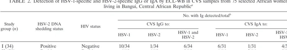
TABLE 2. Detection of HSV-1-specific and HSV-2-specific IgG or IgA by ECL-WB in CVS samples from 75 selected African women
living in Bangui, Central African Republic
aStudy
group (n) shedding statusHSV-2 DNA HIV status
No. with Ig detected/totalb
CVS IgG to: CVS IgA to:
HSV-1 HSV-2 HSV-1 andHSV-2 HSV-1 HSV-2 HSV-1 andHSV-2
I (34)
Positive
Negative
10/34
1/34
6/34
6/31
1/31
4/31
II (20)
Positive
Positive
4/19
4/19
9/19
1/16
2/16
8/16
III (23)
Negative
Negative
7/22
3/22
8/22
2/21
4/21
6/21
aAll women were seropositive for HSV-1 and HSV-2.
bAs assessed by ECL-WB. The denominator indicates the number of CVS lavage samples tested.
on August 17, 2020 by guest
http://cvi.asm.org/
tion, the specific activity of HSV neutralization in CVS was
substantially higher in both HIV-seronegative and
HIV-sero-positive HSV-2 DNA shedders (mean
⫾
standard error, 53.8
⫾
17.5) than in HSV-2 DNA nonshedders (mean
⫾
standard
error, 9.6
⫾
6.7) (Table 4) (
P
⬍
0.04). Moreover, the
distribu-tion of the specific activities of CVS HSV-specific neutralizing
antibodies and CVS levels of HSV-2 DNA (as estimated from
the OD
450of hybridized HSV PCR products) in the 10 women
shedding HSV-2 DNA in their CVS showed a highly significant
correlation (
r
2⫽
0.91;
P
⬍
0.007). The higher the relative level
of DNA, the higher the specific activity of neutralizing activity
to HSV-2 (data not shown).
DISCUSSION
In this study, only African women infected with both HSV-1
and HSV-2 were selected for genital antibody studies. Rates of
HSV-1 and HSV-2 seroprevalence are very high in Central
Africa. HSV-1 is usually acquired in early childhood. HSV-2 is
highly endemic in this population and is generally acquired
after sexual debut (19). All study women were closely
exam-ined to exclude those with herpes-related lesions, and none was
treated with antiherpetic or antiretroviral drugs. Subjects had
no evidence of genital infection with other STDs or clinically
apparent genital inflammation. Specimens with semen traces
were excluded so that our data were not affected by vaginal
contamination with HSV or antibodies from a male sexual
partner. These inclusion criteria allowed for an evaluation of
genital samples in women shedding or not shedding HSV-2
and with only HIV infection as a possible confounder of local
antibody.
HSV-specific binding antibodies were detected in CVS by a
highly sensitive Western blot (ECL-WB) method at rates of
nearly 70% (IgG) and 51% (IgA). These observations confirm
previous reports of IgG and IgA to HSV in genital secretions
(1, 2, 9, 14, 21, 25). Furthermore, as we have reported
previ-ously for women from the United States, HSV-1-specific
anti-bodies, as well as HSV-2 antianti-bodies, were readily detected in
CVS (1).
The distribution of CVS HSV-specific antibodies by
refer-ence to the virus types was highly heterogeneous, with a slight
but statistically insignificant predominance of IgG to HSV-1
over IgG to HSV-2, whereas the frequency of detectable IgA
to HSV-1 was similar to that of IgA to HSV-2. These
obser-vations may reflect higher titers of transudated serum IgG to
HSV-1 and higher levels of mucosal, dimeric IgA to HSV-2. By
Western blotting, persons with dual positivity nearly always
have a clear predominance of HSV-1 antibodies. Much of the
IgG and monomeric IgA in CVS is likely from serum
transu-dation, as has been suggested by studies of cervical antibodies
appearing to parenteral HSV subunit vaccines (4) and by
stud-ies of antibody responses to other pathogens (5, 8). The
rela-tively higher frequency of IgA to HSV-2 than to HSV-1
sug-gests that either (i) more monomeric IgA to HSV-2 is
produced and transduced from sera than occurs with HSV-1 or
(ii) IgA is produced locally in responses to HSV-2 shedding.
Our studies did not discriminate monomeric from dimeric IgA
in CVS, nor did we compare serum versus CVS profiles of
protein targets of IgG or IgA. Previous studies have
demon-strated identical IgG profiles in the two compartments and
distinctly different IgA profiles, implying some level of local
IgA production (2). It should be mentioned that we cannot
rule out the possibility that HSV-2-specific IgG is also be
produced locally at the genital level (7) in response to local
replication of HSV-2.
HSV shedders were less likely than nonshedders to have
either IgG or IgA to HSV-2 in CVS. This could be explained
TABLE 3. Distribution of antibodies to 1 and 2 of the IgG and IgA isotypes as evidenced by type-specific ECL-WB and
HSV-specific neutralizing activity in CVS lavage samples from 77 selected African women
IgA antibody type
No. of samples with indicated IgG antibody statusa
Total no. of samples Negative specificHSV-1 specificHSV-2 HSV-2 specificHSV-1 and determinedNot
Negative
19 (1)
8
4
3 (1)
0
34
HSV-1 specific
0
9
0
0
0
9
HSV-2 specific
0
3
2 (2)
2
0
7
HSV-1 and HSV-2 specific
0
0
1 (1)
17 (7)
0
18
Not determined
4
1
0
2 (1)
2
9
Total
23
21
7
24
2
77
aNumbers in parentheses represent numbers of CVS lavage samples harboring HSV-specific neutralizing activity.
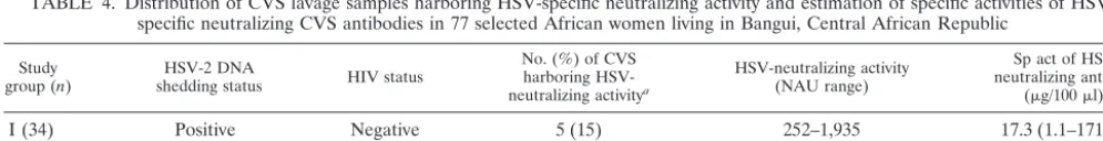
TABLE 4. Distribution of CVS lavage samples harboring specific neutralizing activity and estimation of specific activities of
HSV-specific neutralizing CVS antibodies in 77 selected African women living in Bangui, Central African Republic
Study
group (n) shedding statusHSV-2 DNA HIV status
No. (%) of CVS harboring HSV-neutralizing activitya
HSV-neutralizing activity (NAU range)
Sp act of HSV-neutralizing antibody
(g/100l)b
I (34)
Positive
Negative
5 (15)
252–1,935
17.3 (1.1–171.3)
II (20)
Positive
Positive
5 (25)
503–2,117
33.6 (17.9–100.4)
III (23)
Negative
Negative
3 (13)
435–522
3.1 (0.8–3.6)
aAs assessed by reduction of infectivity in BHKICP6LacZ-5 (ELVIS) cells.
bMedian and range (in parentheses) of specific activities calculated as NAU/(total IgG⫹total IgA). The medians were calculated for only CVS harboring
HSV-neutralizing activity.
on August 17, 2020 by guest
http://cvi.asm.org/
by in vivo binding between type-specific antibodies and virus
being shed into genital secretions. Such an event would prevent
antibody binding in our ECL-WB test but not affect DNA
detection by PCR. As a group, HIV-infected women had the
highest frequency of HSV IgG in CVS. This might reflect
higher antibody titers or greater transudation of serum
anti-body (5).
Neutralizing activity against HSV was demonstrated in 17%
of CVS samples: nearly always (92%) in the presence of
de-tectable antibody binding to HSV-1 or HSV-2. The fact that
most CVS with HSV IgG or IgA lacked neutralizing activity
may be due to insufficient sensitivity of the neutralizing assay
due to inherently low local antibody titers compounded by the
dilution effect of CVS lavage (4, 27).
The single CVS with neutralizing activity that lacked binding
antibodies may reflect the presence of nonimmune inhibitory
factors in CVS. Alternatively, neutralization could be mediated
by F(ab) fragments directed to HSV, since CVS fluid contains
large amounts of F(ab) derived from locally produced
secre-tory IgA or from IgG (15). Such fragments would not be bound
efficiently by the secondary antibody in the ECL-WB assay.
The specific activity of HSV-specific neutralizing activity was
fivefold higher in HSV-2 DNA shedders than in nonshedders,
a finding that appears contradictory to the lower frequency of
IgG or IgA to HSV-2 in HSV-2 shedders than nonshedders.
Our data might reflect the fact that HSV-1 and HSV-2
anti-bodies are highly cross-reactive (11, 28). In our assay, HSV-1
antibodies are very effective at neutralizing HSV-2 (3). Most of
the CVS (9 of 13 [69%]) with neutralizing activity also had
CVS antibodies to HSV-1.
A direct relationship was observed between the specific
ac-tivity of CVS neutralizing antibodies to HSV-2 and the level of
HSV-2 DNA in the CVS. One interpretation of these data is
that HSV-2 shedding in the genital tract induces production of
HSV-specific neutralizing antibodies. Prospective studies that
track CVS antibodies, neutralization of HSV shedding, and
symptoms over time would be useful to prove a cause-effect
hypothesis. Analyses to identify bound antibodies, to
discrim-inate dimeric versus monomeric IgA, and to compare serum
and CVS antibody profiles to individual viral proteins would be
critical to interpreting such prospective data.
Our previous studies have demonstrated HSV-specific
re-sponses to genital infections (1, 2) and to vaccines (4). The in
vitro and in vivo functions of the antibodies in genital
secre-tions are ill defined. Further prospective evaluasecre-tions of virus
shedding, including genital, humoral, and cellular immune
re-sponses to HSV-2, and of the function of HSV-2-specific IgG
and IgA (monomeric and dimeric) in CVS are needed to assess
the possible role of local antibody in HSV-2 genital shedding in
women.
ACKNOWLEDGMENTS
We thank all of the staff of the Unite´ de De´pistage Anonyme, in
particular Ce´cile Bary, Juliette Kapita, and Donoh Rufin, for excellent
contributions in counseling; Samory Aliou for technical assistance with
HIV serological testing; and the authorities of the Ministry of Public
Health and Population of the Central African Republic. We also thank
the attendees of the Centre National de Re´fe´rence des Maladies
Sex-uellement Transmissibles et du SIDA of Bangui for their consent to
receive HIV testing and to assist with the fight against the HIV
epi-demic.
Financial support for F.-X. Mbopi-Ke´ou was provided by a Visiting
Research Scientist Fellowship at Children’s Hospital and Regional
Medical Center, University of Washington, Seattle, and by a research
award from GlaxoWellcome. This work was supported by Central
Public Health Laboratory Services, Colindale, United Kingdom;
Na-tional Institute of Allergy and Infectious Diseases grant AI-30731;
GlaxoWellcome Research and Development, United Kingdom;
Agence Nationale de Recherches sur le SIDA, France (ANRS 12-12);
and the Institut National de la Sante´ et de la Recherche Me´dicale
(INSERM), France.
REFERENCES
1. Ashley, R.., A. Wald, and L. Corey.1994. Cervical antibodies in patients with oral herpes simplex virus type 1 (HSV-1) infection: local anamnestic re-sponses after genital HSV-2 infection. J. Virol.8:5284–5286.
2. Ashley, R. L., L. Corey, J. Dalessio, P. Wilson, M. Remington, G. Barnum, and P. Trethewey.1994. Protein-specific antibody responses to primary gen-ital herpes simplex virus type 2 infections. J. Infect. Dis.170:20–26. 3. Ashley, R. L., J. Dalessio, and R. E. Sekulovish.1997. A novel method to
assay herpes simplex virus neutralizing antibodies using BHKICP6LacZ-5 (ELVIS) cells. Viral Immunol.4:213–220.
4. Ashley, R. L., F. M. Crisostomo, M. Doss, R. E. Sekulovich, R. L. Burke, M. Shaughnessy, L. Corey, N. L. Polissar, and A. G. Langenberg.1998. Cervical antibody responses to a herpes simplex virus type 2 glycoprotein subunit vaccine. J. Infect. Dis.178:1–7.
5. Belec, L., T. Dupre´, T. Prazuck, C. Te´vi-Be´nissan, J. M. Kanga, O. Pathey, X. S. Lu, and J. Pillot.1995. CVS overproduction of specific IgG to human immunodeficiency virus (HIV) contrasting with normal or impaired local response in HIV infection. J. Infect. Dis.172:691–697.
6. Belec, L., D Meillet, M. Levy, A. J. Georges, C. Tevi-Benissan, and J. Pillot.
1995. Dilution assessment of CVS secretions obtained by vaginal washing for immunological assays. Clin. Diagn. Lab. Immunol.2:57–61.
7. Berneman, A., L. Belec, V. A. Fischetti, and J. P. Bouvet.1998. The specific patterns of human immunogloblin G antibodies in serum differ from those in autologous secretions. Infect. Immun.66:4163–4168.
8. Brandtzaeg, P.1992. Humoral immune response patterns of human muco-sae: induction and relation to bacterial respiratory tract infections. J. Infect. Dis.165(Suppl. 1):S167–S176.
9. Coughlan, B. M., and G. R. Skinner.1977. Antibody activity to type 1 and type 2 herpes simplex virus in human cervical mucus. Br. J. Obstet. Gynaecol.
84:622–629.
10. Dalessio, J., and R. L. Ashley.1992. Highly sensitive enhanced chemilumi-nescence immunodetection method for herpes simplex virus type 2 Western immunoblot. J. Clin. Microbiol.4:1005–1007.
11. Eberle, R., and R. J. Courteney.1989. Topological distribution of virus-specific and cross-reactive antigenic determinants on the gB glycoprotein of herpes simplex viruses. J. Med. Virol.27:309–316.
12. Espy, M. J., J. Aslanzadeh, and T. F. Smith.1993. PCR detection of herpes simplex virus DNA sequences in cerebrospinal fluid, p. 332–336.InD. H. Persing, T. F. Smith, F. C. Tenover, and T. J. White (ed.), Diagnostic molecular microbiology: principles and applications. American Society for Microbiology, Washington, D.C.
13. Gopal, R., T. Gibbs, M. J. Slomka, J. Whitworth, L. M. Carpenter, A. Vyse, and D. W. Brown.2000. A monoclonal blocking EIA for herpes simplex virus type 2 (HSV-2) antibody: validation for seroepidemiological studies in Af-rica. J. Virol. Methods87:71–80.
14. Gronroos, M., and E. Honkonen.1983. Cervical and serum IgA and serum IgG antibodies toChlamydia trachomatisand herpes simplex virus in threat-ened abortion: a prospective study. Br. J. Obstet. Gynecol.90:167–170. 15. Hocini, H., A. Barra, L. Belec, S. Iscaki, J. L. Preud’homme, J. Pillot, and
J. P. Bouvet.1995. Systemic and secretory humoral immunity in the normal human vaginal tract. Scand. J. Immunol.42:269–274.
16. Koelle, D. M., M. Schomogyi, and L. Corey.2000. Antigen-specific T cells localize to the uterine cervix in women with genital herpes simplex virus type 2 infection. J. Infect. Dis.182:662–670.
17. Koelle, D. M., H. B. Chen, M. A. Gavin, A. Wald, W. W. Kwok, and L. Corey.
2001. CD8 CTL from genital herpes simplex lesions: recognition of viral tegument and immediate early proteins and lysis of infected cutaneous cells. J. Immunol.166:4049–4058.
18. Mantero, G., A. Zonaro, A. Albertini, P. Bertolo, and D. Primi.1991. DNA enzyme immunoassay: general method for detecting products of polymerase chain reaction. Clin. Chem.37:422–429.
19. Mbopi-Keou, F. X., G. Gresenguet, P. Mayaud, H. A. Weiss, R. Gopal, M. Matta, J. L. Paul, D. W. Brown, R. J. Hayes, D. C. Mabey, and L. Belec.2000. Interactions between herpes simplex virus type 2 and HIV infection in African women: opportunities for intervention. J. Infect. Dis.182:1090–1096. 20. McDermott, M. R., L. J. Brais, and M. J. Evelegh.1990. Mucosal and systemic antiviral antibodies in mice inoculated intravaginally with herpes simplex virus type 2. J. Gen. Virol.71:1497–1504.
21. Merriman, H., S. Woods, C. Winter, A. Fahnlander, and L. Corey.1984.
on August 17, 2020 by guest
http://cvi.asm.org/
Secretory IgA antibodies in CVS secretions from women with genital infec-tion due to herpes simplex virus. J. Infect. Dis.149:505–510.
22. Mostad, S. B., J. K. Kreiss, A. J. Ryncarz, K. Mandaliya, B. Chohan, J. Ndinya-Achola, J. J. Bwayo, and L. Corey.2000. Cervical shedding of herpes simplex virus in human immunodeficiency virus-infected women: effects of hormonal contraception, pregnancy, and vitamin A deficiency. J. Infect. Dis.
181:58–63.
23. Murphy, J. F., D. F. Murphy, S. Barker, M. L. Mylotte, B. M. Coughlan, and G. R. B. Skinner.1985. Neutralizing antibody against type 1 and 2 herpes simplex virus in cervical mucus of women suffering with intra-epithelial neoplasia. Med. Microbiol. Immunol.174:73–80.
24. Parr, E. L., and M. B. Parr.1997. Immunoglobulin G is the main protective antibody in mouse vaginal secretions after vaginal immunization with atten-uated herpes simplex virus type 2. J. Virol.71:8109–8115.
25. Persson, E., P. Eneroth, and S. Jeansson.1988. Secretory IgA against herpes simplex virus in cervical secretions. Genitourin. Med.64:373–377. 26. Posavad, C. M., D. M. Koelle, and L. Corey.1998. Tipping the scales of
herpes simplex virus reactivation: the important responses are local. Nat. Med.4:381–382.
27. Quesnel, A., S. Cu-Uvin, D. Murphy, R. L. Ashley, T. Flanigan, and M. R. Neutra.1997. Comparative analysis of methods for collection and
measure-ment of immunoglobulins in cervical and vaginal secretions of women. J. Im-munol. Methods202:153–161.
28. Rouse, B. T., and M. Gierynska.2001. Immunity to herpes simplex virus: a hypothesis. Herpes8(Suppl. 1):2A–5A.
29. Si-Mohamed, A., M. D. Kazatchkine, I. Heard, C. Goujon, T. Prazuck, G. Aymard, G. Cessot, Y. H. Kuo, M. C. Bernard, B. Diquet, J. E. Malkin, L. Gutmann, and L. Belec.2000. Selection of drug-resistant variants in the female genital tract of human immunodeficiency virus type 1-infected women receiving antiretroviral therapy. J. Infect. Dis.18:112–122. 30. Stabell, E. C., and P. D. Olivo.1992. Isolation of a cell line for rapid and
sensitive histochemical assay for the detection of herpes simplex virus. J. Vi-rol. Methods38:195–202.
31. Vogel, J. U., B. Weber, and H. W. Doerr.1994. Typing and strain differen-tiation of clinical herpes simplex virus type 1 and 2 isolates by polymerase chain reaction and subsequent fragment length polymorphism analysis. Zentbl. Bakteriol.281:502–512.
32. Wald, A., J. Zeh, S. Selke, R. L. Ashley, and L. Corey.1995. Virological characteristics of subclinical and symptomatic genital herpes infections. N. Engl. J. Med.333:770–775.
33. Wald, A., J. Zeh, S. Selke, T. Warren, R. Ashley, and L. Corey.2002. Genital shedding of herpes simplex virus among men. J. Infect. Dis186(Suppl. 1):S34–S39.