ABSTRACT
ARAMBULA, SHERYL ELIZABETH. Sex-Specific Impact of Early-Life Bisphenol A (BPA) Exposure on Brain Development. (Under the direction of Dr. Heather B. Patisaul).
Bisphenol A (BPA) is a well-recognized endocrine disruptor that is commonly used as a component of polycarbonate plastics and epoxy resins, and found in a variety of household products. Thus, human exposure to BPA is widespread, with levels higher in children than adults. Extensive experimental and epidemiological evidence supports associations between developmental BPA exposure and sex-specific socioemotional behavioral outcomes including hyperactivity, anxiety, aggression, and cognitive deficits. However, the molecular underpinnings of these behavioral outcomes remain poorly understood. The studies within this dissertation were conducted as part of the CLARITY-BPA (Consortium Linking Academic and Regulatory Insights on CLARITY-BPA Toxicity) research program and examined the impact of early-life BPA exposure on the hippocampal,
hypothalamic, and amygdalar transcriptomes of neonates and the sexually dimorphic brain nuclei of juveniles. NCTR-Sprague Dawley rats (NCTR-SD) were exposed to wide range of BPA doses (2.5 - 25,000 µg/kg body weight (bw)/day) pre- and/or postnatally. The
hippocampus, hypothalamus, and amygdala were microdissected from postnatal day 1 (PND 1) brains and RNA-sequencing and qRT-PCR were used to assess gene expression. In addition, unbiased stereology was used to quantify the volume of the sexually dimorphic nucleus (SDN), the anteroventral periventricular nucleus (AVPV), the posterodorsal portion of the medial amygdala (MePD), and the locus coeruleus (LC) at PND 28. Overall, early-life BPA exposure induced sex-, brain region-, and dose-specific effects. In the neonate brain, gene expression analysis revealed further evidence for disruption of estrogen receptor
Sex-Specific Impact of Early-Life Bisphenol A (BPA) Exposure on Brain Development
by
Sheryl Elizabeth Arambula
A dissertation submitted to the Graduate Faculty of North Carolina State University
in partial fulfillment of the requirements for the degree of
Doctor of Philosophy
Biology
Raleigh, North Carolina 2017
APPROVED BY:
_______________________________ _______________________________
Heather Patisaul Scott Belcher
Committee Chair
_______________________________ _______________________________
DEDICATION
BIOGRAPHY
“Biography is a system in which the contradictions of a human life are unified.” - José Ortega y Gasset
ACKNOWLEDGMENTS
First and foremost, I would like to thank Dr. Heather Patisaul, whose constant support has been integral to my success as a graduate student. I couldn’t have asked for a better advisor. Thank you for giving me ample opportunities to learn and develop professionally outside of the lab and classroom, while setting an inspiring example. Additionally, I would like to express my deepest gratitude to my undergraduate advisor at Missouri Southern State University, Dr. James R. Jackson, for his continued support, mentorship, advice, and
friendship. You are an inspiration and by far one of the most caring and genuine people I have had the pleasure to meet. Thank you to Dr. Catherine Barrett, Dr. Larry Young, and the Center for Behavioral Neuroscience for giving me my first laboratory research experience, which led me to pursue a career in neuroscience. Joelle Fuchs and Shima Idries, thank you for working extensively with me during the past two years - I have enjoyed every moment. I would like to offer a special thanks to the 2017 SPINES (Summer Program in Neuroscience Excellence and Success) cohort and to all of the SPINES family. Their support, willingness to share their stories and struggles, and enthusiasm for diversity in STEM were
TABLE OF CONTENTS
LIST OF TABLES ... ix
LIST OF FIGURES ... xi
CHAPTER 1 - Endocrine Disrupting Chemicals and Behavior (Review) ... 1
Abstract ... 2
Keywords ... 2
Introduction ... 3
BPA: a landmark endocrine disrupting chemical ... 4
Sexually dimorphic behaviors as indicators of endocrine disruption . 6 Evidence from animal models ... 7
Evidence from epidemiological studies ... 12
Conclusions ... 15
Remaining challenges and future directions ... 15
References ... 18
Chapter 1 Table ... 37
CHAPTER 2 - Impact of Low Dose Oral Exposure to Bisphenol A (BPA) on the Neonatal Rat Hypothalamic and Hippocampal Transcriptome: a CLARITY-BPA Consortium Study 38 Abstract ... 38
Introduction ... 38
Materials and methods ... 40
Results ... 43
Summary and conclusions ... 51
Acknowledgements ... 51
References ... 51
Supplemental tables ... 134
Supplemental figure ... 184
CHAPTER 3 - Prenatal Bisphenol A (BPA) Exposure Alters the Transcriptome of the Neonate Rat Amygdala In a Sex- Specific Manner: a Clarity-BPA Consortium Study ... 55
Grant support ... 55
Disclosure statement ... 55
Highlights ... 56
Keywords ... 56
Abstract ... 57
Introduction ... 58
Materials and methods ... 61
Results ... 69
Discussion ... 74
Conclusions ... 82
Acknowledgements ... 83
References ... 84
Chapter 3 tables ... 101
Chapter 3 figures ... 106
Supplementary figures ... 215
CHAPTER 4 - Effects of Perinatal Bisphenol A Exposure on the Volume of Sexually-Dimorphic Nuclei of Juvenile Rats: A CLARITY-BPA Consortium Study ... 110
Abstract ... 110
Keywords ... 110
Introduction ... 110
Materials and methods ... 111
Results ... 113
Discussion ... 115
Conclusions ... 117
Funding information ... 117
Disclosure statement ... 117
Acknowledgements ... 117
References ... 117
CHAPTER 5 - Conclusions ... 120
References ... 128
APPENDICES ... 134
APPENDIX 1 - Chapter 2 supplementary tables and figures ... 134
LIST OF TABLES
CHAPTER 1 - Endocrine Disrupting Chemicals and Behavior: a Review
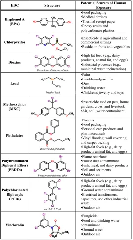
Table 1. Structures and potential sources of endocrine disrupting
chemicals with published behavioral effects ...37 CHAPTER 2 - Impact of Low Dose Oral Exposure to Bisphenol A (BPA) on the Neonatal Rat Hypothalamic and Hippocampal Transcriptome: a CLARITY-BPA Consortium Study Table 1. Candidate and novel genes assessed with qRT-PCR ...40 Table 2. Differentially expressed genes for which there was a significant
sex by exposure interaction at 2500 µg BPA/kg bw/day in males ...44 Table 3. qRT-PCR outcomes and descriptive statistics for genes found
to be significantly altered by BPA exposure ...45 Supplemental Table 1. RNA-seq analysis of differentially expressed hippocampal
genes ...134 Supplemental Table 2. RNA-seq analysis of differentially expressed hypothalamic
genes ...135 Supplemental Table 3. Overlapping significantly (padj ≤ 0.05) altered hypothalamic
genes in males exposed to BPA ...173 Supplemental Table 4. RNA-seq analysis of sexually dimorphic hippocampal and
Table 1. Rationale for genes of interest selected a priori ...101 Table 2. qRT-PCR outcomes and descriptive statistics for genes found
to be significantly altered by BPA or EE2 exposure ...102 Table 3. Differentially expressed genes identified by RNAseq within
selected canonical pathways identified by IPA analysis in exposed females (normalized to male vehicle; adjusted p-value
≤ 0.05) ...103 Supplementary Table 1. Male transcriptomic datasets (normalized to male vehicle;
adjusted p-value ≤ 0.05) ...185 Supplementary Table 2. Female transcriptomic datasets (normalized to female vehicle;
adjusted p-value ≤ 0.05) ...188 Supplementary Table 3. Significant (adjusted p-value ≤ 0.05) sex-differences in gene
expression identified by RNAseq (female vehicle compared to male vehicle) ...214 CHAPTER 4 - Effects of Perinatal Bisphenol A Exposure on the Volume of
Sexually-Dimorphic Nuclei of Juvenile Rats: A CLARITY-BPA Consortium Study
Table 1. Effect of sex and perinatal BPA or EE2 exposure on the volume
LIST OF FIGURES
CHAPTER 2 - Impact of Low Dose Oral Exposure to Bisphenol A (BPA) on the Neonatal Rat Hypothalamic and Hippocampal Transcriptome: a CLARITY-BPA Consortium Study Figure 1. Anatomical representation of regions extracted via micropunch
and sample sizes for RNA-seq and qRT-PCR ... 41 Figure 2. Effects of gestational BPA or EE on neonatal hippocampal ER expression ... 46 Figure 3. Effects of gestational BPA or EE exposure on neonatal
hippocampal expression of selected genes ... 47 Figure 4. Effects of gestational BPA or EE exposure on neonatal
hypothalamic ER expression ... 48 Figure 5. Effects of gestational BPA or EE exposure on hypothalamic
expression of selected genes ... 49 Figure 6. Sex differences in hippocampal and hypothalamic expression
of selected genes ... 50 Supplemental Figure 1. Unsupervised principal component analysis of hippocampal
and hypothalamic RNA-seq data ... 184 CHAPTER 3 - Prenatal Bisphenol A (BPA) Exposure Alters the Transcriptome of the Neonate Rat Amygdala In a Sex- Specific Manner: a Clarity-BPA Consortium Study Figure 1. Effects of gestational BPA or EE2 on neonatal amygdalar
Figure 2. Top canonical pathways enriched by differentially expressed genes ... 107 Figure 3. Prenatal exposure to BPA and EE2 result in common and
unique differently expressed genes ... 108 Figure 4. Sex differences in amygdala expression of selected genes . 109 Supplementary Figure 1. Anatomical representation of regions extracted via micropunch ... 215 Supplementary Figure 2. Unsupervised principal component analyses (PCA) for
RNAseq data ... 216 CHAPTER 4 - Effects of Perinatal Bisphenol A Exposure on the Volume of Sexually-Dimorphic Nuclei of Juvenile Rats: A CLARITY-BPA Consortium Study
Figure 1. Effect of perinatal BPA or EE2 exposure on the volume of the
sexually dimorphic nucleus (SDN) ... 113 Figure 2. Effect of perinatal BPA or EE2 exposure on the volume of the
anteroventral periventricular nucleus (AVPV) ... 114 Figure 3. Effect of perinatal BPA or EE2 exposure on the volume of the
left and right medial posterodorsal amygdala (MePD) ... 114 Figure 4. Effect of perinatal BPA or EE2 exposure on the average
volume of medial posterodorsal amygdala (MePD) ... 115 Figure 5. Effect of perinatal BPA or EE2 exposure on the volume of the
CHAPTER 5 - Conclusions
CHAPTER 1
Endocrine Disrupting Chemicals and Behavior
Sheryl E. Arambula*†, Heather B. Patisaul*†§
*Department of Biological Sciences, North Carolina State University, Raleigh, North Carolina 27695; †Keck Center for Behavioral Biology, North Carolina State University,
Raleigh, North Carolina 27695; §Center for Human Health and the Environment, North Carolina State University, Raleigh, North Carolina 27695
Corresponding author contact information: Heather B Patisaul, PhD
Professor
Department of Biological Sciences Raleigh, NC 27695, USA
ABSTRACT
Endocrine disrupting chemicals (EDCs) are a diverse group of compounds that interfere with the organizational or activational effects of hormones. There is growing concern that early-life exposure to EDCs may be contributing to the increasing prevalence of sex-biased neurodevelopmental disorders by multiple mechanisms. While it is difficult to make causal links, extensive experimental and epidemiological evidence supports associations between early-life exposure to environmental contaminants and sex-specific neurobehavioral outcomes. This review provides an overview of the neurobiological and behavioral consequences of developmental exposure to EDCs with an emphasis on bisphenol A, high volume production chemical found in a variety of commonly used products.
KEYWORDS
1. INTRODUCTION
Inexorably connected, the endocrine system and nervous system work in tandem to regulate the development and expression of behavior. This relationship is best exemplified by the early actions of steroid hormones on the mammalian brain, which induce enduring changes in the brain that impact sexually dimorphic behavior [31, 36, 65, 81]. Although brain
development is a tightly regulated and orchestrated process, it is vulnerable to exogenous substances that interfere with the action of natural hormones.
Endocrine disrupting chemicals (EDCs) are a diverse group of exogenous compounds that have been found to interfere with the endocrine system and produce adverse health effects in exposed individuals or their offspring. Numerous classes of chemicals including plasticizers, flame-retardants, fungicides, pesticides, pharmaceuticals, heavy metals, and even naturally occurring compounds such as phytoestrogens are endocrine disrupting (Table 1). These and other EDCs dampen, block, or potentiate the action of endogenous hormones through a variety of direct and indirect mechanisms. For example, they may agonize or antagonize hormone receptors, interfere with hormone biosynthesis, or alter the number of hormone receptors [48]. The long-term consequences of EDCs depend on a variety of factors including the genetic susceptibility of the organism and the dose, duration, and developmental window of exposure.
Since the term was first coined nearly three decades ago, EDCs have received
in the prevalence of sex-biased neurodevelopmental disorders, most notably attention-deficit hyperactivity disorder and autism spectrum disorder, which cannot be fully explained by genetic factors alone. Although the etiology of these disorders are not well understood, there is increasing concern that developmental exposure to EDCs may enhance risk by disrupting sexual differentiation of the brain. Indeed, extensive experimental and epidemiological evidence supports associations between early-life exposure to environmental contaminants and sex-specific neurobehavioral outcomes.
The goal of the current review is to examine the evidence for altered behavior as a consequence of EDC exposure. Although hundreds of suspected EDCs have been identified, bisphenol A (BPA) is arguably one of the most widely used and extensively studied. Thus for illustrative purposes, we will concentrate on the effects of early exposure to this notorious chemical.
2. BPA: A LANDMARK ENDOCRINE DISRUPTING CHEMICAL
neuroendocrine disrupting agents. In humans, BPA has been detected in fetal plasma [59], amniotic fluid [40], fetal liver [87] and placenta tissue [109], demonstrating the capacity for significant gestational exposure. Moreover, the fetal brain is particularly susceptible to environmental exposures because the blood-brain barrier is not fully formed, and thus provides limited protection [3, 96]. Accordingly, levels of chemical exposures that have no obvious effects on the adult nervous system can pose a significant risk when exposure occurs developmentally.
specific changes in DNA methylation patterns in the brain that are accompanied by decreased expression of ERs [69, 70]. These alternative modes of action emphasize the complex, multi-modal routes by which EDCs can impact brain and behavior across the lifespan.
3. SEXUALLY DIMORPHIC BEHAVIORS AS INDICATORS OF ENDOCRINE DISRUPTION
Sexual differentiation is the process by which the brain becomes structurally and functionally different between males and females. Sexual differentiation of the brain was once thought to hinge almost entirely upon gonadal hormones: generally, the brain develops as male in the presence of these hormones, and as female in their absence. However, recent evidence indicates this process is more nuanced and involves multiple sex-specific hormonal, genetic, and epigenetic factors that influence sexually dimorphic physiology and behavior thorough a variety of mechanisms [82, 83, 110].
Disruption of sexually dimorphic behavior is a common outcome of developmental exposure to EDCs, particularly BPA. Indeed, sex-specific behavioral impacts of BPA have been demonstrated in numerous animal models and human epidemiological studies.
4. EVIDENCE FROM ANIMAL MODELS
The majority of experimental studies on neurobiological and behavioral consequences of developmental BPA exposure (gestational and/or neonatal) employ rodent models. Here we provide a summary of these findings and limit our discussion to studies that use BPA doses at or lower than the current FDA NOAEL (5 mg/kg bw/day).
4.1 Exploratory and Affective Behaviors
Experimental animal studies in rodents provide compelling evidence that developmental BPA exposure can increase the expression of anxiety-related and exploratory behaviors. However, effects vary across sex, animal model, and age at testing. Studies examining developmental BPA exposure in juveniles typically demonstrate sex-specific effects, but results are inconsistent. For example, two studies on juvenile C57BL/6J mice conclude that gestational and/or neonatal BPA exposure increased anxiety in males but had no effect in females [33, 80], while another study with CD-1 mice reported the opposite [45].
Additionally, a recent study in juvenile Sprague Dawley rats found no effects of perinatal BPA exposure on exploratory or anxiety activity, in either sex [101].
Studies in adults are more consistent and in general, females display more robust
anxiogenic effects following early-life BPA exposure compared to males [46, 54, 106, 114]. For example, neonatal exposure to 10 µg/kg of BPA was found to decrease exploratory-behavior and increase anxiety-like exploratory-behavior in adult female CD-1 mice, resulting in a
studies that show developmental BPA exposure can decrease or eliminate sex differences typically observed in adult rodents, on a number of behavioral paradigms used to assess anxiety [45, 54, 62, 66, 69]. Experiments using other animal models (including zebrafish, voles and other alternative rodent species, and non-human primates) provide further evidence that developmental BPA exposure induces anxiety-related behaviors [64, 69, 88, 93, 114]. This consistency across studies and varying animal models prompted the World Health Organization to conclude that there is some concern about impacts of developmental BPA exposure on brain and behavior [43]. While the underlying mechanisms remain unclear, this behavioral disruption is commonly associated with perturbation of ER-related gene
expression in the hypothalamus and the amygdala [4, 21-23, 69, 93, 100, 102]. Moreover, a recent study found that prenatal BPA exposure induced sex-specific effects on anxiety-like behaviors in adult BALB/c mice that corresponded to changes in DNA methylation and mRNA levels of ERα in the hypothalamus [69]. These data provide intriguing evidence that BPA-induced disruption of anxiety behavior may be mediated through an epigenetic
mechanism.
impulsivity, and aggression [17, 27, 37, 51, 56, 74, 77]. While not an EDC in the classic sense, lead and other metals can be endocrine disrupting in some circumstances [28, 53, 55, 85].
4.2 Learning and Memory
Impairments in cognitive abilities have also been observed following developmental exposure to BPA. Under normal conditions, male rodents typically perform significantly better than females on spatial learning and memory tasks and, interestingly, early-life BPA exposure has repeatedly been shown to reduce this sex difference [24, 62, 130]. Several studies in rats and mice suggest that gestational and/or neonatal exposure to BPA can negatively impact spatial memory in both juvenile and adult males [68, 71, 78, 118, 130-132]. As an example, exposure to BPA (0.5 and 5 mg/kg bw/day) during the perinatal period significantly impaired spatial memory in juvenile and adult male ICR mice [132]. In
contrast, data on the effects of BPA on spatial learning and memory in females is sparse and mixed results have been reported [24, 78, 130]. Notably, the described changes in spatial memory were associated with BPA-induced alterations in dendritic spine density and morphology, as well as reduced expression of N-methyl-d-aspartic acid (NMDA) glutamatergic receptors and ERβ [39, 78, 118, 131] in the hippocampus.
exposure to PDBE slowed motor skill development in adolescent and adult CD-1 mice [10]. Similarly, a series of experiments in mice found neonatal exposure to multiple PBDE
congeners caused significant adult learning and memory deficits that corresponded to inhibition of the hippocampal cholinergic system [122, 123].
4.3 Paternal, Social, and Sexual Behaviors
In rodents, changes in parental, social, and sexual behaviors have been reported after
developmental exposure to BPA, but evidence is sparse and inconsistent [1, 2, 44, 54, 61, 86, 98, 99, 107, 114, 129]. To date only two studies have examined the relationship between developmental BPA exposure and subsequent maternal behavior. One of these found that prenatal exposure to BPA 10 µg/kg bw/day decreased the amount of time female CD-1 mice spent huddling over or nursing their offspring [90]. The second study, which was conducted in Wistar rats, reported similar effects of gestational and lifelong BPA exposure to 5 µg/kg bw [9]. The impact of developmental BPA exposure on paternal behavior is unknown. This is likely due to the fact that traditional rodent models used in toxicology do not display bi-parental care.
juvenile Sprague-Dawley rats (males were not assessed) [99]. A study in prairie voles, a more prosocial animal model than laboratory rats or mice, found sex- and age-specific effects on social behavior. Neonatal exposure to 5 and 50 µg/kg bw/day decreased social
investigation in juvenile males and slightly inhibited partner preference formation in adult females. These behavioral outcomes were accompanied by sex-dependent changes in the number of dopaminergic-, oxytocin-, and vasopressin neurons in the paraventricular nucleus of the hypothalamus and dopaminergic neurons in the bed nucleus of stria terminals [114].
Some published data suggests early-life BPA exposure can induce subtle changes in adult sexual behavior, but supporting evidence is mixed [1, 2, 44, 61, 86, 98, 107]. Two studies in rodents have found a slight impairment in male sexual performance in terms of latency, intromission, and ejaculation [44, 61], where others have found none [98]. Female data is similarly mixed but generally indicates female sexual behavior is unaffected by
developmental BPA exposure. In rodents, female proceptive and receptive behaviors are often determined by hopping and darting and the lordosis response. Exposure to 0.05 mg/kg of BPA during the neonatal period decreased hopping and darting rate in adult female Wistar rats, while lordosis behavior was unaffected [86]. Another study, conducted in Sprague-Dawley rats, observed a modest increase in lordosis behavior in adult females following perinatal exposure to 40 µg/kg of BPA [44]. Other studies have observed no effects of developmental exposure on proceptive or receptive behaviors in females [1, 2, 107].
phytoestrogens [91, 92, 94], and chlorpyrifos [35, 119, 120] on parental, social, and sexual behaviors. For instance, a number of studies in rodents provide evidence that PCBs can adversely impact sociosexual behavior and, in general, suggest that females may be more vulnerable to disruption than males. In rats gestational and neonatal exposure reduces receptive and proceptive sexual behaviors such as lordosis and likelihood to mate [29, 113, 115].
5. EVIDENCE FROM EPIDEMIOLOGICAL STUDIES
Although the health impacts of developmental BPA exposure remain controversial, during the last decade several epidemiological studies have reported adverse behavior in children developmentally exposed to BPA [6, 11, 12, 16, 26, 42, 52, 57, 79, 97, 104].
particularly in girls [12]. Of note, two subsequent studies on the HOMES cohort found no associations between maternal levels of gestational BPA and autistic behaviors in children 4 to 5 years old [14] or visual spatial ability in children 7 years old [15]. This outcome
highlights that while chemical exposures can produce measureable and meaningful
decrements in behavior and cognition that can have life-long implications, manifestation of a clinically defined disorder, such as autism, is highly unlikely and would be notoriously difficult to prove.
Collectively these epidemiological studies strongly suggest that developmental BPA exposure may have adverse neurobehavioral effects in children, which may differ between boys and girls. This conclusion is concordant with the abundant animal data although the mechanisms of action remain poorly understood. In general, the conclusions from the described studies are strengthened by their use of large mother-child cohorts, BPA measurements across several gestational time points, and multiple observed behavioral outcomes. However, it is important to recognize the limitations inherent to longitudinal cohort studies that may have contributed to the discrepant results, particularly demographic differences across cohorts. Additionally, differences in neuropsychological assessments and substantial within-person variation in urinary BPA concentrations may also contribute to the heterogeneity in the literature [13, 116].
continuously, combined effects of multiple exposures are a significant and growing concern.
6. CONCLUSIONS
The experimental evidence summarized above supports the hypothesis that developmental exposure to BPA, even at doses below the current NOAEL, may interfere with some aspect of sexual differentiation of the nervous system, thereby resulting in disruption of both reproductive and non-reproductive behaviors. During fetal and child development, the brain is particularly susceptible to environmental stressors such as EDCs. A recent study found that children of parents who were concerned about EDCs had decreased urinary
concentrations of BPA, which suggests that by exercising precaution we may be able to reduce our exposure to chemicals [95]. While developmental exposure to BPA and other EDCs may contribute to neural disorders in children, it should be emphasized that available literature does not provide direct causal evidence. Further mechanistic and epidemiological studies are needed to clarify the relationship between EDC exposure and human health. Greater information is also needed about the effect of mixtures and repeated exposures over multiple critical periods.
7. REMAINING CHALLENGES AND FUTURE DIRECTIONS
neurodevelopmental insult and the manifestation of a resulting behavioral dysfunction can be very long. During this period, which may take years or decades in humans, behavior is concomitantly influenced by other factors including genetics, experience, and lifestyle. Consequently, ascertaining the contribution of single chemical exposure is extraordinarily difficult. Moreover, humans are exposed to varying amounts and mixtures of EDCs throughout their lifetime. Already an area of increased interest, modeling “real world” human exposure (i.e., chronic, low-dose mixtures) will greatly enhance the translational value of animal studies in the EDC field.
In humans, studies on the neurobehavioral changes following early-life EDC exposure are constrained by practical and ethical limitations. An obvious limitation is the relative inaccessibility of the human brain. Both in vivo and in vitro models can be used to identify peripheral biomarkers of EDC exposure and associated diseases, which can be incorporated into new and existing epidemiological studies. Reliable biomarkers could also have
important implications for identifying at-risk populations.
REFERENCES
1. Adewale, H.B., et al., Neonatal bisphenol-a exposure alters rat reproductive
development and ovarian morphology without impairing activation of gonadotropin
releasing hormone neurons. Biology of Reproduction, 2009. 81(4): p. 690-699.
2. Adewale, H.B., et al., The impact of neonatal bisphenol-a exposure on sexually
dimorphic hypothalamic nuclei in the female rat. Neurotoxicology, 2011. 32(1): p.
38-49.
3. Adinolfi, M., The development of the human blood-csf-brain barrier. Developmental
Medicine & Child Neurology, 1985. 27(4): p. 532-7.
4. Arambula, S.E., et al., Impact of low dose oral exposure to bisphenol a (bpa) on the
neonatal rat hypothalamic and hippocampal transcriptome: A clarity-bpa consortium
study. Endocrinology, 2016: p. en20161339.
5. Belcher, S.M. and A. Zsarnovszky, Estrogenic actions in the brain: Estrogen,
phytoestrogens, and rapid intracellular signaling mechanisms. Journal of
Pharmacology and Experimental Therapeutics, 2001. 299(2): p. 408-14.
6. Bellinger, D.C., et al., Dental amalgam restorations and children's neuropsychological
function: The new england children's amalgam trial. Environmental Health
7. Blair, R.M., et al., The estrogen receptor relative binding affinities of 188 natural and
xenochemicals: Structural diversity of ligands. Toxicological Sciences, 2000. 54(1):
p. 138-53.
8. Boucher, O., G. Muckle, and C.H. Bastien, Prenatal exposure to polychlorinated
biphenyls: A neuropsychologic analysis. Environmental Health Perspectives, 2009.
117(1): p. 7-16.
9. Boudalia, S., et al., A multi-generational study on low-dose bpa exposure in wistar
rats: Effects on maternal behavior, flavor intake and development. Neurotoxicology
and Teratology, 2014. 41: p. 16-26.
10. Branchi, I., E. Alleva, and L.G. Costa, Effects of perinatal exposure to a
polybrominated diphenyl ether (pbde 99) on mouse neurobehavioural development.
Neurotoxicology, 2002. 23(3): p. 375-84.
11. Braun, J.M., et al., Prenatal bisphenol a exposure and early childhood behavior.
Environmental Health Perspectives, 2009. 117(12): p. 1945-52.
12. Braun, J.M., et al., Impact of early-life bisphenol a exposure on behavior and
executive function in children. Pediatrics, 2011. 128(5): p. 873-82.
13. Braun, J.M., et al., Variability of urinary phthalate metabolite and bisphenol a
concentrations before and during pregnancy. Environmental Health Perspectives,
14. Braun, J.M., et al., Gestational exposure to endocrine-disrupting chemicals and
reciprocal social, repetitive, and stereotypic behaviors in 4- and 5-year-old children:
The home study. Environmental Health Perspectives, 2014. 122(5): p. 513-20.
15. Braun, J.M., et al., Prenatal phthalate, triclosan, and bisphenol a exposures and child
visual-spatial abilities. Neurotoxicology, 2017. 58: p. 75-83.
16. Braun, J.M., et al., Associations of prenatal urinary bisphenol a concentrations with
child behaviors and cognitive abilities. Environmental Health Perspectives, 2017.
125(6): p. 067008.
17. Burright, R.G., W.J. Engellenner, and P.J. Donovick, Postpartum aggression and
plasma prolactin levels in mice exposed to lead. Physiology & Behavior, 1989. 46(5):
p. 889-93.
18. Bushnik, T., et al., Lead and bisphenol a concentrations in the canadian population.
Health Reports, 2010. 21(3): p. 7-18.
19. Calafat, A.M., et al., Urinary concentrations of bisphenol a and 4-nonylphenol in a
human reference population. Environmental Health Perspectives, 2005. 113(4): p.
391-5.
20. Calafat, A.M., et al., Exposure of the U.S. Population to bisphenol a and
4-tertiary-octylphenol: 2003-2004. Environmental Health Perspectives, 2008. 116(1): p. 39-44.
21. Cao, J., et al., Neonatal bisphenol a exposure alters sexually dimorphic gene
22. Cao, J., et al., Prenatal bisphenol a exposure alters sex-specific estrogen receptor
expression in the neonatal rat hypothalamus and amygdala. Toxicological Sciences,
2013. 133(1): p. 157-73.
23. Cao, J., et al., Sex-specific esr2 mrna expression in the rat hypothalamus and
amygdala is altered by neonatal bisphenol a exposure. Reproduction, 2014. 147(4): p.
537-54.
24. Carr, R., et al., Effect of neonatal rat bisphenol a exposure on performance in the
morris water maze. J ournal of Toxicology and Environmental Health A, 2003.
66(21): p. 2077-88.
25. Casas, M., et al., Exposure to brominated flame retardants, perfluorinated
compounds, phthalates and phenols in european birth cohorts: Enrieco evaluation,
first human biomonitoring results, and recommendations. International Journal of
Hygiene and Environmental Health, 2013. 216(3): p. 230-42.
26. Casas, M., et al., Exposure to bisphenol a during pregnancy and child
neuropsychological development in the inma-sabadell cohort. Environmental
Research, 2015. 142: p. 671-9.
27. Cervantes, M.C., et al., Lead exposure alters the development of agonistic behavior in
golden hamsters. Developmental Psychobiology, 2005. 47(2): p. 158-65.
28. Chen, L., et al., Transgenerational endocrine disruption and neurotoxicity in zebrafish
larvae after parental exposure to binary mixtures of decabromodiphenyl ether
29. Chung, Y.W. and L.G. Clemens, Effects of perinatal exposure to polychlorinated
biphenyls on development of female sexual behavior. Bulletin of Environmental
Contamination and Toxicology, 1999. 62(6): p. 664-70.
30. Colborn, T., F.S. vomSaal, and A.M. Soto, Developmental effects of
endocrine-disrupting chemicals in wildlife and humans. Environmental Health Perspectives,
1993. 101(5): p. 378-384.
31. Cooke, B., et al., Sexual differentiation of the vertebrate brain: Principles and
mechanisms. Frontiers in Neuroendocrinology, 1998. 19(4): p. 323-62.
32. Costa, L.G. and G. Giordano, Developmental neurotoxicity of polybrominated
diphenyl ether (pbde) flame retardants. Neurotoxicology, 2007. 28(6): p. 1047-67.
33. Cox, K.H., et al., Gestational exposure to bisphenol a and cross-fostering affect
behaviors in juvenile mice. Hormones and Behavior, 2010.
34. Dalsenter, P.R., et al., Phthalate affect the reproductive function and sexual behavior
of male wistar rats. Human & Experimental Toxicology, 2006. 25(6): p. 297-303.
35. De Felice, A., et al., Prenatal exposure to a common organophosphate insecticide
delays motor development in a mouse model of idiopathic autism. PLoS One, 2015.
10(3): p. e0121663.
36. De Vries, G.J., Minireview: Sex differences in adult and developing brains:
Compensation, compensation, compensation. Endocrinology, 2004. 145(3): p.
37. Delville, Y., Exposure to lead during development alters aggressive behavior in
golden hamsters. Neurotoxicology and Teratology, 1999. 21(4): p. 445-9.
38. Dodds, E.C. and W. Lawson, Synthetic estrogen agents without the phenanthrene
nucleus. Nature, 1936. 137: p. 996.
39. Eilam-Stock, T., et al., Bisphenol-a impairs memory and reduces dendritic spine
density in adult male rats. Behavioral Neuroscience®, 2012. 126(1): p. 175-85.
40. Engel, S.M., et al., Xenobiotic phenols in early pregnancy amniotic fluid.
Reproductive Toxicology, 2006. 21(1): p. 110-2.
41. Eriksson, P. and A. Fredriksson, Developmental neurotoxicity of four
ortho-substituted polychlorinated biphenyls in the neonatal mouse. Environmental
Toxicology and Pharmacology, 1996. 1(3): p. 155-65.
42. Evans, S.F., et al., Prenatal bisphenol a exposure and maternally reported behavior in
boys and girls. Neurotoxicology, 2014. 45: p. 91-9.
43. FAO/WHO, Toxicological and health aspects of bisphenol a: Report of joint fao/who
expert meeting and report of stakeholder meeting on bisphenol a. 2011, World Health
Organization.
44. Farabollini, F., et al., Effects of perinatal exposure to bisphenol a on sociosexual
behavior of female and male rats. Environmental Health Perspectives, 2002. 110
45. Gioiosa, L., et al., Developmental exposure to low-dose estrogenic endocrine
disruptors alters sex differences in exploration and emotional responses in mice.
Hormones and Behavior, 2007. 52(3): p. 307-16.
46. Gioiosa, L., et al., The effects of bisphenol a on emotional behavior depend upon the
timing of exposure, age and gender in mice. Hormones and Behavior, 2013. 63(4): p.
598-605.
47. Gomez-Gimenez, B., et al., Sex-dependent effects of developmental exposure to
different pesticides on spatial learning. The role of induced neuroinflammation in the
hippocampus. Food and Chemical Toxicology, 2017. 99: p. 135-148.
48. Gore, A.C., et al., Edc-2: The endocrine society's second scientific statement on
endocrine-disrupting chemicals. Endocrine Reviews, 2015. 36(6): p. E1-E150.
49. Grandjean, P. and P.J. Landrigan, Developmental neurotoxicity of industrial
chemicals. Lancet, 2006. 368(9553): p. 2167-78.
50. Gray, L.E., Jr., et al., Methoxychlor induces estrogen-like alterations of behavior and
the reproductive tract in the female rat and hamster: Effects on sex behavior, running
wheel activity, and uterine morphology. Toxicology and Applied Pharmacology,
1988. 96(3): p. 525-40.
51. Hahn, M.E., R.G. Burright, and P.J. Donovick, Lead effects on food competition and
predatory aggression in binghamton het mice. Physiology & Behavior, 1991. 50(4): p.
52. Harley, K.G., et al., Prenatal and early childhood bisphenol a concentrations and
behavior in school-aged children. Environmental Research, 2013. 126: p. 43-50.
53. Henson, M.C. and P.J. Chedrese, Endocrine disruption by cadmium, a common
environmental toxicant with paradoxical effects on reproduction. Experimental
Biology and Medicine (Maywood), 2004. 229(5): p. 383-92.
54. Hicks, K.D., et al., Interaction of bisphenol a (bpa) and soy phytoestrogens on
sexually dimorphic sociosexual behaviors in male and female rats. Hormones and
Behavior, 2016. 84: p. 121-6.
55. Hirsch, H.V., D. Possidente, and B. Possidente, Pb2+: An endocrine disruptor in
drosophila? Physiology & Behavior, 2010. 99(2): p. 254-9.
56. Holloway, W.R., Jr. and D.H. Thor, Low level lead exposure during lactation
increases rough and tumble play fighting of juvenile rats. Neurotoxicology and
Teratology, 1987. 9(1): p. 51-7.
57. Hong, S.B., et al., Bisphenol a in relation to behavior and learning of school-age
children. Journal of Child Psychology and Psychiatry, 2013. 54(8): p. 890-9.
58. Hotchkiss, A.K., et al., Fifteen years after "wingspread"--environmental endocrine
disrupters and human and wildlife health: Where we are today and where we need to
go. Toxicological Sciences, 2008. 105(2): p. 235-59.
59. Ikezuki, Y., et al., Determination of bisphenol a concentrations in human biological
fluids reveals significant early prenatal exposure. Human Reproduction, 2002. 17(11):
60. Jacobson, J.L. and S.W. Jacobson, Intellectual impairment in children exposed to
polychlorinated biphenyls in utero. The New England Journal of Medicine, 1996.
335(11): p. 783-9.
61. Jones, B.A., J.J. Shimell, and N.V. Watson, Pre- and postnatal bisphenol a treatment
results in persistent deficits in the sexual behavior of male rats, but not female rats, in
adulthood. Hormones and Behavior, 2011. 59(2): p. 246-51.
62. Jones, B.A. and N.V. Watson, Perinatal bpa exposure demasculinizes males in
measures of affect but has no effect on water maze learning in adulthood. Hormones
and Behavior, 2012. 61(4): p. 605-10.
63. Kakeyama, M. and C. Tohyama, Developmental neurotoxicity of dioxin and its
related compounds. Industrial Health, 2003. 41(3): p. 215-30.
64. Kinch, C.D., et al., Low-dose exposure to bisphenol a and replacement bisphenol s
induces precocious hypothalamic neurogenesis in embryonic zebrafish. Proceedings
of the National Academy of Sciences of the United States of America, 2015.
65. Knudsen, E.I., Sensitive periods in the development of the brain and behavior.
Journal of Cognitive Neuroscience, 2004. 16(8): p. 1412-25.
66. Kubo, K., et al., Low dose effects of bisphenol a on sexual differentiation of the brain
and behavior in rats. Neuroscience Research, 2003. 45(3): p. 345-56.
67. Kuiper, G.G., et al., Interaction of estrogenic chemicals and phytoestrogens with
68. Kumar, D. and M.K. Thakur, Perinatal exposure to bisphenol-a impairs spatial
memory through upregulation of neurexin1 and neuroligin3 expression in male mouse
brain. PLoS One, 2014. 9(10): p. e110482.
69. Kundakovic, M., et al., Sex-specific epigenetic disruption and behavioral changes
following low-dose in utero bisphenol a exposure. Proceedings of the National
Academy of Sciences U S A, 2013. 110(24): p. 9956-61.
70. Kundakovic, M., et al., DNA methylation of bdnf as a biomarker of early-life
adversity. Proceedings of the National Academy of Sciences U S A, 2015. 112(22): p.
6807-13.
71. Kuwahara, R., et al., Perinatal exposure to low-dose bisphenol a impairs spatial
learning and memory in male rats. Journal of Pharmacological Sciences, 2013.
123(2): p. 132-9.
72. Lai, T.J., et al., A cohort study of behavioral problems and intelligence in children
with high prenatal polychlorinated biphenyl exposure. Archives of General
Psychiatry, 2002. 59(11): p. 1061-6.
73. LaKind, J.S. and D.Q. Naiman, Temporal trends in bisphenol a exposure in the united
states from 2003-2012 and factors associated with bpa exposure: Spot samples and
urine dilution complicate data interpretation. Environmental Research, 2015. 142: p.
74. Laughlin, N.K., P.J. Bushnell, and R.E. Bowman, Lead exposure and diet:
Differential effects on social development in the rhesus monkey. Neurotoxicology
and Teratology, 1991. 13(4): p. 429-40.
75. Lee, K.Y., et al., Diverse developmental toxicity of di-n-butyl phthalate in both sexes
of rat offspring after maternal exposure during the period from late gestation through
lactation. Toxicology, 2004. 203(1-3): p. 221-38.
76. Levin, E.D., et al., Persistent behavioral consequences of neonatal chlorpyrifos
exposure in rats. Developmental Brain Research, 2001. 130(1): p. 83-9.
77. Li, W., et al., Lead exposure potentiates predatory attack behavior in the cat.
Environmental Research, 2003. 92(3): p. 197-206.
78. Liu, Z.H., et al., Early developmental bisphenol-a exposure sex-independently
impairs spatial memory by remodeling hippocampal dendritic architecture and
synaptic transmission in rats. Scientific Reports, 2016. 6: p. 32492.
79. Maserejian, N.N., et al., Dental composite restorations and psychosocial function in
children. Pediatrics, 2012. 130(2): p. e328-38.
80. Matsuda, S., et al., Effects of perinatal exposure to low dose of bisphenol a on anxiety
like behavior and dopamine metabolites in brain. Progress in
Neuro-Psychopharmacology and Biological Psychiatry, 2012. 39(2): p. 273-9.
81. McCarthy, M.M., Estradiol and the developing brain. Physiological Reviews, 2008.
82. McCarthy, M.M. and B.M. Nugent, Epigenetic contributions to hormonally-mediated
sexual differentiation of the brain. Journal of Neuroendocrinology, 2013. 25(11): p.
1133-40.
83. McCarthy, M.M., Multifaceted origins of sex differences in the brain. Philosophical
Transactions of the Royal Society of London, 2016. 371(1688): p. 20150106.
84. McLachlan, J.A., Environmental signaling: From environmental estrogens to
endocrine-disrupting chemicals and beyond. Andrology, 2016. 4(4): p. 684-94.
85. Miao, W., et al., Effects of titanium dioxide nanoparticles on lead bioconcentration
and toxicity on thyroid endocrine system and neuronal development in zebrafish
larvae. Aquatic Toxicology, 2015. 161: p. 117-26.
86. Monje, L., et al., Neonatal exposure to bisphenol a alters estrogen-dependent
mechanisms governing sexual behavior in the adult female rat. Reproductive
Toxicology, 2009. 28(4): p. 435-42.
87. Nahar, M.S., et al., Fetal liver bisphenol a concentrations and biotransformation gene
expression reveal variable exposure and altered capacity for metabolism in humans. J
Journal of Biochemical and Molecular Toxicology, 2013. 27(2): p. 116-23.
88. Nakagami, A., et al., Alterations in male infant behaviors towards its mother by
prenatal exposure to bisphenol a in cynomolgus monkeys (macaca fascicularis)
89. Palanza, P., et al., Ethological methods to study the effects of maternal exposure to
estrogenic endocrine disrupters: A study with methoxychlor. Neurotoxicology and
Teratology, 2002. 24(1): p. 55-69.
90. Palanza, P.L., et al., Exposure to a low dose of bisphenol a during fetal life or in
adulthood alters maternal behavior in mice. Environmental Health Perspectives, 2002.
110 Suppl 3: p. 415-22.
91. Patisaul, H.B. and H.B. Adewale, Long-term effects of environmental endocrine
disruptors on reproductive physiology and behavior. Frontiers in Behavioral
Neuroscience, 2009. 3: p. 10.
92. Patisaul, H.B. and W. Jefferson, The pros and cons of phytoestrogens. Frontiers in
Neuroendocrinology, 2010. 31(4): p. 400-19.
93. Patisaul, H.B., et al., Anxiogenic effects of developmental bisphenol a exposure are
associated with gene expression changes in the juvenile rat amygdala and mitigated
by soy. PLoS One, 2012. 7(9): p. e43890.
94. Patisaul, H.B., Endocrine disruption by dietary phyto-oestrogens: Impact on
dimorphic sexual systems and behaviours. Proceedings of the Nutrition Society,
2017. 76(2): p. 130-144.
95. Pell, T., et al., Parental concern about environmental chemical exposures and
children's urinary concentrations of phthalates and phenols. J Pediatr, 2017. 186: p.
96. Perera, F. and J. Herbstman, Prenatal environmental exposures, epigenetics, and
disease. Reproductive Toxicology, 2011. 31(3): p. 363-73.
97. Perera, F., et al., Prenatal bisphenol a exposure and child behavior in an inner-city
cohort. Environmental health perspectives, 2012. 120(8): p. 1190-4.
98. Picot, M., et al., Vulnerability of the neural circuitry underlying sexual behavior to
chronic adult exposure to oral bisphenol a in male mice. Endocrinology, 2014.
155(2): p. 502-12.
99. Porrini, S., et al., Early exposure to a low dose of bisphenol a affects socio-sexual
behavior of juvenile female rats. Brain Res Bull, 2005. 65(3): p. 261-6.
100. Rebuli, M.E., et al., Investigation of the effects of subchronic low dose oral exposure
to bisphenol a (bpa) and ethinyl estradiol (ee) on estrogen receptor expression in the
juvenile and adult female rat hypothalamus. Toxicological Sciences, 2014. 140(1): p.
190-203.
101. Rebuli, M.E., et al., Impact of low-dose oral exposure to bisphenol a (bpa) on juvenile
and adult rat exploratory and anxiety behavior: A clarity-bpa consortium study.
Toxicological Sciences, 2015. 148(2): p. 341-54.
102. Rebuli, M.E. and H.B. Patisaul, Assessment of sex specific endocrine disrupting
effects in the prenatal and pre-pubertal rodent brain. Journal of Steroid Biochemistry
103. Ribas-Fito, N., et al., Polychlorinated biphenyls (pcbs) and neurological development
in children: A systematic review. Journal of Epidemiology and Community Health,
2001. 55(8): p. 537-46.
104. Roen, E.L., et al., Bisphenol a exposure and behavioral problems among inner city
children at 7-9 years of age. Environmental Research, 2015. 142: p. 739-45.
105. Rubin, B.S., Bisphenol a: An endocrine disruptor with widespread exposure and
multiple effects. Journal of Steroid Biochemistry and Molecular Biology, 2011.
127(1-2): p. 27-34.
106. Ryan, B.C. and J.G. Vandenbergh, Developmental exposure to environmental
estrogens alters anxiety and spatial memory in female mice. Hormones and Behavior,
2006. 50(1): p. 85-93.
107. Ryan, B.C., et al., In utero and lactational exposure to bisphenol a, in contrast to
ethinyl estradiol, does not alter sexually dimorphic behavior, puberty, fertility, and
anatomy of female le rats. Toxicological Sciences, 2010. 114(1): p. 133-48.
108. Schantz, S.L., J. Moshtaghian, and D.K. Ness, Spatial learning deficits in adult rats
exposed to ortho-substituted pcb congeners during gestation and lactation.
Fundamental and Applied Toxicology, 1995. 26(1): p. 117-26.
109. Schonfelder, G., et al., Parent bisphenol a accumulation in the human
110. Schwarz, J.M. and M.M. McCarthy, Steroid-induced sexual differentiation of the
developing brain: Multiple pathways, one goal. J Neurochem, 2008. 105(5): p.
1561-72.
111. Seo, B.W., et al., Learning and memory in rats gestationally and lactationally exposed
to 2,3,7,8-tetrachlorodibenzo-p-dioxin (tcdd). Neurotoxicology and Teratology, 1999.
21(3): p. 231-9.
112. Simmons, S.L., et al., Exposure to pcb 77 affects the maternal behavior of rats.
Physiology and Behavior, 2005. 84(1): p. 81-6.
113. Steinberg, R.M., T.E. Juenger, and A.C. Gore, The effects of prenatal pcbs on adult
female paced mating reproductive behaviors in rats. Hormones and Behavior, 2007.
51(3): p. 364-72.
114. Sullivan, A.W., et al., A novel model for neuroendocrine toxicology:
Neurobehavioral effects of bpa exposure in a prosocial species, the prairie vole
(microtus ochrogaster). Endocrinology, 2014. 155(10): p. 3867-81.
115. Suzuki, M., et al., Effects of methoxychlor exposure during perinatal period on
reproductive function after maturation in rats. Journal of Reproduction and
Development, 2004. 50(4): p. 455-61.
116. Thayer, K.A., et al., Pharmacokinetics of bisphenol a in humans following a single
oral administration. Environ Int, 2015. 83: p. 107-15.
117. Thomas, P. and J. Dong, Binding and activation of the seven-transmembrane estrogen
endocrine disruption. Journal of Steroid Biochemistry and Molecular Biology, 2006.
102(1-5): p. 175-9.
118. Tian, Y.H., et al., Prenatal and postnatal exposure to bisphenol a induces anxiolytic
behaviors and cognitive deficits in mice. Synapse, 2010. 64(6): p. 432-9.
119. Venerosi, A., et al., Prenatal chlorpyrifos exposure alters motor behavior and
ultrasonic vocalization in cd-1 mouse pups. Environmental Health, 2009. 8: p. 12.
120. Venerosi, A., et al., Sex dimorphic behaviors as markers of neuroendocrine disruption
by environmental chemicals: The case of chlorpyrifos. Neurotoxicology, 2012. 33(6):
p. 1420-6.
121. Viberg, H., A. Fredriksson, and P. Eriksson, Neonatal exposure to polybrominated
diphenyl ether (pbde 153) disrupts spontaneous behaviour, impairs learning and
memory, and decreases hippocampal cholinergic receptors in adult mice. Toxicology
and Applied Pharmacology, 2003. 192(2): p. 95-106.
122. Viberg, H., et al., Neurobehavioral derangements in adult mice receiving
decabrominated diphenyl ether (pbde 209) during a defined period of neonatal brain
development. Toxicological Sciences, 2003. 76(1): p. 112-20.
123. Viberg, H., et al., Neonatal exposure to higher brominated diphenyl ethers, hepta-,
octa-, or nonabromodiphenyl ether, impairs spontaneous behavior and learning and
124. vom Saal, F.S., et al., Chapel hill bisphenol a expert panel consensus statement:
Integration of mechanisms, effects in animals and potential to impact human health at
current levels of exposure. Reproductive Toxicology, 2007. 24(2): p. 131-8.
125. von Goetz, N., et al., Bisphenol a: How the most relevant exposure sources contribute
to total consumer exposure. Risk Analysis, 2010. 30(3): p. 473-87.
126. Waller, C.L., et al., Ligand-based identification of environmental estrogens. Chemical
Research in Toxicology, 1996. 9(8): p. 1240-8.
127. Wang, X.Q., et al., Developmental exposure to polychlorinated biphenyls affects
sexual behavior of rats. Physiol Behav, 2002. 75(5): p. 689-96.
128. Winneke, G., Developmental aspects of environmental neurotoxicology: Lessons
from lead and polychlorinated biphenyls. Journal of the Neurological Sciences, 2011.
308(1-2): p. 9-15.
129. Wolstenholme, J.T., et al., Gestational exposure to low dose bisphenol a alters social
behavior in juvenile mice. PLoS One, 2011. 6(9): p. e25448.
130. Xu, X., et al., Perinatal bisphenol a affects the behavior and src-1 expression of male
pups but does not influence on the thyroid hormone receptors and its responsive gene.
Neuroscience Research, 2007. 58(2): p. 149-55.
131. Xu, X.H., et al., Perinatal exposure to bisphenol-a changes n-methyl-d-aspartate
receptor expression in the hippocampus of male rat offspring. Environmental
132. Xu, X.H., et al., Perinatal exposure to bisphenol-a impairs learning-memory by
concomitant down-regulation of n-methyl-d-aspartate receptors of hippocampus in
male offspring mice. Hormones and Behavior, 2010. 58(2): p. 326-33.
133. Zoeller, R.T., Environmental chemicals as thyroid hormone analogues: New studies
indicate that thyroid hormone receptors are targets of industrial chemicals? Molecular
CHAPTER 1 TABLES Table 1
EDC Structure Potential Sources of Human Exposure
Bisphenol A (BPA)
•Food packaging •Medical devices •Thermal receipt paper •Epoxy resins and polycarbonate plastics
Chlorpyrifos
•Insecticide in agricultural and commercial settings
•Reside on fruits and vegetables
Dioxins
•High fat food (e.g., dairy products, animal fat, and eggs) •Industrial processes (e.g., municipal waste incineration)
Lead
•Paint
•Lead-based gasoline •Dust
•Drinking water
•Children's jewelry and toys
Methoxychlor (MXC)
•Insecticide used on pets, home gardens, crops, and livestock •Air, soil, water contaminant
Phthalates
•Plastics •Food packaging
•Personal care products and pharmaceuticals
•Vinyl flooring, wall covering, and carpet backing
•High-fat foods (e.g., dairy products animal fat, and eggs)
Polybrominated Diphenyl Ethers
(PBDEs)
•Flame retardants •House dust contaminant •Fish, meat, and dairy products •Soil and sediments
•Outdoor air
Polychlorinated Biphenyls
(PCBs)
•High-fat foods (e.g., dairy products animal fat, and eggs) •Ground water contaminant •Electrical transformers, capacitors, and other industrial waste
•Outdoor air
Vinclozolin
•Fungicide
•Food and drinking water contaminant
•Ground water •Outdoor air
Table 1. Structures and potential sources of endocrine disrupting chemicals with published behavioral effects.
Pentabromodiphenyl ether Tetrachlorodibenzo-p-dioxin
Triethyl lead
Benzyl butyl phthalate
CHAPTER 3
Prenatal bisphenol A (BPA) exposure alters the transcriptome of the neonate rat amygdala in a sex-specific manner: a CLARITY-BPA consortium study
Sheryl E. Arambula a, b, Dereje Jima c, d, and Heather B. Patisaul a, b, c
a Department of Biological Sciences, North Carolina State University, Raleigh, NC 27695,
USA;
b WM Keck Center for Behavioral Biology, North Carolina State University, Raleigh, North
Carolina 27695, USA;
c Center for Human Health and the Environment, North Carolina State University, Raleigh,
NC 27695, USA
d Bioinformatics Research Center, North Carolina State University, Raleigh, NC 27695
Corresponding author and person to whom reprint requests should be addressed:
Heather B Patisaul, Ph.D.
Department of Biological Sciences North Carolina State University Raleigh, NC 27695, USA Phone: 919-513-7567 Email: hbpatisa@ncsu.edu
Grant Support: This work was supported by NIEHS P30ES025128 (to NCSU) and NIEHS U011ES020929 (to HP).
HIGHLIGHTS
• Epidemiological data links prenatal BPA exposure to adverse behavior in children. • Prenatal BPA exposure was hypothesized to alter the PND 1 amygdalar transcriptome.
• Female amygdala appears more sensitive to BPA during fetal development.
• Oxt, Avpr1a, Esr2, Ar, Camk4, and Grm5 were altered in sex-specific manner. • Prenatal BPA may alter pathways for synaptic transmission and neurodevelopment.
ABSTRACT
Bisphenol A (BPA) is a widely recognized endocrine disruptor prevalent in many household items. Because experimental and epidemiological data suggest links between prenatal BPA exposure and altered affective behaviors in children, even at levels below the current US FDA No Observed Adverse Effect Level (NOAEL) of 5 mg/kg body weight (bw)/day, there is concern that early life exposure may alter neurodevelopment. The current study was conducted as part of the CLARITY-BPA (Consortium Linking Academic and Regulatory Insights on BPA Toxicity) program and examined the full amygdalar transcriptome on postnatal day (PND) 1, with the hypothesis that prenatal BPA exposure would alter the expression of genes and pathways fundamental to sex-specific affective behaviors. NCTR Sprague-Dawley dams were gavaged from gestational day 6 until parturition with BPA (2.5, 25, 250, 2500, or 25000 µg/kg bw /day), a reference estrogen (0.05 or 0.5 µg ethinyl estradiol (EE2)/kg bw/day), or vehicle. PND 1 amygdalae were microdissected and gene expression
was assessed with qRT-PCR (all exposure groups) and RNAseq (vehicle, 25 and 250 µg BPA, and 0.5 µg EE2 groups only). Our results demonstrate that that prenatal BPA exposure
1. INTRODUCTION
Bisphenol A (BPA) is a widely recognized endocrine disruptor and ubiquitous environmental contaminant prevalent in many household items including food and beverage containers, medical equipment, plastic water pipes, and thermal receipt paper. In
industrialized countries greater than 90% of individuals have detectable levels of BPA in their bodies, with exposure occurring primarily through diet [1-5]. Additionally, levels of BPA have been detected in placental tissue, amniotic fluid, and maternal and fetal plasma [6-8], which is of particular concern because it is well-established that exposure to chemicals during the critical period of fetal brain development can cause long-term impairments to brain function [9]. Moreover, throughout this period of rapid growth, the blood-brain barrier is immature and provides limited protection against neurotoxic and neuroendocrine
disrupting agents [10, 11]. Here we extended on prior work in the hypothalamus and hippocampus [12], conduced as part of a uniquely constructed research consortium, to test the hypothesis that prenatal BPA exposure produces sex-specific transcriptomic changes in the neonatal rat amygdala.
Animal and human data suggest that early-life BPA exposure may disrupt
neurodevelopmental processes and contribute to, at least in part, the increasing incidence of sex-biased neurobehavioral and mood disorders [13, 14]. Extensive experimental and epidemiological evidence supports associations between developmental BPA exposure and sex-specific socioemotional behavioral outcomes including hyperactivity, anxiety,
Administration No Observed Adverse Effect Level (NOAEL) of 5 mg/kg body weight (bw)/day [13, 15-21]. Furthermore, a published report by the Food and Agriculture
Organization of the United Nations and the World Health Organization identified “changes in anxiety and convergence of anatomical brain sex differences” as a potential human-relevant health risk of developmental BPA exposure [22]. The mammalian amygdala plays an integral part in the regulation of socioemotional behaviors, particularly those related to anxiety and fear [23-27], and is thus a conceivable target of prenatal BPA exposure.
Previous work has revealed that early-life exposure to low doses of BPA can induce structural, molecular and functional changes in the amygdala that are associated with altered behaviors [28]. For example, perinatal BPA exposure was found to alter synaptic
transmission and plasticity in the basolateral amygdala of juvenile rats [29]. An additional study found that prenatal and lactational exposure to BPA disrupted levels of the
neurotransmitters GABA and glutamate in the amygdala of adult mice in a sex-specific manner [30]. Moreover, studies from our group and others have observed modified
expression of genes encoding DNA methyltransferase 1 [31], estrogen receptors (ERs) [15], AMPA and NMDA receptor subunits [32], and vasopressin [33], in juvenile and adult rats and mice developmentally exposed to BPA.
transcriptome in the amygdala on postnatal day (PND) 1, with the hypothesis that prenatal BPA exposure alters the expression of genes and pathways fundamental to sex-specific socioemotional behaviors including anxiety.
Because few published studies are designed for the specific purpose of informing human risk assessment and thus fail to meet the strict criteria for inclusion, recent reviews of the BPA literature by regulatory agencies exclude most studies from consideration and maintain the position that BPA is safe at current exposure levels (documents available for download here: https://www.fda.gov/NewsEvents/PublicHealthFocus/ucm064437.htm). As part of a collaborative research program known as the consortium linking academic and regulatory insights on BPA toxicity (CLARITY-BPA), the present study, and the other published and forthcoming studies encompassed in the program, fill a critical data communication gap because they were specifically designed to resolve controversies surrounding the design and interpretation of BPA toxicity studies and to be informative for risk assessment [34-37]. CLARITY-BPA studies incorporate research recommendations published by the WHO and others for enhancing robustness and reproducibility of endocrine disrupting chemical (EDC) studies [22, 38-41]. This includes strict use of blinding,
Reduction of Animals in Research (NC3Rs) to maximize reproducibility and utility for systematic review.
Using tissues from a complementary, previously published study [12], a
transcriptome-wide approach was used for the first time to identify genes and pathways targeted by low levels of BPA during fetal development in the amygdala. Pregnant NCTR Sprague Dawley rats (NCTR-SD) were exposed to a wide range of BPA doses (2.5, 25, 250, 2500, and 25,000 µg/kg bw/day), ethinyl estradiol (EE2; 0.05- or 0.5-µg/kg bw/day), or
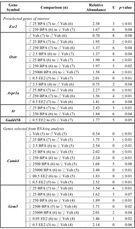
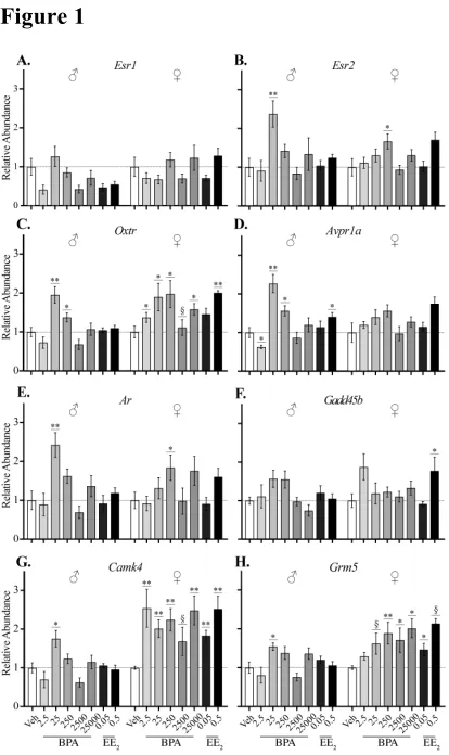
vehicle from gestational day (GD) 6 to parturition through oral gavage. Quantitative real-time PCR (qRT-PCR) was used to evaluate the expression of six candidate genes pre-selected because of their (1) role in socioemotional behaviors, (2) sex-biased expression pattern in the amygdala, (3) sensitivity to BPA or estrogen and/or, (4) importance in sexual differentiation of the amygdala (Table 1). Additionally, RNA sequencing (RNAseq) and enrichment analysis were used to characterize the neonatal amygdala transcriptome of four exposure groups (vehicle, BPA 25, BPA 250, and 0.05 EE2) and to probe for evidence of
previously unidentified modes of action. Additional genes were also subsequently analyzed by qRT-PCR to validate the RNAseq analysis.
2. MATERIALS AND METHODS
2.1 Animal Care:
advance by the National Center for Toxicological Research Institutional Animal Care and Use Committee (NCTR-IACUC). PND 1 pups were obtained from litters produced for the CLARITY-BPA program [35, 37]. Methods for animal husbandry, diet, breeding, dose preparation and administration, and necropsy are described in detail elsewhere [34]; therefore, only relevant methods are reviewed below.
Sprague-Dawley rats from the NCTR colony (NCTR-SD strain code 23) were housed in solid-bottomed polysulfone caging with hardwood chip bedding at 23 ± 3°C with a relative humidity level of 50 ± 20% on a 12:12h light/dark cycle (0600-1800). Food (soy- and
alfalfa-free diet verified casein diet 10 IF 5K96; Cat. 1810069; Purina Mills, Richmond, IN) and Millipore-filtered water in glass water bottles with silicone stoppers (#7721 clear, The Plasticoid Co., Elkton, MD) were provided for ad libitum consumption. Extracts of each diet lot were analyzed for BPA and myco/phytoestrogens (genistein, daidzein, zearalenone, and coumestrol) by liquid chromatography and mass spectrometry [42] and all had levels below the average analytical method blanks [34]. Drinking water, polysulfone cage leachates, and bedding extracts were also found to have BPA levels below the level of the average
analytical method blanks [34].
2.2 Reagents and Dosing:
The BPA (CAS # 80-05-7, catalog # B0494, TCI America, Portland, OR) and EE2
Aldrich, St. Louis, MO). The EE2 groups were included to serve as the “reference estrogen”
and to determine if BPA-related effects were consistent with an estrogenic mode of action. Two weeks before mating, dams were randomized to one of eight exposure groups stratified by body weight to produce approximately equal mean body weights in each group. Sires were randomly assigned subject to the constraint that no sibling or first cousin mating was permitted, as previously described [42]. Mating was confirmed by the presence of a sperm plug or sperm-positive vaginal cytology [defined as GD 0]. To model the exposure route used to establish the NOAEL, dams were gavaged daily with vehicle (0.3% CMC/kg bw/day), BPA (2.5, 25, 250, 2500, or 25000 µg BPA/kg bw/day), or EE2 (0.05 or 0.5 µg
EE2/kg bw/day) from GD6 until the day of parturition [postnatal day (PND) 0]. Dams and
pups were left undisturbed on PND0. On PND 1, pups (one per sex per litter) were weighed and euthanized by rapid decapitation. Heads were collected, snap frozen, and shipped coded (blinded) to the Patisaul lab where they were stored at -80°C until processing.
2.3 Tissue Collection and Preparation:
were collected at the same time we collected hypothalamic and hippocampal studies for a prior, published study [12] with the intention of performing the amygdalar assessment as a follow-up (secondary analysis) if any significant observations were found in the other brain regions. The outcomes of that prior study informed the selection of the dose groups and primary genes of interest for the present study.
2.4 Quantitative real-time PCR:
Analysis was performed on eight exposure groups (n = 5-7 for the predetermined genes, n = 3-7 for the validation genes; sample size based on availability of cDNA): vehicle, BPA 2.5, 25, 250, 2500, and 25000 and EE2 0.05 and 0.5. Total RNA was extracted with the Qiagen
RNEasy Miniprep kit. An Agilent 2100 Bioanalyzer with an RNA 6000 Nano Chip was used to determine RNA purity and concentration and each sample had a RIN of 10.
Single-stranded cDNA synthesis was performed with 350 ng of RNA input using the high capacity RNA-to-cDNA kit (Applied Biosystems, Cat. 4387406) and samples were stored at -20°C until use. qRT-PCR was performed as previously published [12] using a TaqMan probe-based protocol and detected on a StepOnePlus™ Real-Time PCR System (Applied