LUIS, JANE MARIAN SA-ONG. Sexual Reproduction and Enhancing the Efficacy of Biocontrol of Aflatoxin Contamination by Aspergillus flavus. (Under the direction of Dr. Peter S. Ojiambo and Dr. Ignazio Carbone).
by
Jane Marian Sa-ong Luis
A dissertation submitted to the Graduate Faculty of North Carolina State University
in partial fulfillment of the requirements for the degree of
Doctor of Philosophy
Plant Pathology
Raleigh, North Carolina 2019
APPROVED BY:
_______________________________ _______________________________ Dr. Peter S. Ojiambo Dr. Ignazio Carbone
Committee Co-Chair Committee Co-Chair
DEDICATION
BIOGRAPHY
ACKNOWLEDGEMENTS
I would like to express my deepest gratitude to many people who made my PhD life both full of intensive learning and enjoyable moments. To my co-advisers Drs. Peter Ojiambo and Ignazio Carbone; committee members Drs. Shuijin Hu and Ralph Dean; and Drs. Gary Payne and Marc Cubeta, who used their expertise in their own fields to guide me in my research and widen my scientific skills. They also offered encouraging words of wisdom, abundant support and valuable insights during the duration of my graduate study. To our collaborators from USDA-ARS SRRC at New Orleans including Drs. Jeffrey Cary, Matthew Lebar, Geromy Moore and Deepak Bhatnagar for their involvement in my study regarding the morphology, secondary metabolite expression and transcriptomic profiles of A. flavus crosses with different levels of female fertility. To our AMCOE collaborators including Drs. Ron Heiniger, Mark Weaver, Thomas Isakeit, and Kira Bowen for sending soil samples and maize kernels for my biocontrol study.
TABLE OF CONTENTS
LIST OF TABLES ... x
LIST OF FIGURES ... xii
CHAPTER 1 ... 1
Literature Review ... 1
1.0. Origin and Importance of Maize ... 1
2.0. Genus Aspergillus ... 3
3.0. Aspergillus flavus ... 7
4.0 Aflatoxin ... 12
5.0 Biological Control of Aflatoxin Contamination in Maize ... 16
6.0 Rationale and Objectives ... 18
Literature Cited ... 22
CHAPTER 2 ... 33
Morphological changes within stromata during sexual reproduction in Aspergillus flavus………… ... 33
ABSTRACT ... 34
INTRODUCTION ... 36
MATERIALS AND METHODS ... 39
RESULTS ... 44
DISCUSSION ... 47
ACKNOWLEDGEMENTS ... 53
LITERATURE CITED ... 54
CHAPTER 3 ... 68
Female fertility influences metabolomic and transcriptomic profiles during sexual reproduction in Aspergillus flavus ... 68
ABSTRACT ... 69
1. Introduction ... 71
2. Materials and Methods ... 74
3. Results ... 79
4. Discussion ... 85
Acknowledgements ... 93
References ... 94
CHAPTER 4 ... 121
Impact of RMb10 on the population dynamics of Aspergillus flavus in southern United States ... …121
ABSTRACT ... 122
INTRODUCTION ... 124
MATERIALS AND METHODS ... 128
RESULTS ... 136
ACKNOWLEDGEMENTS ... 146
REFERENCES ... 147
CHAPTER 5 ... 168
LIST OF TABLES CHAPTER 2
Table 2.1. Fertility of crosses between fungal strains belonging to opposite mating types used to characterize morphological changes during sexual reproduction in
A. flavus ... 59
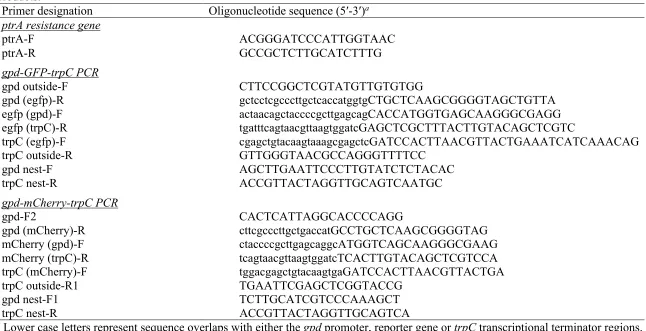
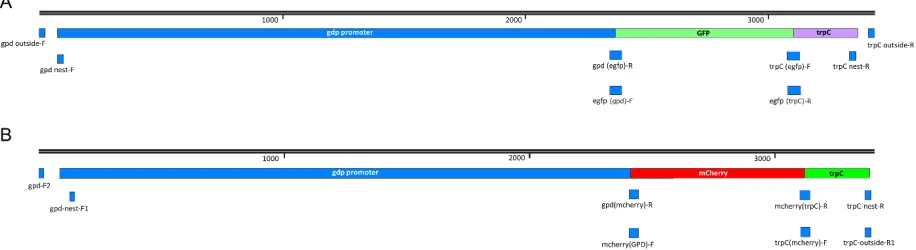
Table S2.1. Physical map of A. flavus 1534-eGFP and m-Cherry A. flavus 1582-mCherry
constructs ... 66
CHAPTER 3
Table 3.1. Fertility of stromata generated from reciprocal crosses involving two
parental strains of A. flavus ... 105 Table 3.2. Aflatoxin B1 (AFB1) production in stromata from high and low fertility
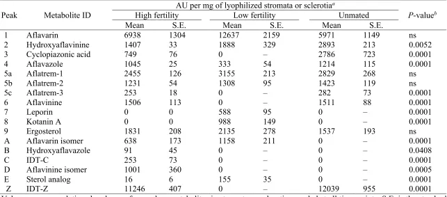
crosses and in sclerotia from an unmated strain of A. flavus ... 106 Table 3.3. Normalized abundance (absorbance per arbitrary units, AU) of 17 secondary
metabolites detected in stromata from high and low fertility crosses and
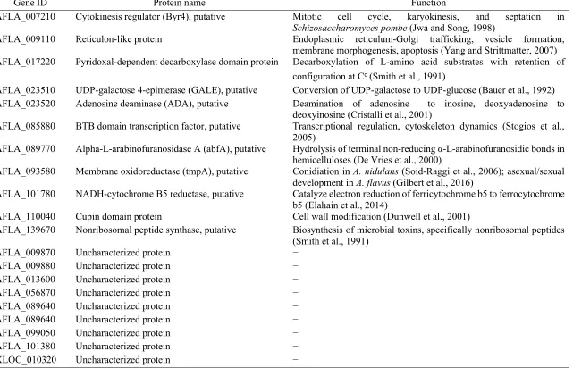
sclerotia from an unmated strain of A. flavus. ... 107 Table 3.4. Genes involved in sexual reproduction in A. flavus as identified from gene
ontology (GO) functional and K-means cluster analyses ... 108 Table 3.5. Differential expression of genes involved in growth and development in
Aspergillus species as identified in stromata from the high fertility cross ... 109
Table 3.6. Differential expression of backbone genes encoding the A. flavus secondary
metabolism gene clusters as identified in stromata from high fertility cross.... ... 110
CHAPTER 4
Table 4.1. Preliminary assessment of candidate biocontrol strains for reducing aflatoxin
contamination in maize ... 154 Table 4.2. Daily mean, maximum, minimum air temperatures, and accumulated rainfall
recorded at the field sites in Alabama, Mississippi, North Carolina and Texas
in April to July of 2016 and 2017 ... 155 Table 4.3. Soil population densities of A. flavus prior to application of RMb10 and
Afla-Guard® biocontrol strains, and at harvest in maize fields in southern
Table 4.4. Number of unique multi-locus haplotypes (MLHs) inferred from populations of A. flavus in maize fields in southern United States treated with RMb10
and Afla-Guard® biocontrol strains in 2016 and 2017 ... 157 Table 4.5. Frequency of the mating type (MAT) genes among A. flavus isolates in maize
fields in southern United States treated with RMb10 and Afla-Guard®
biocontrol strains in 2016 and 2017 ... 158 Table 4.6. Frequency of aflatoxin gene cluster types among A. flavus isolates in maize
fields in southern United States treated with RMb10 and Afla-Guard®
biocontrol strains in 2016 and 2017 ... 160 Table 4.7. Parameter estimates of A. flavus populations in maize fields in southern
United States treated with RMb10 and Afla-Guard® biocontrol strains in
2016 and 2017 ... 161 Table 4.8. Mean estimates of nucleotide diversity (p) and mutation rates (q) of A. flavus
populations in maize fields in southern United States treated with RMb10
LIST OF FIGURES CHAPTER 2
Figure 2.1. Unmated sclerotium of A. flavus strain NRRL 29507 and fertilized stroma
of A. flavus strain NRRL 29507 ... 60 Figure 2.2. Confocal images (10X to 40X) showing the different phases of interaction
between conidia (NRRL 21882) and sclerotia (NRRL 29507) of sexually
compatible strains of A. flavus ... 61 Figure 2.3. Low magnification (90X to 130X) scanning electron micrographs showing
morphological changes in the development of fertilized stromata and unmated sclerotia of A. flavus strains NRRL 29507 and NRRL 21882
from time of crossing (T0) until 8 weeks of incubation (T4) ... 62 Figure 2.4. High magnification (500X to 8500X) scanning electron micrographs
showing morphological changes in the development of fertilized stromata and unmated sclerotia of A. flavus strains NRRL 29507 and NRRL 21882
from time of crossing (T0) until 8 weeks of incubation (T4) ... 63 Figure 2.5. Scanning electron micrographs (1000X to 8500X) showing sexual
reproductive structures within the stromata of A. flavus ... 65 Figure S2.1. Physical map of A. flavus 1534-eGFP and A. flavus 1582-mCherry
constructs ... 67
CHAPTER 3
Figure 3.1. Aflatoxin B1 (AFB1) content produced over time in stromata from low and
high fertility crosses and in sclerotia from an unmated strain of A. flavus ... 111 Figure 3.2. Multivariate analysis of secondary metabolites detected in stromata from
low and high fertility crosses and in sclerotia from an unmated strain of
A. flavus ... 112
Figure 3.3. Hierarchical cluster analysis of secondary metabolites detected in stromata from low and high fertility crosses and in sclerotia of an unmated strain of
A. flavus ... 113
Figure 3.6. Functional annotation of differentially expressed genes (DEGs) between
stromata from low and high fertility crosses over time ... 116 Figure 3.7. Diagrams of K-means cluster analysis of gene expression between stromata
from low and high fertility crosses over time. ... 119 Figure 3.8. Times-series analysis of differentially expressed genes in stromata from
low and high fertility crosses ... 120
CHAPTER 4
Figure 4.1. Frequency of multi-locus haplotypes (MLHs) recovered as a proportion of the total number of MLHs observed at each sampling period from maize fields in Alabama, Mississippi, North Carolina and Texas in 2016 and 2017 using combined five next generation multi-locus (aflM, aflW, amdS, mfs
and trpC) sequence data ... 163
Figure 4.2. Frequency of the mating type (MAT) genes among A. flavus isolates in maize fields in southern United States treated with RMb10 and Afla-Guard®
biocontrol strains in 2016 and 2017 ... 164 Figure 4.3. Frequency of aflatoxin gene cluster types among A. flavus isolates in maize
fields in southern United States treated with RMb10 and Afla-Guard®
biocontrol strains in 2016 and 2017 ... 165 Figure 4.4. Phylogenetic relationships showing patristic distances of 311 A. flavus
isolates to RMb10 or Afla-Guard® strain ... 166 Figure 4.5. Aflatoxin contamination in harvested grain collected from maize fields in
southern United States treated with RMb10 and Afla-Guard® biocontrol
CHAPTER 1
Literature Review
1.0. Origin and Importance of Maize
Maize (Zea mays L.) is one of the most widely distributed grain crops around the world. It is believed to have been domesticated from a subspecies of the wild grass teosinte, Zea mays ssp. parviglumis, about 9,000 years ago (Doebley 1990; Matsuoka et al. 2002). The wild progenitor of maize which is native to the Central Balsas River Valley in Mexico (Hastorf 2009; Ranere et al. 2009) is characterized by a tall stalk, long lateral branches, broad leaves, and ears consisting of 5 to 12 kernels individually sealed in a hard fruit case (Doebley 2004). Domestication was initiated when ancient farmers in Mexico selected kernels with desirable characteristics to be planted the next season. Continuous selection over many seasons led to the development of modern ears which bear 500 or more exposed kernels attached to the central axis of the cob (Beadle 1939; Doebley 2004). Maize subsequently became a major part of the diet of most native people in Mexico. From Mexico, domesticated maize dispersed northward into southwestern United States and Canada, and southward into Guatemala, South America and the Andes Mountains (Matsuoka et al. 2002). Contact between Native Americans and European colonists in the 15th century paved way for the spread of maize to different parts of the world. With the exemption of Antarctica, maize is now cultivated on all continents from 58o North to 40o South (OGTR 2008; Encyclopedia Britannica 2018; ICGA 2018).
2018). Maize serves as the main staple diet in many countries in sub-Saharan Africa and Latin
America, while it is consumed in substantial quantities in Asia and Europe. In the United States,
high proportion of the produce is used for livestock feed as green chop, dry forage, silage or grain
(OGTR 2008; NCGA 2018; USDA-ERS 2018). Maize also serves as a big component of alcohol
production (fuel ethanol), pharmaceutical supplies (antibiotics, vitamin carriers), and industrial
products (paper, soap, textiles) (OGTR 2008; ISU 2009).
In 2018, maize ranked as the highest produced grain in the world. It was cultivated on 179.6
million hectares of land with a total production of 1.1 billion metric tons (Statista 2018a; Statista
2018b; USDA-FAS 2018). United States (376.6 million metric tons, 34%) is the highest producer,
where more than half of its total production is from the Midwestern states of Iowa, Illinois,
Nebraska and Minnesota. China (215.9 million metric tons, 20%) and Brazil (94.5 million metric
tons, 9%) rank as the second and third highest producers, respectively (NCGA 2018; USDA-NASS
2018).
The average price of maize runs around $3.25 per bushel (NCGA 2018), but the price
received by maize growers and suppliers from food and feed companies is influenced by the quality
and safety of the grain. Contamination with mycotoxins, particularly aflatoxin, can lower the price
or even lead to rejection of contaminated maize lots. Additional costs may be incurred from
mycotoxin testing and potential lawsuits from consumers (Mitchell et al. 2016). Hence, there is a
constant need to develop management strategies that either prevent or limit mycotoxin
2.0. Genus Aspergillus 2.1. Taxonomy
Aspergillus is a large genus belonging to phylum Ascomycota, subphylum
Pezizomycotina, class Eurotiomycetes, order Eurotiales, and family Trichocomaceae. The phylum
Ascomycota (sac fungi) consists of ‘meiosporic’ species that have the ability to reproduce
sexually, either through heterothallic (obligate outbreeding) or homothallic (self-fertile) breeding
mechanisms. Ascomycetes produce four to eight ascospores (teleomorph spores) within a saclike
structure called an ascus. This is in contrast to the ‘mitosporic’ species traditionally classified
under phylum Deuteromycota, which are known to only reproduce asexually (Taylor et al. 1999;
Taylor et al. 2006; Dyer and O’Gormann 2012). The subphylum Pezizomycotina consists of all
filamentous ascomycetes that produce ascospores within a fruiting body (ascoma or ascocarp).
Many form closed fruiting bodies, while some lost the ability to undergo meiosis and no longer
produce asci (Spatofora 2007; Holt and Iudica 2013). Members of the class Eurotiomycetes and
order Eurotiales produce small, evanescent asci scattered within a fully closed, spherical ascocarp
(cleistothecium) rather than gathered in a spore-bearing hymenium (Geiser et al. 2006). Members
of the family Trichocomaceae are most commonly observed in their anamorph state and are
typically recognized by their complex phialide-bearing structures (Aspergillus, Paecilomyces and
Penicillium). Most members have opportunistic lifestyles that rapidly invade uncolonized
substrata. In many species, sexual reproduction typically occurs at the end of their proliferative
stage after fully colonizing their substrata (Malloch and Cain 1972; Malloch 1986). The genus
Aspergillus is comprised of more than 250 recognized species that are collectively called the
‘Aspergilli’ (Geiser et al. 2007; Dyer and O’Gormann 2012). The genus is divided into eight
Circumdati) is one of the most economically important because it contains A. flavus and A.
parasiticus which are the main producers of aflatoxin in harvested produce (Diener et al. 1987;
CAST 2003). The teleomorphs of A. flavus and A. parasiticus are named Petromyces flavus and
P. parasiticus, respectively, to indicate the production of one to multiple ascocarps borne within a
larger structure known as sclerotium. Ascospores are released following natural breakdown of the
ascus wall and outer wall (peridium) of the ascocarp instead of being forcefully discharged
(Malloch and Cain 1972; Dyer and O’Gormann 2012).
2.2. Morphology
Aspergillus was originally described by Pier Antonio Micheli in 1729 (Micheli 1729). He
noted that the spores of the fungus radiated from a central structure similar to an ‘aspergillum’
used by Roman Catholic clergy to sprinkle holy water in the church (Ainsworth 1976). This
structure is now referred to as the vesicle and is the defining characteristic of the genus (Bennet
2010).
The vegetative mycelium of Aspergillus consists of a network of septate hyphae that can
be hyaline, bright colored, light brown, or colored in localized areas. The formation of a conidial
structure starts with the enlargement of a thick-walled hyphal cell (foot cell). A septate or aseptate
conidiophore (stalk) branch out perpendicularly from the foot cell and the surface of the substrate.
When the conidiophore reaches its maximum height, the tip swells to form a globose,
hemispherical, elliptical, or long clavate vesicle that provides expanded surface for the attachment
of reproductive cells. In uniseriate species, the vesicle gives rise to a single layer of
conidium-producing sterigmata (phialides), while in biseriate species, the vesicle gives rise to a base layer
sterigmata successively produce unbranched chains of conidia to craft globose, radiate, or
columnar shaped conidial heads (Raper and Fennel 1965).
Sclerotia are produced by some Aspergillus species, but are lacking in many. These
structures are hardened, normally darkly pigmented hyphal masses that are capable of remaining
dormant in the soil for long periods of time during which they can survive harsh environmental
conditions with limited nutritional need (Dyer and O’Gormann 2012; Chang et al. 2014). Sclerotia
are composed of polyhedral, thick-walled cells that assume globose to subglobose shapes when
mature (Raper and Fennel 1965).
Ascocarp formation in the majority of ascosporic Aspergilli is initiated by the formation of
a terminally coiled ascogone (Raper and Fennell 1965). The ascocarps of different species
similarly arise from ascocarp initials but differ in gross appearance. A. glaucus (teleomorph:
Eurotium herbariorum) produces small, yellow, naked ascocarps borne on a single stalk-like
hypha that are loosely suspended in a network of pigment-encrusted mycelia (Rapper and Fennell
1965). The ascocarps of A. nidulans (Emericella nidulans) are dark-purple and produced in a
nest-like arrangement that is completely enveloped by or associated with thick-walled hϋlle cells
(Fennell and Warcup 1959; Rapper and Fennell 1965; Sharma 2005). A. fischeri (Neosartorya
fischeri) produces globose, white to cream ascocarps covered by abundant sterile hyphae that
develop within a cottony mycelial matrix (Rapper and Fennell 1965; Sarbhoy 1985). A. alliaceus
(Petromyces alliaceus) produces one to several, variably-sized, globose ascocarps within a
dark-walled and persistently hard, sclerotium-like body (Rapper and Fennell 1965; Sarbhoy 1985).
Unlike the above species, A. athecius (Eurotium athecium) fails to develop walled ascocarps and
produce asci in naked clusters on undifferentiated mycelium (Rapper and Fennel 1965; Sharma
ascogone (Raper and Fennell 1965). The nuclei from a heterothallic partner, or nucleus in a
homothallic species, normally form the maternal tissues and ascocarp wall while the compatible
partner provides the fertilizing nucleus that passes into the ascogenous hyphae (Dyer and
O’Gormann 2012). Fusing of compatible hyphae gives rise to crozier tips and non-linear asci
(Raper and Fennell 1965). The ascocarps are usually thin walled and produce asci and ascospores
within a few weeks. Conversely, ascocarps that are borne within sclerotia necessitate longer time
(usually months) for maturation. In the latter case, the ascocarps become embedded in a matrix of
vegetative hyphae (stroma) consisting of pseudoparenchymatous hyphae (stromal matrix).
Flattened cells make up the outer wall of the ascocarp (peridium). The ascocarps may contain up
to 100,000 asci that individually enclose, with very rare exceptions, eight ascospores (Dyer and
O’Gormann 2012). Ascospores are hyaline in all species except A. nidulans for which they are
red-brown through purple-red to violet and A. ornatus in which they are red-brown shades when
mature. Mature ascospores resemble a double convex lens. Upon germination of ascospores, the
two valves part completely and allow the growth of a germ tube (Raper and Fennell 1965).
2.3 Distribution and Significance
Aspergillus species are ubiquitous in nature. They can survive in a wide range of
temperature (6 to 55oC) and feed on a large variety of plant, animal and man-made substrates
(Krijgsheld et al. 2013). Their effective dispersal mechanism through massive conidia production
makes them the most dominant fungal structures in the air. Air movement, and possibly insects,
transports them over short and long distances where they can establish new colonies (Hedayati et
climates, and hence they are particularly common in agricultural soils of subtropical and warm
temperate regions (Horn 2003).
The ability of many Aspergillus species to secrete high amounts of enzymes and organic
acids makes them beneficial in food and industrial processes. For example, kojic acid produced by
A. oryzae is used in food fermenting processes, while cellulases and hemicellulases produced by
A. niger are utilized in textile, pulp and paper production (Polizeli et al. 2016). On the other hand,
many Aspergillus species pose a high health and economic risk as opportunistic pathogens and
toxin producers. A. fumigatus is the most predominant cause of allergic bronchopulmonary
aspergillosis in immunocompromised patients (Chaudhary and Marr 2011). Many species grow on
fresh and processed plant produce, and are thus, known as common food spoilage molds
(Krijgsheld et al. 2013). A. flavus causes Aspergillus ear rot in maize and the fungus can also
contaminate maize kernels with toxic secondary metabolites such as aflatoxin (Smart et al. 1990).
3.0. Aspergillus flavus
3.1. Distinguishing characteristics
A. flavus produces B aflatoxins (AFB1 and AFB2) and cyclopiazonic acid (CPA), but not
G aflatoxins (AFG1 and AFG2). The inability to produce G aflatoxins is due to a deletion in its
cypA gene that is predicted to encode a cytochrome P450 monooxygenase in the aflatoxin gene
cluster (Ehrlich et al. 2004).
Conidia of A. flavus are globose to subglobose when mature and their size can range from
3.5 to 4.5 μm in diameter. Its conidial wall is relatively thin with fine to moderately roughened
ornamentations. This is in contrast to the conidia of A. parasiticus that are more spherical in shape
composed of yellow-green to green or brown sporulation and goldish to red-brown reverse. The
conidiophores are hyaline, usually roughened but can vary from smooth to coarsely roughened.
Sterigmata can be uniseriate or biseriate, with both conditions normally seen within the same strain
or vesicle. A. flavus produces sclerotia that are dark red-brown to purple-brown or black at
maturity, and vary in shape as globose, subglobose or vertically elongate (Raper and Fennell 1965).
3.2. Diversity within A. flavus
A. flavus has two sclerotia morphotypes and different chemotypes. Strains that produce
sclerotia are classified as either “S” or “L” morphotype strain based on production of small or large
sized sclerotia, respectively. The L morphotype strain, that is also most common, typically
produces sclerotia that are greater than 400 μm in diameter. The S morphotype strain, which is less
common, produces relatively more abundant sclerotia that are lesser than 400 μm in diameter and
fewer conidial heads than the L morphotype strains (Cotty 1989; Geiser et al. 2000; Chang et al.
2014). Mycotoxin production in A. flavus is highly variable and can range from atoxigenic to
potent producers of AFB1, AFB2 and CPA. The atoxigenic populations are normally found
coexisting with toxigenic populations in plant tissues and in soil (Payne and Widstrom 1992; Horn
2003). For example, an intensive field sampling conducted by Horn and Dorner (1999) showed
that 12% of the A. flavus L strains along a wide transect within the United States were
non-aflatoxigenic, while 88% were toxigenic. L morphotype strains in the United States exist in high
densities in the southern, wetter regions and are more variable in aflatoxin and CPA production. S
morphotype strains exist in high densities in the northern, drier regions next to the Sahara Dessert
produce B aflatoxins only (SB), while those from Argentina, West Africa, Southeast Asia and
Australia produce both B and G aflatoxins (SBG) (Horn and Dorner 1999).
Isolates of A. flavus can also be categorized into vegetative compatibility groups (VCGs),
also called heterokaryon compatibility groups. Vegetative compatibility is indicated by the
formation of a zone of dense growth and heavy sporulation at the intersection of the two colonies
(Horn and Greene 1995). Vegetative compatibility functions as a multi-locus measure of genetic
diversity because the multiple, unlinked heterokaryon incompatibility (het) loci distributed across
the genome of the paired colonies must all be identical for anastomosis to occur (Horn and Greene
1995). Isolates within the same VCG are clonal, thereby, are morphologically similar and variation
in mycotoxin profile is lesser within a VCG than between VCGs (Bayman and Cotty 1993). The
diversity of VCGs in a given location is high and the populations of VCGs can change even over
a short period of time (Scheidegger and Payne 2003).
Phylogenetic analysis based on restriction fragment length polymorphisms (RFLPs) of
nuclear-coding genes and DNA sequences identified two reproductively isolated clades within A.
flavus that cannot be readily separated by morphology and metabolite production (Geiser et al.
2000). Group I includes L sclerotia that produce B aflatoxins (LB) and SB isolates, while Group II
includes SB and SBG isolates. Group I is further subdivided into lineages IA, IB and IC. Lineage
IA contains SB and LB isolates. Lineage IB contains non-aflatoxigenic L isolates and A. oryzae.
Lineage IC contains L isolates that produce and do not produce aflatoxins (Geiser et al. 2000).
Multi-locus analysis using 21 regions in the aflatoxin gene cluster of A. flavus confirms that lineage
IB contains only non-aflatoxigenic species with complete or partial deletions in their aflatoxin
gene cluster. This lineage also includes NRRL 21882, which is the active ingredient of the
non-aflatoxigenic isolates and share a more recent common ancestor with A. parasiticus than lineage
IB. Lineage IC includes NRRL 18543, which is the active ingredient of AF36 (Moore et al. 2009).
Observed diversity in morphology, toxicity and genetic composition within A. flavus has
been attributed to several factors. These factors include movement of individuals between
locations, current practices of intensive agriculture, dominant crops grown in the area, mutation,
and sexual recombination between compatible genotypes (Bayman and Cotty 1991; Horn 2007;
Olarte et al. 2012).
3.3. Life cycle and reproduction
The ubiquitousness of A. flavus in the soil led to the conclusion that soil is its primary
habitat (Horn 2007). The fungus spends a large portion of its life cycle as soil saprobe, where it
plays an important role as a decomposer and nutrient recycler of plant and animal debris (Hedayati
et al. 2007). Under certain circumstances, A. flavus can become an opportunistic pathogen of
developing seeds (Payne 2016). It produces a large array of degrading enzymes to utilize
temporarily available plant materials including cellulose, pectin, lignin, tannins, cutin, starch, lipid
and proteins (Horn 2007).
A. flavus produces numerous conidia that are easily dispersed aerially by wind and insects
over short and long distances. During adverse conditions such as lack of adequate water or
nutrients, the fungus produces resistant structures called sclerotia (Hedayati et al. 2007).
Populations in the soil (conidia, hyphae, sclerotia) serve as the primary inocula for infection when
conidia land on the surface of susceptible maize (Horn 2003). The susceptibility of maize to
infection by the fungus commences at the emergence of silks. Conidia of A. flavus that land on the
insects into the ear (Payne and Widstrom 1992). Populations that infect the ear replenish the
populations in the soil when infected maize ears fall on the soil surface during the growing season
or at harvest. Pre-infection of the ears before dropping on the ground gives the fungus a
competitive advantage over other microorganisms in the soil. Secondary infection can occur when
conidia from southern regions of Northern America is wind dispersed to crops in the northern
areas. The fungus overwinters as mycelium or sclerotium in soil or infected corncobs, which later
serve as source of primary inoculum for infection of next season’s crop (Hedayati et al. 2007; Horn
2007).
A. flavus was originally considered to be strictly asexual. This was thought to explain the
strong clonal structure of populations, in which vegetative compatibility was believed to be a
strong barrier of genetic exchange (Chang et al. 2014; Ehrlich 2014). However, populations of A.
flavus show considerable genetic, morphological and chemotype diversity within vegetative
compatibility groups. Populations are also dynamic and the dominant VCG group in an area can
shift from year to year (Bayman and Cotty 1991). This observed diversity was first attributed to
parasexuality, which involves the exchange of genetic material between two haploid strains and
formation of heterokaryons. However, the parasexual cycle has only been observed under
laboratory conditions and hyphal fusion would primarily occur between genetically similar
individuals of the same VCG (Chang et al. 2014).
The discovery of the sexual stage of A. flavus showed that VCG is not a strong barrier to
genetic exchange and outcrossing among VCGs leads to formation of new VCGs (Olarte et al.
2012). A. flavus was identified to be heterothallic, where each individual contain one of the two
mating type idiomorphs, MAT1-1 or MAT1-2, at a single locus. MAT1-1 encodes an α-domain
factor (Ramirez-Prado et al. 2008). Successful crosses between individuals of opposite mating
types and different VCGs lead to the formation of ascospore-bearing ascocarps embedded within
the matrix of the sclerotia after an extended incubation of five to seven months in dark conditions
(Horn et al. 2014). A. flavus was also identified to be hermaphroditic with respect to male and
female roles during sexual reproduction (Horn et al. 2016). The sclerotium functions as the female
by providing nutrient sources and bearing the ascocarps and ascospores, while the conidium
functions as the male by fertilizing the sclerotium. The degree of fertility or sterility of the cross
is dictated by the parental source of conidium and sclerotium (Horn et al. 2016). Recombinant
progenies are produced through independent assortment of chromosomes and crossing over within
the aflatoxin gene cluster or other portions of the genome (Olarte et al. 2012).
4.0. Aflatoxin
4.1. Background and description
Aflatoxins are hepatotoxic, carcinogenic and immunosuppressive secondary metabolites
produced mainly by A. flavus and A. parasiticus (Diener et al. 1987; CAST 2003). These toxins
are chemically composed of dihydrofuran or tetrahydrofuran moieties that are fused to a coumarin
ring (Hussein and Brasel 2001). The four major types of aflatoxins (AFB1, AFB2, AFG1, AFG2)
are identified based on their blue (B) or green (G) fluorescence under ultraviolet light and relative
chromatographic mobility (1 or 2). Among the four, AFB1 is the most toxic when studied on
experimental animals and clinical patients (Chu 2003). AFB2 and AFG2 are hydroxyl-derivatives
of AFB1 and AFB2, respectively, and remain relatively nontoxic unless metabolically oxidized in
lactating animals ingest contaminated feed. AFM1 is metabolized by the hepatic system of animals
and becomes absorbed in their milk (Ghanem and Orfi 2009).
Aflatoxin continues to be the most problematic and heavily regulated mycotoxin in the
United States and other parts of the world (Mitchell et al. 2016). Awareness of the harmful effects
of aflatoxin started in the mid-1960s when Brazilian peanut meals infected by A. flavus and
contaminated with high levels of aflatoxin were identified as the causative agents of Turkey “X”
disease. This epidemic killed 100,000 turkey poults that were being raised around 500 farms in
England (Wannop 1961; Kensler et al. 2011). Continuous consumption of food or feed products
with high aflatoxin concentrations have also been associated with high mortality in other avian
flocks, reduced milk production in cattle, lesser egg yield in poultry, stunted growth in children,
and hepatocellular carcinoma (liver cancer) in elder adults (CAST 2003; Wild 2007).
Due to the above health risks, over 100 nations have established regulatory limits for
aflatoxin contamination. The United States Food and Drug Administration established a maximum
level of 100 to 300 parts per billion (ppb) total aflatoxin content for maize and peanut products
intended for beef cattle, swine and mature poultry consumption; 20 ppb for human and immature
animals; and, 0.5 ppb AFM1 in milk (FDA 2000). European countries established a much stricter
cut-off of 4 ppb total aflatoxin content in all cereals and dried fruits intended for direct human
consumption or use as ingredients in foodstuffs; and, 0 ppb in milk or milk-based products (FAO
2004; FAO 2006).
4.2. Aflatoxigenic species of Aspergillus
A. flavus and A. parasiticus are the two major producers of aflatoxin on a number of
commonly found aflatoxin-producing fungus in maize, peanuts, cottonseed and tree nuts. In
contrast, A. parasiticus produces AFB and AFG but not CPA. It occurs more frequently in peanuts
than other crops but is typically outcompeted by A. flavus when both fungi are present (Payne and
Widstrom 1992; Payne 1998; Horn and Dorner 1999).
Aflatoxin production necessitates a substantial expenditure of energy that involves around
25 genes arranged in a complex biosynthetic cluster. These genes encode for the proteins necessary
for the oxidative, reductive, and regulatory processes in aflatoxin biosynthesis. The aflatoxin gene
cluster in A. flavus is located within a 70kb DNA region in the subtelomeric region of chromosome
III (Yu et al. 2004). Since polymorphism for aflatoxin production is maintained by balancing
selection (Carbone et al. 2007), it has been postulated that aflatoxin production provides some
benefits to the fungus. Some of the proposed benefits include competitive mechanism (Bilgrami
et al. 1988), feeding deterrents against arthropods for resource competition or protection from
fungivory (Drott et al. 2017), pathogenicity mechanism (Horn 2007), adaptation to ecological
niche (Horn and Dorner 2002), and alleviation of environmental stress (Fountain et al. 2014).
Other Aspergillus species that produce aflatoxins have been reported. These include A.
nomius, A. pseudonomius, A. bombycis, A. pseudotamarii, A. pseudocaelatus, A.
ochraceoroseus and A. rambellii. A. nomius is often found in soil and on dead or diseased insects.
It is morphologically similar to A. flavus and produces AFB and AFG but not CPA. A.
pseudonomius was isolated from insects and soil in the United States and produces AFB (Varga et
al. 2011). A. bombycis, which was isolated along with A. nomius from insect frass in
silkworm-rearing houses in Japan and Indonesia, also produces AFB and AFG but not CPA (Peterson et al.
2001). A. pseudotamarii, collected from a tea field soil in Japan and Brazil, produces AFB (Ito et
AFG and CPA (Varga et al. 2014). A. ochraceoroseus and A. rambellii were isolated from soil
detritus in Africa and produce AFB1 (Moore et al. 2015). All the abovementioned species
infrequently contaminate agricultural crops, and thus they are not considered to pose a significant
economic risk.
4.2. Impact to the maize industry in the United States
Aflatoxin has been detected in maize kernels at different stages of development and
maturity from late milk stage until harvest (Anderson et al. 1975). Drought stress accompanied by
elevated temperatures during seed development promotes A. flavus invasion and subsequent
aflatoxin contamination. More rapid accumulation of aflatoxin occurs when kernels are naturally
wounded or damaged by insects (Payne and Widstrom 1992; Horn and Dorner 1999). Not all
kernels need to be contaminated with aflatoxin before losses due to contamination are incurred. A
single heavily contaminated kernel can bring the average concentration of 10,000 seeds to a
dangerous level and even lead to the rejection of the whole lot (Schroeder 1969). Contamination
can ensue during postharvest when inadequately dried grains are stored or where storage
conditions favor the growth of the fungus and production of aflatoxin (Ojiambo et al. 2018).
Annually, a large acreage of maize in the United States is under threat of aflatoxin
contamination. This acreage ranges from Texas to Oklahoma in the south and Alabama to North
Carolina in the southeast (Mitchell et al. 2016; Reus 2016). During intense drought conditions, the
threat of aflatoxin contamination can extend northward into the midwestern states of Kansas,
Missouri and Illinois (Ingwersen 2013). Extremely severe losses occurred in Mississippi,
Louisiana and Texas in 1998 (Cardwell et al. 2001), which forced growers to either destroy or sell
the potential for maize to be used as a rotational crop in Arizona and Corpus Christi, Texas.
Aflatoxin contamination in the Corn Belt states of Indiana, Illinois, Iowa, Missouri, Nebraska and
Kansas arises sporadically but can be extreme when droughts occur (Cardwell et al. 2001). The
regional differences in aflatoxin contamination has been attributed to climatic conditions and
agricultural practices that increase the susceptibility of the crop to A. flavus invasion (Horn and
Dorner 1999).
The annual loss due to aflatoxin contamination in maize in the United States is estimated
to be around $163 million. Around $31 million comes from maize rejected for food and around
$132 million from maize rejected for feed and through livestock losses (Wu 2006). Losses to the
maize industry can even reach up to $1.68 billion annually if climate changes causes more regular
aflatoxin contamination in the maize-growing states in the Corn Belt (Mitchell et al. 2016).
5.0. Biological Control of Aflatoxin Contamination in Maize
Several management strategies ranging from pre-harvest to storage options have been
suggested to prevent or minimize the possibility of aflatoxin contamination in maize. Some
pre-harvest strategies include biological control, host resistance, plant density, and good agricultural
practices such as crop rotation, pesticide application, soil amendment and moisture management
(Ojiambo et al. 2018). Among these, biological control, which involves application of atoxigenic
strains of A. flavus to competitively outcompete toxigenic strains, has one of the greatest impacts
(Ojiambo et al. 2018). The other strategies are either still in their early stage of development, do
not reduce the risk of A. flavus invasion and resultant aflatoxin contamination significantly relative
to the regulatory limits, or may not be practical in areas where resources are limited. Some
(<14%), alteration of storage conditions to reduce fungal activity, and cleaning and sorting of grain
prior to storage (Ojiambo et al. 2018).
Two commercially available biocontrol strains, AF36 and Afla-Guard®, are currently used
in the United States to reduce aflatoxin contamination. A. flavus strain NRRL 18543 used as the
active ingredient in AF36 was isolated from an upland cotton in Arizona (Cotty 1989) and is
currently registered for use in cotton (Arizona, California, Texas), maize (Arizona, Texas) and
pistachio (Arizona, California, New Mexico, Texas) (EPA 2015). Despite having a full aflatoxin
gene cluster, it does not produce aflatoxin due to a point mutation in its polyketide synthase (pksA)
gene that introduces a premature stop codon to prevent formation of norsolorinic acid and aflatoxin
accumulation (Ehrlich and Cotty 2004). Conversely, NRRL 18543 produces the mycotoxin
cyclopiazonic acid (CPA) that was linked as a probable co-contaminant of the peanut meal
associated with the Turkey “X” disease in the mid-1960s (King et al. 2011). A. flavus strain NRRL
21882 used as active ingredient in Afla-Guard® was isolated from peanut in Georgia (Dorner 2005)
and is currently registered for use in maize and peanut in the United States (EPA 2013). This strain
does not produce aflatoxin nor CPA due to complete deletion of its entire aflatoxin and CPA gene
clusters (Chang et al. 2005; King et al. 2011).
Application of these commercial biocontrol strains has been reported to reduce aflatoxin
contamination by up to 98% (Ehrlich 2014). High concentrations of atoxigenic strain of A. flavus
are coated in nutrient-supplying carriers, such as barley for Afla-Guard® and wheat for AF36, then
applied in high densities to the plant canopy or soil surface. The treated grain absorbs moisture
and allows the atoxigenic strain to sporulate profusely. The spores disperse to the developing maize
prior to occupation of toxigenic strains, thus physically excluding toxigenic strains from infecting
exclusion’ (Chang et al. 2014; Damann 2015). In order to maintain efficacy, the biocontrol product
needs to be applied annually (Lewis et al. 2019). Further studies are needed to determine whether
the biocontrol strains truly displace the native aflatoxin-producing population in the field (Moore
et al. 2013a). Another mechanism called ‘thigmo-down regulation of aflatoxin synthesis’ (Huang
et al. 2011) proposes that aflatoxin is inhibited only when appropriate atoxigenic and toxigenic
isolates, with certain specificities, come in contact in the first day of growth. Such interaction
initiate an unknown signaling pathway, perhaps related to specific and consistent component of
programmed development, that prevents or down regulates the biosynthesis of aflatoxin which
normally develops after three days of fungal growth (Huang et al. 2011).
6.0. Rationale and Objectives 6.1. Sexual reproduction in A. flavus
While useful information regarding mycelium growth, sclerotium production, and the
teleomorphic stage of A. flavus is available in literature (Horn et al. 2009; Wu et al. 2014; Horn et
al. 2016), comprehensive details on the actual process of sexual reproduction are still lacking. For
example, there is no information about the mechanism on how conidia fertilize the sclerotia during
sexual reproduction in A. flavus. Additional research is also needed to determine how the
interaction between conidia and sclerotia leads to the formation of ascocarps, asci and ascospores.
Unlike other heterothallic fungi, no receptor nor specialized structures for fertilization have been
observed on the surface of the A. flavus sclerotia (Horn et al. 2016).
As conidia and sclerotia come in contact during sexual reproduction, the sclerotia are also
transitioning from asexual to sexual structures. This transition can affect the survival and genetic
including aflatoxin, produced by the fungus. To the best of our knowledge, there is little to no
information available on changes in morphology, secondary metabolism, and transcription during
the fertilization of sclerotia and the formation of sexual structures. In addition, while the degree of
female fertility is dependent upon the sclerotial strain, it is not known how female fertility could
influence morphological, metabolomic, and transcriptional changes during sexual reproduction in
A. flavus. Understanding the biology and sexual cycle of A. flavus could be useful in identifying
additional biological strategies to control toxigenic strains of A. flavus or facilitate design of novel
biocontrol agents. For example, depending on the prevailing genetic structure of the native
population, a highly fertile or a low fertility biocontrol strain would be more effective in reducing
aflatoxin contamination. A highly fertile MAT1-2 biocontrol strain could be applied in a field
whose native population is primarily composed of MAT1-1 toxigenic strains (or vice versa) so as
to encourage recombination and drive the population towards atoxigenecity. In populations with
mixed chemotypes and mating types, the density of the predominant non-aflatoxigenic native
strain could be inflated by applying a low fertility biocontrol strain of the same mating type. An
increased density of non-aflatoxigenic population would lead to decreased levels of aflatoxin in
the crop (Cotty 1994). In such a case, information on the metabolomic and transcriptional changes
could be useful in screening and rapid selection of biocontrol strains with the desired level of
female fertility.
6.2. A novel strain for biocontrol of aflatoxin contamination in maize
Despite the persistent risk of aflatoxin contamination in the maize-growing areas of the
United States, only two commercial biocontrol products (AF36 and Afla-Guard®) are available in
Afla-Guard® as the solitary option in other states. Currently, there is an increasing interest to
identify additional strains and develop sustainable biocontrol approaches to successfully reduce
aflatoxin contamination. In addition, both AF36 and Afla-Guard® were developed on the premise
that members of different VCGs cannot recombine. With the discovery of sexual reproduction in
A. flavus, it is important to select and identify biocontrol strains whose recombination potential
with native populations will avoid production of high frequency of aflatoxigenic progeny in their
area of use.
AF36, Afla-Guard®, and all reported biocontrol strains are of the mating type MAT1-2
(Moore et al. 2013b), giving the implication that biocontrol strains should be MAT1-2. In 2015,
we identified a MAT1-1 strain (RMb10), which has a comparable ability as Afla-Guard® to reduce
aflatoxin contamination when co-inoculated with NRRL 3357 in preliminary greenhouse and field
trials (J.M. Luis et al., unpublished). This strain was isolated from a maize field in Rocky Mount,
North Carolina in 2012. Previous studies showed that mitigation of aflatoxin contamination
through biocontrol tends to be more persistent when the applied strain matches the genetic and
ecological structure of the native population of A. flavus in the soil (Lewis et al. 2019). Availability
of a MAT1-1 biocontrol strain diversifies the choice of biocontrol products that can be used in the
field. The goal of this study is to test the effect of RMb10 in changing the distribution of native A.
flavus populations when applied in large-scale field trials in southern United States.
6.3. Research objectives
Given the abovementioned considerations, the overall goal of this dissertation is to
understand the biology of sexual reproduction in A. flavus and to improve the efficacy of biocontrol
1. Determine morphological changes that occur in fertilized stromata and unmated sclerotia
during sexual reproduction in A. flavus as influenced by the degree of female fertility
(Chapter 2).
2. Characterize differences in secondary metabolite production and gene expression between
crosses with high and low levels of female fertility during sexual reproduction in A. flavus
(Chapter 3).
3. Examine the impact of RMb10 on the population dynamics of native A. flavus in maize
Literature Cited
Anderson H.W., Nehring E.W., Wichser W.R. 1975. Aflatoxin contamination of corn in the field.
J. Agric. Food Chem. 23: 775-782.
Ainsworth G.C. 1976. Introduction to the history of mycology. Cambridge: Cambridge University
Press, pp. 1-351.
Bayman P., Cotty P.J. 1993. Genetic diversity in Aspergillus flavus: association with aflatoxin
production and morphology. Can. J. Bot. 71: 23-31.
Bayman P., Cotty P.J. 1991. Vegetative compatibility and genetic diversity in the Aspergillus
flavus population of a single field. Can. J. Bot. 69: 1707-1711.
Beadle G.W. 1939. Teosinte and the origin of maize. J. Hered. 30: 245-247.
Bennett J.W. 2010. An overview of the genus Aspergillus. In: Aspergillus: molecular biology and
genomics. Machida M., Gomi K. (Eds). Portland: Caiser Academic Press, pp. 1-17.
Bilgrami K.S., Sinha S.P., Jeswal P. 1988. Loss of toxigenicity of Aspergillus flavus strains during
subculturing - a genetic interpretation. Curr. Sci. 57: 551-552.
Carbone I., Jakobek J.L., Ramirez-Prado J.H., Horn B.W. 2007. Recombination, balancing
selection and adaptive evolution in the aflatoxin gene cluster of Aspergillus parasiticus.
Mol. Ecol. 16: 4401-4417.
Cardwell K.F., Desjardins A., Henry S.H., Munkvold G., Robens J. 2001. Mycotoxins: the cost of
achieving food security and food quality. Retrieved Oct 3, 2018. Available from
http://www.apsnet.org/publications/apsnetfeatures/Pages/Mycotoxins.aspx.
CAST. 2003. Mycotoxins: risks in plant, animal, and human systems. Council for Agricultural
Chang P.K., Horn B.W., Abe K., Gomi K. 2014. Aspergillus. In: Encyclopedia of food
microbiology. Vol 1. Batt C.A., Tortorello M.L. (Eds). Elsevier Ltd, Academic Press, pp.
77-82.
Chang P., Horn B.W., Dorner J.W. 2005. Sequence breakpoints in the aflatoxin biosynthesis gene
cluster and flanking regions in nonaflatoxigenic Aspergillus flavus isolates. Fungal Genet.
Biol. 42: 914-923.
Chaudhary N., Marr K.A. 2011. Impact of Aspergillus fumigatus in allergic airway diseases. Clin.
Transl. Allergy 1: 4.
Chu F.S. 2003. Mycotoxins | Toxicology. In: Encyclopedia of food sciences and nutrition.
Caballero B. (Ed). Oxford: Academic Press, pp. 4096-4108.
Cotty P.J. 1994. Influence of field application of an atoxigenic strain of Aspergillus flavus on the
populations of A. flavus infecting cotton bolls and on the aflatoxin content of cottonseed.
Phytopathology 84: 1270-1277.
Cotty P.J. 1989. Virulence and cultural characteristics of two Aspergillus flavus strains pathogenic
on cotton. Phytopathology 79: 808-814.
Damann K.J. 2015. Atoxigenic Aspergillus flavus biological control of aflatoxin contamination:
what is the mechanism? World Mycotoxin J. 8: 235-244.
Diener U.L., Cole R.J., Sanders T.H., Payne G.A., Lee L.S., Klich M.A. 1987. Epidemiology of
aflatoxin formation by Aspergillus flavus. Annu. Rev. Phytopathol. 25: 249-270.
Doebley J. 2004. The genetics of maize evolution. Annu. Rev. Genet. 38: 37-59.
Doebley J. 1990. Molecular evidence and the evolution of maize. Econ. Bot. 44: 6-27.
Dorner J.W. 2005. Biological control of aflatoxin crop contamination. In: Aflatoxin and food
Drott M.T., Lazzaro B.P., Brown D.L., Carbone I., Milgroom M.G. 2017. Balancing selection for
aflatoxin in Aspergillus flavus is maintained through interference competition with, and
fungivory by insects. Proc. Biol. Sci. 284: 20172408.
Dyer P.S., O'Gorman C.M. 2012. Sexual development and cryptic sexuality in fungi: insights from
Aspergillus species. FEMS Microbiol. Rev. 36: 165-192.
Ehrlich K.C. 2014. Non-aflatoxigenic Aspergillus flavus to prevent aflatoxin contamination in
crops: advantages and limitations. Front. Microbiol. 5: 50.
Ehrlich K.C., Chang P., Yu J., Cotty P.J. 2004. Aflatoxin biosynthesis cluster gene is required for
aflatoxin formation. Appl. Environ. Microbiol. 70: 6518-6524.
Ehrlich K., Cotty P. 2004. An isolate of Aspergillus flavus used to reduce aflatoxin contamination
in cottonseed has a defective polyketide synthase gene. Appl. Microbiol. Biotechnol. 65:
473-478.
Encyclopedia Britannica. 2018. Corn | History, cultivation, uses, & description. Retrieved Sep 25,
2018. Available from https://www.britannica.com/plant/corn-plant.
EPA. 2015. Aspergillus flavus AF36 Prevail. United States Environmental Protection Agency.
Retrieved Oct 1, 2018. Available from https://www3.epa.gov/pesticides/chem_
search/ppls/071693-00002-20150625.pdf.
EPA. 2013. Afla-Guard(R) GR. United States Environmental Protection Agency. Retrieved Oct
17, 2018. Available from
https://www3.epa.gov/pesticides/chem_search/ppls/000100-01469-20130805.pdf.
FAO. 2006. Commission Regulation No 1881/2006 setting maximum levels for certain
contaminants in foodstuffs. Food and Agriculture Organization of the United Nations.
FAO. 2004. Worldwide regulations for mycotoxins in food and feed in 2003. Food and Agriculture
Organization of the United Nations. Food Nutr. Pap. 81: 1-171.
FDA. 2000. Guidance for industry: action levels for poisonous or deleterious substances in human
food and animal feed. United States Food and Drug Administration. Retrieved Sep 25,
2018. Available from https://www.fda.gov/food/guidanceregulation/ucm077969.htm#afla.
Fennell D.I., Warcup J.H. 1959. The ascocarps of Aspergillus alliaceus. Mycologia 51: 409-415.
Fountain J.C., Scully B.T., Ni X., Kemerait R.C., Lee R.D., Chen Z.Y., Guo B. 2014.
Environmental influences on maize-Aspergillus flavus interactions and aflatoxin
production. Front. Microbiol. 5: 40.
Geiser D.M., Klich M.A., Frisvad J.C., Peterson S.W., Varga J., Samson R.A. 2007. The current
status of species recognition and identification in Aspergillus. Stud. Mycol. 59: 1-10.
Geiser D.M., Gueidan C., Miadlikowska J., Lutzoni F., Kauff F., Hofstetter V., Fraker E., Schoch
C.L., Tibell L., Untereiner W.A., Aptroot A. 2006. Eurotiomycetes: Eurotiomycetidae and
Chaetothyriomycetidae. Mycologia 98: 1053-1064.
Geiser D.M., Dorner J.W., Horn B.W., Taylor J.W. 2000. The phylogenetics of mycotoxin and
sclerotium production in Aspergillus flavus and Aspergillus oryzae. Fungal Genet. Biol.
31: 169-179.
Ghanem I., Orfi M. 2009. Aflatoxin M1 in raw, pasteurized and powdered milk available in the
Syrian market. Food Control 20: 603-605.
Hastorf C.A. 2009. Rio Balsas most likely region for maize domestication. Proc. Natl. Acad. Sci.
USA 106: 4957-4958.
Hedayati M.T., Pasqualotto A.C., Warn P.A., Bowyer P., Denning D.W. 2007. Aspergillus flavus:
Holt J.R., Iudica C.A. 2013. Hierarchical classification of the phylum Ascomycota. Retrieved Sep
28, 2018. Available from https://comenius.susqu.edu/biol/202/fungi/ascomycota/
ascomycota-taxonomy.html.
Horn B.W., Gell R.M., Singh R., Sorensen R.B., Carbone I. 2016. Sexual reproduction in
Aspergillus flavus sclerotia: acquisition of novel alleles from soil populations and
uniparental mitochondrial inheritance. PLoS One 11: e0146169.
Horn B.W., Sorensen R.B., Lamb M.C., Sobolev V.S., Olarte R.A., Worthington C.J., Carbone I.
2014. Sexual reproduction in Aspergillus flavus sclerotia naturally produced in corn.
Phytopathology 104: 75-85.
Horn B.W., Moore G.G., Carbone I. 2009. Sexual reproduction in Aspergillus flavus. Mycologia
101: 423-429.
Horn B.W. 2007. Biodiversity of Aspergillus section Flavi in the United States: a review. Food
Addit. Contam. 24: 1088-1101.
Horn B.W. 2003. Ecology and population biology of aflatoxigenic fungi in soil. J. Toxicol. Toxin
Rev 22: 351-379.
Horn B.W., Dorner J.W. 2002. Effect of competition and adverse culture conditions on aflatoxin
production by Aspergillus flavus through successive generations. Mycologia 94: 741-751.
Horn B.W., Dorner J.W. 1999. Regional differences in production of aflatoxin B1 and
cyclopiazonic acid by soil isolates of Aspergillus flavus along a transect within the United
States. Am. Soc. Microbiol. 65: 1444-1449.
Horn B.W., Greene R.L. 1995. Vegetative compatibility within populations of Aspergillus flavus,
Huang C., Jha A., Sweany R., DeRobertis C., Damann K.E.J. 2011. Intraspecific aflatoxin
inhibition in Aspergillus flavus is thigmoregulated, independent of vegetative compatibility
group and is strain dependent. PLoS One 6: e23470.
Hussein H.S., Brasel J.M. 2001. Toxicity, metabolism, and impact of mycotoxins on humans and
animals. Toxicology 167: 101-134.
ICGA. 2018. Corn facts. Iowa Corn Growers Association. Retrieved Sep 25, 2018. Available from
https://www.iowacorn.org/media-page/corn-facts/.
Ingwersen J. 2013. Grain handlers wary of toxin lingering in ’12 U.S. corn harvest. Reuters News.
Retrieved Dec 17, 2018. Available from
https://www.reuters.com/article/us-usa-crops-
aflatoxin/grain-handlers-wary-of-toxin-lingering-in-12-u-s-corn-harvest-idUSBRE93I11I20130419.
ISU. 2009. Corn. Iowa State University. Retrieved Sep 25, 2018. Available from
http://www.ncga.com/file/317.
Ito Y., Peterson S.W., Wicklow D.T., Goto T. 2001. Aspergillus pseudotamarii, a new aflatoxin
producing species in Aspergillus section Flavi. Mycol. Res. 105: 233-239.
Kensler T.W., Roebuck B.D., Wogan G.N., Groopman J.D. 2011. Aflatoxin: a 50-year odyssey of
mechanistic and translational toxicology. Toxicol. Sci. 120: S48.
King E.D., Bassi A.B.J., Ross D.C., Druebbisch B. 2011. An industry perspective on the use of
“atoxigenic” strains of Aspergillus flavus as biological control agents and the significance
of cyclopiazonic acid. Toxin Rev. 30: 33-41.
Krijgsheld P., Bleichrodt R., van Veluw G.J., Wang F., Muller W.H., Dijksterhuis J., Wosten H.A.
Lewis M.H., Carbone I., Luis J.M., Payne G.A., Bowen K.L., Hagan A.K., Kemerait R., Heiniger
R., Ojiambo P. 2019. Biocontrol strains differentially shift the genetic structure of
indigenous soil populations of Aspergillus flavus. Front. Microbiol. 10: 1738.
Malloch D. 1986. The Trichocomaceae: relationships with other ascomycetes. In: Advances in
Penicillium and Aspergillus systematics. Samson R.A., Pitt J.I. (Eds). Boston, MA:
Springer, pp. 365-382.
Malloch D., Cain R.F. 1972. The Trichocomataceae: Ascomycetes with Aspergillus,
Paecilomyces, and Penicillium imperfect states. Can. J. Bot. 50: 2613-2628.
Matsuoka Y., Vigouroux Y., Goodman M.M., Sanchez J. G., Buckler E., Doebley J. 2002. A single
domestication for maize shown by multilocus microsatellite genotyping. Proc. Natl. Acad.
Sci. USA 99: 6080-6084.
Mejia D. 2003. Maize: post-harvest operations. Food and Agriculture Organization of the United
Nations, INPhO Post-harvest Compendium. Retrieved Sep 24, 2018. Available from
http://www.fao.org/fileadmin/user_upload/inpho/docs/Post_Harvest_Compendium_-_MAIZE.pdf.
Micheli P.A. 1729. Nova plantarum genera. Florentiae: Bernardi Paperninii, pp. 1-234.
Mitchell N.J., Bowers E., Hurburgh C., Wu F. 2016. Potential economic losses to the USA corn
industry from aflatoxin contamination. Food Addit. Contam. 33: 540-550.
Moore G.G., Mack B.M., Beltz S.B. 2015. Draft genome sequences of two closely related
aflatoxigenic Aspergillus species obtained from the Ivory Coast. Genome Biol. Evol. 8:
Moore G.G., Elliot J.L., Singh R., Horn B.W., Dorner J.W., Stone E.A., Chulze S.N., Barros G.G.,
Manjunath K.N., Wright G.C., Hell K., Carbone I. 2013a. Sexuality generates diversity in
the aflatoxin gene cluster: evidence on a global scale. PLoS Pathogens 9: e1003574.
Moore G.G., Mack B.M., Beltz S.B. 2013b. Testing the efficacy of eGFP-transformed Aspergillus
flavus as biocontrol strains. Food Nutr. Sci. 4: 1-11.
Moore G., Singh R., Horn B.W., Carbone I. 2009. Recombination and lineage-specific gene loss
in the aflatoxin gene cluster of Aspergillus flavus. Mol. Ecol. 18: 4870-87.
NCGA. 2018. World of corn 2018. National Corn Growers Association. Retrieved Sep 24, 2018.
Available from http://www.worldofcorn.com/#/.
OGTR. 2008. The Biology of Zea mays L. ssp mays (maize or corn). Office of the Gene
Technology Regulator, DOHA, Australian Gov: 1-80.
Ojiambo P.S., Battilani P., Cary J.W., Blum B.H., Carbone I. 2018. Cultural and genetic
approaches to manage aflatoxin contamination: recent insights provide opportunities for
improved control. Phytopathology 108: 1024-1037.
Olarte R.A., Horn B.W., Dorner J.W., Monacell J.T., Singh R., Stone E.A., Carbone I. 2012. Effect
of sexual recombination on population diversity in aflatoxin production by Aspergillus
flavus and evidence for cryptic heterokaryosis. Mol. Ecol. 21: 1453-1476.
Payne G.A. 2016. Mycotoxins and product safety. In: Peanuts: genetics, processing and utilization.
Stalker T.H., Wilson R.F. (Eds). Elsevier Inc, pp. 347-361.
Payne G.A. 1998. Process of contamination by aflatoxin-producing fungi and their impact on
crops. In: Mycotoxins in agriculture and food safety. Shinha K.K., Bhatnagar D. (Eds).
New York: Marcel Decker Inc, pp. 279-300.
Peterson S.W., Varga J., Frisvad J.C., Samson R.A. 2008. Phylogeny and subgeneric taxonomy of
Aspergillus. In: Aspergillus in the genomic era. Varga J., Samson R.A. (Eds). Wageningen,
Netherlands: Wageningen Academic Publishers, pp. 33-56.
Peterson S.W., Ito Y., Horn B.W., Goto T. 2001. Aspergillus bombycis, a new aflatoxigenic species
and genetic variation in its sibling species, A. nomius. Mycologia 93: 689-703.
Polizeli M., Vici A.C., Scarcella A., Cereia M., Pereira M.G. 2016. Enzyme system from
Aspergillus in current industrial uses and future applications in the production of
second-generation ethanol. In: New and future developments in microbial biotechnology and
bioengineering: Aspergillus system properties and applications. Gupta V.K. (Ed). Elsevier,
pp. 127-140.
Ramirez-Prado J.H., Moore G.G., Horn B.W., Carbone I. 2008. Characterization and population
analysis of the mating-type genes in Aspergillus flavus and Aspergillus parasiticus. Fungal
Genet. Biol. 45: 1292-1299.
Ranere A.J., Piperno D.R., Holst I., Dickau R., Iriarte J. 2009. The cultural and chronological
context of early Holocene maize and squash domestication in the Central Balsas River
Valley, Mexico. Proc. Natl. Acad. Sci. USA 106: 5014-5018.
Raper K.B., Fennell D.I. 1965. The Genus Aspergillus. Baltimore: The Williams & Wilkins Co.,
pp. 1-686.
Reus A. 2016. Infographic: Mycotoxins in 2016 US corn crop. Retrieved Oct 3, 2018. Available
from
https://www.wattagnet.com/articles/28963-infographic-mycotoxins-in-2016-us-corn-crop?v=preview.
Sarbhoy A.K. 1985. Cleistothecial states of Aspergillus and their taxonomic position.
Scheidegger K.A., Payne G.A. 2003. Unlocking the secrets behind secondary metabolism: a
review of Aspergillus flavus from pathogenicity to functional genomics. J. Toxicol. Toxin
Rev. 22: 423-459.
Schroeder H.W. 1969. Factors influencing the development of aflatoxins in some field crops. J.
Stored Prod. Res. 5: 187-190.
Sharma P.D. 2005. Fungi and allied organisms. Oxford: Alpha Science International Ltd., pp.
1-157.
Smart M.G., Wicklow D.T., Caldwell R.W. 1990. Pathogenesis in Aspergillus ear rot of maize:
light microscopy of fungal spread from wounds. Phytopathology 80: 1287-1294.
Spatofora J. 2007. Pezizomycotina. The Tree of Life Web Project. Retrieved Sep 28, 2018.
Available from http://tolweb.org/Pezizomycotina/29296.
Statista. 2018a. Global grain production by type, 2016/2017 | Statistic. The Statistics Portal.
Retrieved Sep 25, 2018. Available from https://www.statista.com/statistics/272536/
acreage-of-grain-worldwide-by-type/.
Statista. 2018b. Grain production worldwide by type, 2017/2018 | Statistic. The Statistics Portal.
Retrieved Sep 25, 2018. Available from
https://www.statista.com/statistics/263977/world-grain-production-by-type/.
Taylor J.W., Spatofora J., Berbee M. 2006. Ascomycota: sac fungi. The Tree of Life Web Project.
Retrieved Sep 28, 2018. Available from http://tolweb.org/Ascomycota.
Taylor J.W., Jacobson D.J., Fisher M.C. 1999. The evolution of asexual fungi: reproduction,
USDA-ERS. 2018. Corn and other feedgrains. United States Department of Agriculture -
Economic Research Service. Retrieved Sep 24, 2018. Available from
https://www.ers.usda.gov/topics/crops/corn-and-other-feedgrains.
USDA-FAS. 2018. Grain: world markets and trade. United States Department of Agriculture -
Foreign Agricultural Service. Retrieved Sep 24, 2018. Available from
https://apps.fas.usda.gov/psdonline/circulars/grain.pdf.
USDA-NASS. 2018. National Statistics for Corn. United States Department of Agriculture -
National Agricultural Statistics Service. Retrieved Sep 24, 2018. Available from
https://www.nass.usda.gov/Statistics_by_Subject/.
Varga J., Frisvad J.C., Samson R.A. 2011. Two new aflatoxin producing species, and an overview
of Aspergillus section Flavi. Stud. Mycol. 69: 57-80.
Wannop C.C. 1961. The histopathology of Turkey "X" disease in Great Britain. Avian Diseases
5: 371-381.
Wild C.P. 2007. Aflatoxin exposure in developing countries: the critical interface of agriculture
and health. Food Nutr. Bull. 28: S380.
Wu X., Zhou B., Yin C., Guo Y., Lin Y., Pan L., Wang B. 2014. Characterization of natural
antisense transcript, sclerotia development and secondary metabolism by strand-specific
RNA sequencing of Aspergillus flavus. PLoS One 9: e97814.
Wu F. 2006. Mycotoxin reduction in Bt corn: potential economic, health, and regulatory impacts.
Transgenic Res. 15: 277-289.
Yu J., Chang P., Ehrlich K.C., Cary J.W., Bhatnagar D., Cleveland T.E., Payne G.A., Linz J.E.,
Woloshuk C.P., Bennet J.W. 2004. Clustered pathway genes in aflatoxin biosynthesis.
CHAPTER 2
Morphological changes within stromata during sexual reproduction in Aspergillus flavus
Submitted to Mycologia
Jane Marian Luisa, Ignazio Carbonea, Gary A. Paynea, Deepak Bhatnagarb, Jeffrey W. Caryb,
Geromy G. Mooreb, Matthew D. Lebarb, Qijian Weib, Brian Mackb, and Peter S. Ojiamboa
a Center for Integrated Fungal Research, Department of Entomology and Plant Pathology, North
Carolina State University, Raleigh, NC 27695