A Review on Abdominal Mass Diagnosis Models
1
Shivshankar Sambhajirao Kore,
2Dr. Ankush B. Kadam
1
Research Scholar, Pacific Academy of Higher Education and Research, University, Udaipur, India
2
Assistant Professor, Jawahar Arts, Science and Commerce College, Andoor. Maharashtra.India
ABSTRACT
An abdominal mass is any localized swelling or enlargement in the abdomen of human. Based on its position,
the it may be occur as an hepatomegaly, a retroperitoneal mass splenomegaly, a pancreatic mass, protruding
kidney, an abdominal aortic aneurysm, or different tumors, like those occurred by abdominal omental
metastasis and carcinomatosis. The diagnosis is based on the reason, and may perhaps range from observant
waiting to radical surgery. In this paper, various kinds of classifiers adopted for classifying the medical images
related to numerous diseases are focused. As there is lack of contributions based on abdominal mass detection,
this paper is focused on other various diseases and their corresponding methodologies. Accordingly, abdominal
mass detection can be detected by means of any of the adopted methodologies in future.
Keywords—Medical Image Classification; Abdominal mass Detection; Features; Classifiers;
Challenges
I
INTRODUCTION
decade, in medical imaging, significant attempts have been dedicated to intend valuable feature representation for mechanical classification. The majority of the researchers concerns on exploiting a variety of feature descriptors, namely, Local Binary Patterns (LBP), Scale-invariant Feature Transform (SIFT), etc., to take out features from medical images. Moreover, feature encoding methods such as locality-constrained linear coding and sparse coding generally integrate histogram representation or bag of words framework and multiple feature combination techniques by aggregating various feature information. As the contribution for abdominal mass detection in medical image is insufficient, this paper has look forwarded for various other diseases in medical imaging classification [4], which can be adopted for abdominal mass diagnosis.
II
LITERATURE
WORKS
2.1
Related Works
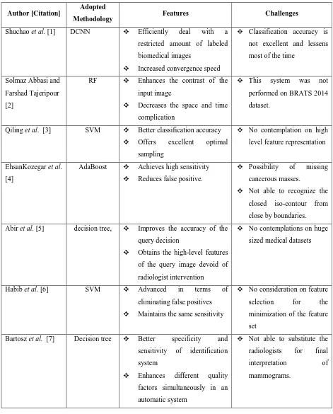
In 2016, Shuchao et al. [1] have introduced a deep convolution neural network (DCNN) for constructing a deep model; and subsequently to extend the deep learning structural design depending on the raw pixels of actual biomedical images by means of supervised training. Accordingly, the feature space was modeled, and it explores an effectual feature vector classifier or segments particular image patches and detection object that were the major technological complicatedness in the implementation of conventional image classification techniques. In addition, there was no requirement to be troubled with whether there were large training sets of interpreted biomedical images to wait for training a ideal deep model that were the major troubles to train DNN for biomedical image classification as noticed in current works.
In 2017, Solmaz Abbasi and Farshad Tajeripour [2] has developed an automatic technique in 3D images for brain tumor detection. In the initial pace, the histogram matching and bias field corrections were utilized for image preprocessing. In the subsequent pace, the region of interest (ROI) was recognized and alienated from the Flair image background. Histogram of orientation gradients (HOG-TOP) and LBP in three orthogonal planes (LBP-TOP) was exploited as the learning features. As 3D images were deployed in this study, the suggestion of LBP in three orthogonal planes for expanding HOG for 3D images was deployed. The random forest (RF) was subsequently utilized to segment the regions of cancer. Moreover, the computation of the proposed scheme on glioma images from BRATS 2013 was evaluated.
recognition of initial candidates that was dependent mainly on isocontours. With the intention of reducing the erroneously created isocontours, certain features like „roundness,' „hypoechoicity‟, „area strength‟ and „contour strength‟ were utilized. Consequently, the successive candidates were further practiced by means of a cascade classifier, and the corresponding base classifiers were „Random Under-Sampling Boosting (RUSBoost)‟ that was initiated for managing with the imbalanced datasets. The design was investigated depending on „Free response Operating Characteristics (FROC)‟ and has revealed its enhanced performance.
In 2018, Abir et al. [5] have introduced a content-dependent image retrieval technique on the basis of med-level descriptors. These descriptors were mechanically produced from low-level image features by utilizing the semantic conceptions depending on the clinician medical-knowledge. Actually, the suggested technique was dependent on three major processes. First is the low-level extraction of features. Next is the med-level model extraction and last is the online retrieval depending on med-level feature vectors. The major contributions exist in the incorporation of clinician medical-knowledge with respect to the med-level features devoid of requiring radiologists interface. The implemented scheme was authenticated on the MIAS dataset, in the framework of mammogram retrieval, and the outcomes confirm its efficiency and its dominance to the distinguished techniques.
In 2017, Habib et al. [6] have suggested a scheme that initially identifies an preliminary set of candidates by means of a Gaussian Matched Filter and subsequently classifies the corresponding set to decrease the number of false positives. In addition, a Tree Ensemble classifier was adopted with a group of 70 features. A novel set from the MESSIDOR dataset of 32 MA ground truths depending on images was established as a public dataset for benchmarking MA detection techniques. The suggested scheme was estimated on this dataset in addition to various public dataset (DIARETDB2) and distinguishes it in opposition to the most excellent obtainable option. Outcomes demonstrate that the suggested classifier was better in eradicating false positive MA detection from the primary set of candidates.
In 2017, Bartosz et al. [7] have introduced a novel advanced technique of image description and an ensemble classifier for identification of mammograms in breast cancer. The non-negative matrix factorization and various techniques of image representation, which were not utilized in the area of mammogram recognition were improved and ensured in the function of investigative features. Ultimate image recognition was made by means of an ensemble classifier. In addition, the novel scheme to the combination of an ensemble was suggested. It relates the weighted majority voting together with the weights described from the optimization task described depending on the region under curve of ROC. The consequences of arithmetical experiments carried out on large database “Digital Database for Screening Mammography” enclosing higher than 10000 mammograms have established higher accuracy in recognizing the abnormal ones from the normal cases. The offered outcome of class recognition goes beyond the most excellent accomplishments for this base reported in the original publications.
shortly they have deployed the threshold value of customized bayes shrink representation. This was made for the inequity of homogeneous regions together with speckle noise that was acquired from the process of filtering. The suggested „Homogeneity MBS (HMBS)‟ has revealed its dominance over other traditional models.
In 2015, G. Mahendran and Dhanasekaran [9] has implemented a technique to identify lesion exudates mechanically with the support of non-dilated retinal funds for supporting ophthalmologists to treat the disease. The low contrast images of the exudates were recognized and localized by means of a neighborhood dependent segmentation method. A probabilistic neural network (PNN) classifiers and SVM were suggested to evaluate the rigorousness of the disease, and the consequences were distinguished with the similar segmentation method. The standard classification accurateness for the PNN and SVM classifiers were found to be 94.76% and 97.89%, correspondingly.
In 2016, PengGu et al. [10] have introduced a mechanized technique for the segmentation of 3D US volumes. The segmentation was categorized into three varieties known as, „fatty tissue,' „mass/cist‟ tissue, and „fibro-glandular tissue.' They have scrutinized the effectiveness in addition to the steadiness of suggested representation by deploying them on a database of 21 test cases of all breast US. Outcomes of the research have offered improved computation of the proposed model by comparing tissues, namely non-fat or fat, and in addition, it outperformed in tissues classification also. On the whole, the design has acquired ideal consistency and potential in characterizing the tissues.
In 2017, Pedro et al. [11] have proposed a new method for extracting features based on radiological density patterns of the brain, called Analysis of Brain Tissue Density (ABTD). The proposed method is a specific approach applied to CT images to identify and classify the occurrence of stroke diseases. The evaluation of the results of the ABTD extractor proposed in this paper were compared with extractors already established in the literature, such as features from Gray-Level Co-Occurrence Matrix (GLCM), Local binary patterns (LBP), Central Moments (CM), Statistical Moments (SM), Hu‟s Moment (HM) and Zernike‟s Moments (ZM).
In 2014, GerardPons et al. [12] have implemented a „deformable part models‟ (DPM) and object detection model (ODM) for the detection of cysts in breast images. They have further adopted a data set of 326 images from a variety of patients. The implemented design has outperformed numerous traditional models regarding lesion detection. Moreover, „False positive detection‟ of 0.28 (per image), high sensitivity of 86% were achieved. In addition, the introduced design has demonstrated its effectiveness concerning the „Malignant lesion detection.'
In 2017, Qi Dou et al. [14] has suggested a 3D DSN scheme, which was proficient in performing volume-to-volume inference and learning that can eradicate superfluous performances and lessen the risk of over-fitting on restricted training data. More significantly, the 3D deep supervision method can efficiently manage with the optimization crisis of gradients fading. The speed of convergence has enhanced the discrimination ability simultaneously. Such a method was introduced by formulating an intended function, which directs the training of both upper and lower layers directly in the network, thus the unfavorable consequences of unbalanced gradient alterations can be frustrated throughout the training process. In addition, an entirely connected conditional random field design was employed as a post-processing pace to purify the segmentation consequences.
In 2018, Alexander et al. [15] have offered a novel quantitative technique for detecting variations in 3D medical images. The variation among shapes was measured as a determination of the attempts it takes to distort one 3D region into another one. The evaluation of isometric and conformal deformations of mappings was major equipment among volumes. Contrasting to the majority of the conventional schemes for shape assessments, the suggested one functions both on tetrahedral and triangular meshes, and as a result can be deployed for volumetric domains homeomorphic to a ball in addition to closed simply connected surfaces with geometrically complex limitations. In addition, the major geometric deformation access to higher dimensions was evaluated in a manner, which permits for managing with spatial data at the maximal, at the entire lower dimensions.
In 2017, Sheng-Chih Yang [16] has established a novel medical image segmentation scheme, which obtains better image segmentation precision. With the intention of demonstrating the finest outcomes distributed by the suggested Progressive Support-pixel Correlation Statistical Method (PSCSM) for actual medical images. Moreover, investigational data were classified as multi-spectral breast magnetic resonance images (MRI), computer-processed images, and actual single-spectral mammograms. At last, the research consequences were distinguished with various renowned conventional and aggressive image segmentation techniques to substantiate the contributions and advantages of the suggested technique.
TABLE I.
R
EVIEW ON STATE OF THE ART MEDICAL IMAGE CLASSIFICATION TECHNIQUESAuthor [Citation] Adopted
Methodology Features Challenges
Shuchao et al. [1] DCNN Efficiently deal with a restricted amount of labeled biomedical images
Increased convergence speed
Classification accuracy is not excellent and lessens most of the time
Solmaz Abbasi and Farshad Tajeripour [2]
RF Enhances the contrast of the input image
Decreases the space and time complication
This system was not performed on BRATS 2014 dataset.
Qiling et al. [3] SVM Better classification accuracy
Offers excellent optimal sampling
No contemplation on high level feature representation
EhsanKozegar et al. [4]
AdaBoost Achieves high sensitivity
Reduces false positive.
Possibility of missing cancerous masses.
Not able to recognize the closed iso-contour from close by boundaries. Abir et al. [5] decision tree, Improves the accuracy of the
query decision
Obtains the high-level features of the query image devoid of radiologist intervention
No contemplations on huge sized medical datasets
Habib et al. [6] SVM Advanced in terms of
eliminating false positives
Maintains the same sensitivity
No consideration on feature selection for the minimization of the feature set
Bartosz et al. [7] Decision tree Better specificity and sensitivity of identification system
Enhances different quality factors simultaneously in an automatic system
ImanElyasi et al. [8]
Bayesian Achieves fast running time
Improves the performance by enhancing the image quality
High smoothing leads to blurring the image.
Complication increases in image registration process. G. Mahendran and
Dhanasekaran [9]
SVM Facilitates ophthalmologists to identify the exudates in a very short time of scrutiny
Low-cost and does not necessitate trained experts
Requirement of features may be increased on the accumulation of the exudates with respect to the distance
PengGu et al. [10] NN Automatically differentiate non-fatty and fatty tissues.
Outperforms in combined imaging design.
Shadow separation from lesion is very complicated.
Necessitates certain manual corrections for automatic segmentation.
Pedro et al. [11] Bayesian Simple to implement
Includes low processing time
Increased accuracy
No contemplation on the exploitation of deep learning techniques to accelerate this scheme GerardPons et al.
[13]
SVM Possible to execute in clinical applications
Lesions are identified accurately.
Cancer detection is not feasible via this design.
Performance is not so promising.
Proper evaluation of bounding box is not possible.
Woo KyungMoon et al. [13]
Fuzzy classifier Contributes constructive information for the prediction of ALN status.
Performance of texture feature set is extremely improved
Could not predict the unidentified regions in image.
Considering extra features may frequently grant worse performance.
Qi Dou et al. [14] CNN Proficiently carry out the dense segmentation in a volume-to-volume approach.
Directly attain equal-sized output as the input data
Develop the discriminative
ability of networks
Alexander et al. [15]
Jacobian Better detection of changes in volumetric models
Can be deployed on both closed simply connected surfaces
Accuracy and distortion measures have to be determined more.
Sheng-Chih Yang [16]
Fuzzy classifier Segments medical images more accurately
Evades statistical error occurred due to large patches of background and noise
Requires more
contemplation on 3D reconstruction, lesion localization, benign or
malignant tumor
determination Khatami et al. [17] DBN Assists to discover the most
practical features from raw data proficiently.
Minimizes the computational time.
Develops the performance of classification
No investigation of other multiclass classifiers for
processing noisy
radiographic and high dimensional imaging data
III
PROBLEM
STATEMENT
1806 | P a g e the feature set. Also, decision tree was suggested in [7], which provides better specificity and sensitivity and it also enhances different quality factors simultaneously in an automatic system. Anyhow, it was not able to substitute the radiologists in final interpretation of mammograms. Similarly, Bayesian classifier was adopted in [8] that achieve fast running time and it also improves the performance by enhancing the image quality. However, high smoothing leads to blurring the image and the complication also increases in image registration process. Similarly, SVM was presented in [9], which facilitates ophthalmologists to identify the exudates in a very short time of scrutiny. It also offers low-cost and does not necessitate trained experts, but the requirement of features may be increased on the accumulation of the exudates with respect to the distance. In addition, NN was presented in [10] that differentiates non-fatty and fatty tissues and also outperforms in combined imaging design. But the Shadow separation from lesion was very complicated and it necessitates certain manual corrections for automatic segmentation. Similarly, Bayesian classifier was adopted in [11] that were simple to execute and it requires only less computing time with increased accuracy. However, there was no contemplation on the exploitation of deep learning techniques to accelerate this scheme. Moreover, SVM was suggested in [12], which was possible to be executed in medical appliances to detect the lesions accurately. Anyhow, cancer detection is not feasible via this design and the performance is not so promising. Moreover, Fuzzy classifier was suggested in [13] that enhances the texture feature set, and provides useful information for the prediction of ALN status. However, the prediction of unknown regions is impossible. In addition, CNN were proposed in [14], which proficiently carries out the dense segmentation in a volume-to-volume approach. Also, it directly attains equal-sized output as the input data and develops the discriminative ability of networks, but slight adjustments have to be done owing to deviations in size and shapes. Similarly, Jacobian based method was suggested in [15], which provides better detection of changes in volumetric models and it can be deployed on both closed simply connected surfaces. However, accuracy and distortion measures have to be determined more. Fuzzy classifier was implemented in [16] that Segments medical images more precisely and evades statistical error occurred due to large patches of background and noise. Anyhow, it requires more contemplation on 3D reconstruction, lesion localization, benign or malignant tumour determination. Furthermore, DBN was suggested in [17] that assist to discover the most practical features from raw data proficiently with minimization of computational time. Also, it develops the performance of classification, but there was no investigation of other multiclass classifiers for processing noisy radiographic and high dimensional imaging data. These limitations have to be considered for improving the abdominal mass diagnosis performance.
3.1
Contribution on Feature Extraction
Fig. 1.
Fig. 2.
Fig. 1 Graphical representation of feature extraction methods
3.2
Medical Disease Diagnosis
The bar chart representation of the medical disease diagnosis is given by Fig. 2. From Fig. 2, 59% contribution is based on breast cancer, 16% contribution is based on brain tumor, 5% contribution is based on diabetes, and 12% contribution is based on heart disease and 6% contribution is based on retinal disorder. Thus the contribution by found to be more than other disorders verified in this paper.
Fig. 2 Bar chart representation of medical disease diagnosis
IV
RESEARCH
GAPS
AND
CHALLENGES
1808 | P a g e systems, which were dependent on local limits or object detection, were considered as less effectual than preceding group of methods. They have reduced flexibility on various kinds of tumor and diverse information. On the other hand, the chief restriction of medical image classification systems is the semantic gap that was the differentiation among high-level semantics and their low-level features of images in a specified circumstance. In addition, RF has been seemed to have more complexity, and hence in several cases, ensemble classifiers and fuzzy based classifiers were adopted. There were several contributions for various diseases as given in the literature, but the contribution with respect to abdominal mass detection was not focused more by the researchers. Hence, this paper mostly concerns on different type of classifiers, which could be adopted for better classification of medical images regarding abdominal mass detection in future.
V
CONCLUSION
This paper has presented a survey on medical image classification for various diseases. In addition, several types of feature extraction techniques and classifiers, which were exploited for classifying the medical images associated with numerous diseases, were focused. Since there was lack of classifications depending on abdominal mass detection, this paper has concerned on other various diseases and their corresponding methodologies. Furthermore, by means of any of the classification schemes mentioned in the literature works, abdominal mass detection can be detected in future.
REFRENCES:
[1] Shuchao Pang , Zhezhou Yu , Mehmet A. Orgun, "A Novel End-to-End ClassiÞer Using Domain Transferred Deep Convolutional Neural Networks for Biomedical Images",Computer Methods and Programs in Biomedicine, 31 December 2016
[2] Solmaz Abbasi, Farshad Tajeripour, “Detection of brain tumor in 3D MRI images using local binary patterns and histogram orientation gradient”, Neurocomputing, vol. 219, pp. 526-535, 5 January 2017. [3] Qiling Tang, Yangyang Liu, Haihua Liu, “Medical image classification via multiscale representation
learning”, Artificial Intelligence in Medicine, vol. 79, pp. 71-78, June 2017.
[4] EhsanKozegar, MohsenSoryani, HamidBehnam, MasoumehSalamati and TaoTan, " Breast cancer detection in automated 3D breast ultrasound using iso-contours and cascaded RUSBoosts", Ultrasonics, vol. 79, pp. 68-80, 2017.
[5] Abir Baâzaoui, Walid Barhoumi, Amr Ahmed, Ezzeddine Zagrouba, “Modeling clinician medical-knowledge in terms of med-level features for semantic content-based mammogram retrieval”, Expert Systems with Applications, vol. 94, pp. 11-20, 15 March 2018.
[6] M.M. Habib, R.A. Welikala, A. Hoppe, C.G. Owen, S.A. Barman, “Detection of microaneurysms in retinal images using an ensemble classifier”, Informatics in Medicine Unlocked, vol. 9, pp. 44-57, 2017.
[8] ImanElyasi, Mohammad AliPourmina and Mohammad-ShahramMoin, " Speckle reduction in breast cancer ultrasound images by using homogeneity modified bayes shrink", Measurement, vol. 91, pp. 55-65, 2016. [9] G. Mahendran, R. Dhanasekaran, “Investigation of the severity level of diabetic retinopathy using
supervised classifier algorithms”,Computers & Electrical Engineering, vol. 45, pp. 312-323, July 2015. [10] PengGu, Won-MeanLee, Marilyn A.Roubidoux, JieYuan, XuedingWang and Paul L.Carson, " Automated
3D ultrasound image segmentation to aid breast cancer image interpretation", Ultrasonics", vol. 65, pp. 51-58, 2016.
[11] Pedro P. Rebouças Filho, Róger Moura Sarmento, Gabriel Bandeira Holanda, Daniel de Alencar Lima, “New approach to detect and classify stroke in skull CT images via analysis of brain tissue densities”, Computer Methods and Programs in Biomedicine, vol. 14, pp. 27-438, September 2017.
[12] GerardPons, RobertMartí, SergiGanau, MelciorSentís and JoanMartí, " Computerized Detection of Breast Lesions Using Deformable Part Models in Ultrasound Images", Ultrasound in Medicine & Biology, vol. 40, no. 9, pp. 2252-2264, 2014.
[13] Woo KyungMoon, Yan-WeiLee, Yao-SianHuang, Su HyunLee, .Min SunBae, AnnYi, Chiun-ShengHuang and Ruey-FengChang, " Computer-aided prediction of axillary lymph node status in breast cancer using tumor surrounding tissue features in ultrasound images", Computer Methods and Programs in Biomedicine, vol. 146, pp. 143-150, 2017.
[14] Qi Dou, Lequan Yu, Hao Chen, Yueming Jin, Pheng-Ann Heng, “3D deeply supervised network for automated segmentation of volumetric medical images”, Medical Image Analysis, vol. 41, pp. 40-54, October 2017.
[15] Alexander Naitsat, Shichao Cheng, Xiaofeng Qu, Xin Fan, Yehoshua Y. Zeevi, “Geometric approach to detecting volumetric changes in medical images”, Journal of Computational and Applied Mathematics, vol. 329, pp. 37-50, February 2018.
[16] Sheng-Chih Yang, “A robust approach for subject segmentation of medical Images: Illustration with mammograms and breast magnetic resonance images”, Computers & Electrical Engineering, vol. 6, pp. 151-1652, August 2017.