FARMER, WILLIAM TAYLOR QUINTON. Detection of Antisense to IGF2R (AIR) RNA in Cattle. (Under the direction of Charlotte Farin.)
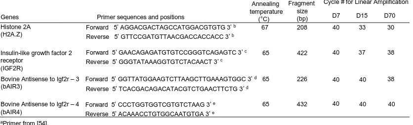
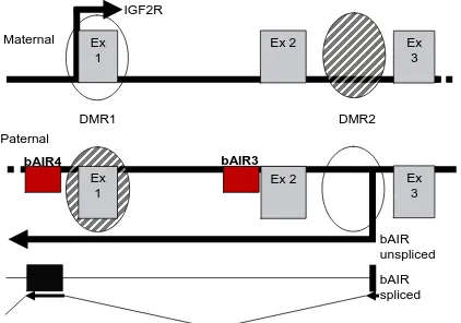
The insulin-like growth factor type 2 receptor (Igf2r/IGF2R) regulates fetal growth by removing Igf2 from circulation, thus preventing overgrowth. In mice, expression of the Igf2r gene is imprinted only after implantation and is associated with expression of the antisense non-coding (nc)RNA, Air. In contrast, the human IGF2R gene is not imprinted and AIR ncRNA does not exist. Because it is known that IGF2R is imprinted in cattle, the objectives of this study were to determine if AIR ncRNA exists in cattle; if so, whether bovine AIR (bAIR) expression changes at developmentally important stages of gestation, and whether method of embryo production affects air expression. For objective 1, primer sets were designed for bAIR based on bovine genomic sequence. The primer set, bAIR3, was used to amplify a region of bAIR corresponding to an antisense segment within intron 1 of IGF2R. Primer set bAIR4 amplified a segment of bAIR
ncRNA corresponding to an antisense region upstream of the 5'-untranslated region of IGF2R.
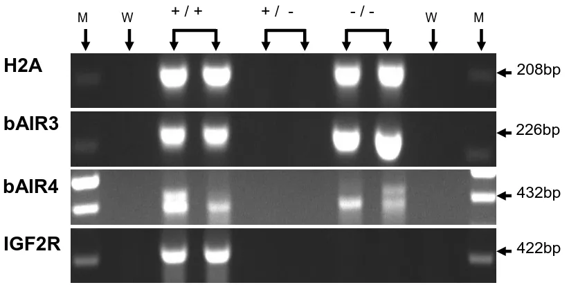
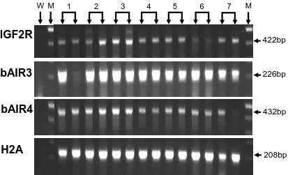
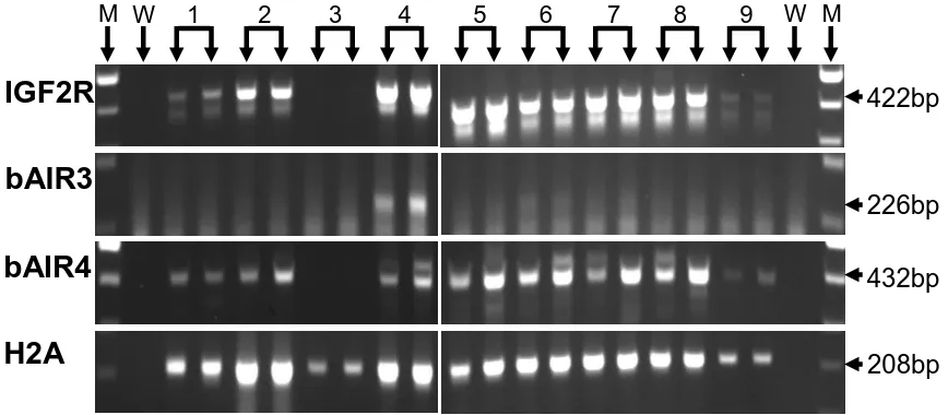
were recovered from cows on day 7 of development and snap frozen for RNA extraction. Semi-quantitative RT-PCR assays were performed to assess levels of IGF2R mRNA, H2A mRNA and bAIR ncRNA. Relative RNA expression was calculated as the ratio of band intensities of the RNA of interest to that ofH2A. Data on levels of expression in fetal liver between IVO and IVP treatment groups were analyzed by Student’s T-test. H2A mRNA was expressed in all day 70
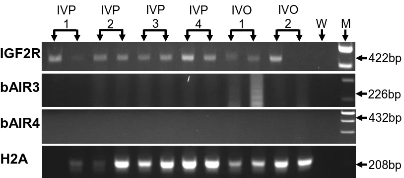
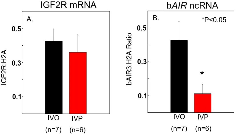
fetal liver samples, day 15 conceptuses, and day 7 blastocyst pools. IGF2R mRNA was expressed in all fetal liver samples, in 8 of 9 day 15 conceptuses, and in all day 7 blastocyst pools. bAIR ncRNA was expressed in 7 of 7 samples of day 70 fetal liver. In contrast, only 1 of 9 conceptuses expressed a bAIR ncRNA signal based on the bAIR3 primer set whereas 8 of 9 conceptuses expressed bAIR ncRNA based on the bAIR4 primer set. No bAIR ncRNA was expressed in any blastocyst pools based on either the bAIR3 or bAIR4 primer sets. Relative levels of bAIR ncRNA were greater (P<0.05) in fetal liver generated from the transfer of in vivo-produced embryos compared to that from in vitro-vivo-produced embryos (IVO: 0.426 ± 0.090 vs. IVP: 0.112 ± 0.098). In summary, the antisense ncRNA AIR exists in cattle and is expressed following implantation. Furthermore, the relative level of bAIR ncRNA can be altered by method of embryo production. These observations are consistent with data from the mouse and suggest that bAIR may be involved in regulating imprinted expression of IGF2R in cattle.
by
William Taylor Quinton Farmer
A thesis submitted to the Graduate Faculty of North Carolina State University
in partial fulfillment of the requirements for the Degree of
Master of Science
Animal Science
Raleigh, North Carolina 2008
APPROVED BY:
___________________________ _____________________________ Dr. Peter W. Farin Dr. Jorge A. Piedrahita Co-Chair of Advisory Committee
BIOGRAPHY
William Taylor Quinton Farmer
Education
MS Animal Science, 2008, North Carolina State University, Raleigh, NC BS Animal Science, 2003, North Carolina State University, Raleigh, NC
Honors and Awards
First Place award, International Embryo Transfer Society, Student Research Competition, January, 2008
Professional Affiliations
ACKNOWLEDGEMENTS
I would like to thank my advisor, mentor and friend, Dr. Charlotte Farin, for all her expertise, advice, guidance and patience throughout my graduate program. I would also like to thank my co-advisor, Dr. Peter Farin, and my thesis committee member Dr. Jorge
Piedrahita for contributing their wealth of knowledge and insight. I would especially like to thank Dr. Peter Farin for all his assistance with embryo transfer and production of
conceptuses and fetuses. I am sincerely grateful for the supporting efforts made by my colleagues Steve Bischoff, Eric Alexander and Lauren Kuchenbrod.
I will forever be indebted to all of my friends for their support and encouragement. Special thanks are given to my good friends and non-academic mentors, Kevin Jones and Dr. Richard Cochrane, for their advice and unwavering friendship. I would also like to thank my parents, Rev. Dr. Terry and Marian Farmer, my parents-in-law, Dr. Hugh Powell and
TABLE OF CONTENTS
page
LIST OF TABLES..………. viii
LIST OF FIGURES..……… ix
LITERATURE REVIEW………. 1
ABNORMAL OFFSPRING SYNDROME………. 1
Phenotypes of AOS...……… 1
Potential Mechanisms of AOS………. 3
IMPRINTED GENES……….. 7
DNA Methylation………. 8
Dnmt1 and Dnmt1o……….. 9
Dnmt3a, Dnmt3b and Dnmt3L………. 9
Histone Modifications……….. 10
Histone Methylation………. 12
Histone Acetylation……….. 12
Non-Coding RNAs………... 12
EPIGENETIC REPROGRAMMING OF IMPRINTED GENES……… 15
Erasure of Imprints……… 15
Acquisition of Imprints………. 16
Maintenance of Imprints………... 18
page
IGF Ligands………. 20
IGF Receptors……….. 21
IGF Binding Proteins………... 22
IGF2R/AIR CLUSTER……….………... 23
Imprinting Control in the Igf2r/Air Cluster……….. 24
Acquisition of Gametic and Somatic Imprints………. 26
Antisense to Igf2r (Air)………. 27
EVOLUTION OF IGF2R IMPRINTING……… 30
Evolutionary Pressure for Imprinted Expression of Igf2r/IGF2R…………. 30
Species Differences in Requirements for Imprinted Expression of Igf2r/IGF2R………... 32
IMPLICATIONS OF GENOMIC IMPRINTING DYSREGULATION IN AOS... 36
Disruption of DNA Methylation………... 37
Perturbed Histone Modifications……….. 38
Contribution of Imprinted Expression of IGF2R to AOS……… 39
STATEMENT OF THE PROBLEM……… 43
INTRODUCTION……… 45
MATERIALS AND METHODS………. 47
In Vivo Embryo Production………. 47
In Vitro Embryo Production………. 48
page
Transfer of Embryos………. 49
Embryos Transferred to Produce Day 70 Fetuses………. 49
Embryos Transferred to Produce Day 15 Conceptuses………. 49
Recovery of Fetuses and Conceptuses………. 49
Recovery of Day 70 Fetuses………. 49
Recovery of Day 15 Conceptuses………. 50
RNA Extraction……… 50
Day 70 Fetal Liver………. 50
Day 15 Conceptuses……….. 51
Day 7 Blastocysts……….. 51
cDNA Synthesis……… 51
Day 70 Fetal Liver………. 51
Day 15 Conceptuses……….. 52
Day 7 Blastocysts……….. 52
Bovine AIR Primers……….……….. 52
Semi-Quantitative RT-PCR……….. 53
Day 70 Fetal Liver………. 53
Day 15 Conceptuses……….. 56
Day 7 Blastocysts……….. 56
page
Day 70 Fetal Liver……… 56
Day 15 Conceptuses……….. 56
Day 7 Blastocysts……….. 57
Statistics……… 57
RESULTS………. 57
Detection and Validation of bAir……….. 57
Effect of Stage of Development on bAir Expression……… 58
Post-Implantation Stage (Day 70 Bovine Fetal Liver)……….. 58
Peri-Implantation Stage (Day 15 Bovine Conceptuses)……… 60
Pre-Implantation Stage (Day 7 Bovine Blastocyst Pools)…………. 60
Effect of Method of Embryo Production on bAir Expression……….. 60
DISCUSSION………... 66
REFERENCES………. 72
APPENDICES……….. 98
Appendix A. Response Curves for the Determination of the Linear Phase of Amplification……….. 99
LIST OF TABLES
LIST OF FIGURES
page DETECTION OF ANTISENSE TO IGF2R (AIR) RNA IN CATTLE
Figure 1. Regulation of Igf2r expression in the mouse……… 54
Figure 2. Ethidium bromide-stained agarose gel of H2A, IGF2R, bAIR3 and bAIR4 Amplification products from bovine fetal liver at Day 70 of gestation………… 59
Figure 3. Post-implantation-stage RNA expression in Day 70 bovine fetal liver………… 61
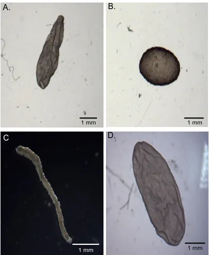
Figure 4. Bovine conceputses at Day 15 of gestation……….. 62
Figure 5. Peri-implantation-stage RNA expression in Day 15 bovine conceptuses……… 63
Figure 6. Pre-implantation-stage RNA expression in Day 7 bovine blastocysts…………. 64
Figure 7. Expression of IGF2R mRNA in bovine fetal liver at Day70 of gestation derived from the transfer of either in vivo-(IVO) or in vitro produced (IVP) embryos… 65 Figure 8. Potential splice variants for bovine AIR……….………... 69
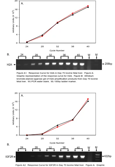
APPENDIX A Figure A1. RT-PCR Linear Response curve for H2A mRNA (Day 70)..………... 99
Figure A2. RT-PCR Linear Response curve for IGF2R mRNA (Day 70).………... 99
Figure A3. RT-PCR Linear Response curve for bAIR3 ncRNA (Day 70)……….. 100
Figure A4. RT-PCR Linear Response curve for bAIR4 ncRNA (Day 70)……….. 100
Figure A5. RT-PCR Linear Response curve for H2A mRNA (Day 15)…..………... 101
Figure A6. RT-PCR Linear Response curve for IGF2R mRNA (Day 15)……….. 101
page APPENDIX B
Figure B1. RNA expression in Day 70 bovine fetal liver derived from the transfer of in vitro-produced embryos………..……….………. 104 Figure B2. RNA expression in Day 70 bovine fetal liver derived from the transfer of in
The transfer of in vitro manipulated and, to a lesser extent, in vitro produced (IVP) bovine embryos results in fetuses, placentas, and offspring that exhibit abnormalities [1-3]. Abnormalities observed following the transfer of IVP or somatic cell nuclear transfer (SCNT) embryos are stochastic in occurrence and duplication of all phenotypes have not been consistently linked with any single gene or mechanism [4-6]. Initially it was observed that offspring created from IVP embryos were larger than normal and were more susceptible to dystocia at birth [7]. The common occurrence of an overgrowth phenotype along with increased perinatal death, longer gestation lengths, congenital deformities, and abnormalities of placental vasculature resulting from the transfer of IVP embryos became known as Large Offspring Syndrome (LOS) [1, 7]. These developmental abnormalities occur at a higher incidence in fetuses, placentas, and offspring following the transfer of SCNT embryos compared to embryos produced in vitro [3, 8, 9]. However not all transfers of IVP or SCNT embryos result in excessive growth [10-14]. Therefore, the term abnormal offspring
syndrome (AOS), better accommodates the known abnormalities that have been documented [5].
Phenotypes of AOS
IVP- or SCNT- derived fetuses, placentas and offspring exhibit abnormalities that are highly variable and may range from apparently normal with subtle phenotypes to the most obvious [5]. Unusually large or heavy offspring have been observed following the transfer of IVP or SCNT embryos in cattle [1, 9, 13, 15-17] and sheep [7, 18-20]. Increased fetal
in sheep where lambs twice the normal birthweight have been observed [7]. Conceptus and fetal overgrowth has also been detected as early as days 17 and 21 of gestation, respectively [21]. The increase in birthweights is also associated with a higher rate of dystocia and delivery by cesarean section [13]. Other characteristics of this syndrome include a longer gestation period [18], increased perinatal losses in the first half of pregnancy [22], and altered energy metabolism [23]. In addition, gross abnormalities have been observed in several organs. These include increased muscle mass and altered muscle fiber composition [24, 25]. Other abnormalities include skeletal malformations [7], facial malformations [7, 19, 26] and cerebellar dysplasia [27]. In cattle and sheep, placental abnormalities such as
polyhydramnios, hydrallantois, and alterations in placentome number, and placental
morphology have also been reported [13, 26, 28-32]. Defects in placental growth exhibited by cloned mice and cloned ruminants have been proposed to contribute to increased perinatal mortality by failure to initiate an adequate blood supply [15, 33].
or may not occur, and the resulting calves have severely altered clinical, hematological, and biochemical parameters resulting in death at or near the time of parturition [5]. In contrast, in Type IV AOS full term fetuses and placentas are present and exhibit only moderate
abnormalities as a result of compensatory responses from the fetus and placenta [5]. Potential mechanisms of AOS in cattle
The occurrence of AOS phenotypes appears to be associated with the environmental conditions by which embryos are created [34]. Bovine IVP embryos are created from immature oocytes that are matured in vitro, fertilized in culture, and then maintained in culture medium to the blastocyst stage [34]. Cloned bovine embryos are created by maturing an oocyte to metaphase II in vitro, extracting its genetic material and then electrically fusing a donor cell with this karyoplast [35]. The resulting zygote is cultured to the blastocyst stage and transferred into a recipient animal. Both in vitro production and cloning techniques expose the developing bovine zygote to inadequate culture conditions that result in aberrant imprinted gene expression [35]. Embryos likely exhibit a high level of plasticity and an ability to adapt to stress considering all of the negative conditions associated with culture they must over come in order to develop to the blastocyst stage [36, 37]. Some of the obstacles the early embryo must combat include exposure to culture medium, serum, and coculture with other cell types [21, 38]. In vitro embryo development is also influenced by factors in the physical environment including temperature variation, embryo density,
constraints such as the stability of the incubator environment and the ability of technicians handling the embryos [4, 40].
In vitro maturation of oocytes and generation of embryos by in vitro production (IVP) or nuclear transfer result in embryos with obvious differences compared to in vivo-produced embryos. IVP embryos show differences in gross morphology when exposed to serum [4]. These differences include incomplete compaction, a darkened appearance, and less
organization of the inner cell mass [4, 41, 42]. In contrast, IVP embryos not exposed to serum exhibit more complete compaction, have a lighter appearance, and their inner cell mass is more organized [4, 42]. Blastocysts generated in the presence of serum may also have a higher lipid content and incomplete junctional complexes between the inner cell mass and the trophectoderm [43-46]. Embryo fragmentation can result from high concentrations of serum in culture [20]. In addition, IVP embryos, regardless of their exposure to serum, have a more rapid development of male embryos [47, 48], increased intracellular lipid content [49, 50], fewer surface microvilli [49, 51], and altered cytoplasmic mitochondrial density [51] compared to in vivo-produced embryos [4]. Embryos created by nuclear transfer are subjected to the physical traumas of enucleation, electrofusion and reconstruction [52] as well as factors associated with the in vitro culture environment as discussed previously.
pre-implantation embryos that results in fetal and placental abnormalities [53-55]. Gene expression varies with the type of IVP system and SCNT protocols used suggesting that imprinted gene expression in the pre-implantation embryo is sensitive to alterations in epigenetic modifications [37, 56, 57]. The pre-implantation developmental program is governed by epigenetic mechanisms that, if altered or delayed, result in improper gene expression leading to abnormalities [58]. Epigenetic reprogramming errors likely occur during critical periods of pre-implantation development as a result of exposure to the culture environment during in vitro production [59, 60]. Similarly, epigenetic reprogramming errors in nuclear transfer-derived embryos likely occur from a failure to properly reprogram the donor nucleus and exposure to in vitro culture environments [59-61]. Some of the physiological aberrations associated with AOS are, in part, a result of aberrant DNA methyltransferase-1 (Dnmt1/DNMT1) expression [4, 62]. This enzyme regulates maintenance of methyation patterns at imprinted loci [4]. In mice, disruption of Dnmt1
and imprinted genes are aberrantly expressed in fetuses, placentas and offspring following the transfer of in vitro-manipulated embryos further supports this suggestion [53, 54, 61, 68, 69].
An alternative explanation for abnormal fetal and placental development is that exposure of an embryo to culture conditions results in aberrant causal pathways [36]. Based on the aberrant causal pathway hypothesis, critical transcriptional signaling molecules are inappropriately activated during developmentally important periods of embryonic growth, such as the pre-implantation stage [36]. Improper activation of these signaling molecules is proposed to result as a response to the environment [36]. Therefore, the environment induces activation of signaling molecules inappropriately and subsequent epigenetic modifications mediate altered imprinted and non-imprinted gene expression [36] .
IMPRINTED GENES
DNA Methylation
Imprinting control regions (ICRs) control the imprinting of genes within in a cluster by acquiring differential DNA methylation on the parental alleles during gametogenesis [77]. The methylation imprint is a modification of the DNA involving the addition of methyl
groups to cytosine residues at CpG islands [74]. The term, CpG island, refers to a series of cytosine-guanine dinucleotide repeats within a genome that are ≥ 500bp in length and have a GC content ≥ 55% [80].
DNA methylation can regulate stable transcriptional repression by preventing transcription factors from binding or by recruiting methyl-DNA binding domain proteins (MBDs), histone deacteylases (HDACs) and chromatin remodeling complexes that function to alter the accessibility of DNA [81]. DNA methylation is thought to only regulate
expression in a small number of genes, but may also be involved in preventing the
propagation of repetitive DNA sequences and in regulating X chromosome inactivation [74]. DNA methylation imprints are acquired at differentially methylated domains (DMDs) in a sex-specific-manner during gametogenesis and are completed prior to fertilization [82]. These new patterns of DNA methylation on both imprinted and non-imprinted genes are established by de novo DNA methyltransferases (DNMTs) that methylate cytosine residues where both DNA strands are unmethylated [82]. After fertilization, genome wide
Dnmt1 and Dnmt1o. Maintenance of DNA methylation occurs through the action of methyltransferases such as Dnmt1 and Dnmt1o. Dnmt1 maintains methylation marks on hemi-methylated DNA and methylates newly replicated DNA strands [83]. Dnmt1 null mice exhibited perturbed imprinting and died at embryonic Day 11, demonstrating the importance of this methyltransferases activity during development [63, 84]. In addition to Dnmt1, Dnmt1o is an oocyte specific maintenance methyltransferase that is expressed only in oocytes and preimplantation embryos [65]. In the mouse, Dnmt1o has been shown to translocate from the cytoplasm to the nucleus specifically at the 8-cell stage apparently to methylate imprinted genes and parasitic sequences [85]. The movement of Dnmt1o from the cytoplasm to the nucleus indicates that maintenance of methylated imprints at this stage is important [65]. The observation that deletion of the Dnmt1o promoter resulted in loss of methylation at imprinted loci but had no effect on the establishment of imprints indicates the importance of methylation maintenance by Dnmt1o in early embryonic development [65, 86].
Dnmt3a, Dnmt3b, and Dnmt3L. After implantation, a second wave of genome-wide
during embryonic development [87]. The Dnmt3a null mutation is lethal in mice by 4 weeks after birth and the Dnmt3b null mutation is lethal late in gestation [87]. The Dnmt3a de novo methyltransferase appears to be particularly important for establishing methylation imprints during gametogenesis [88]. Deletion of Dnmt3a in female PGCs prevented the establishment of maternal methylation imprints [88]. Similarly, Dnmt3a deletion in male PGCs resulted in loss of paternally methylated imprints [88].
A third member of the de novo methyltransferase family is Dnmt3L which is also required for methylation of imprinted genes [89, 90]. Dnmt3L does not have the catalytic site for methyltransferase activity [89]; however, it does associate with Dnmt3a and 3b apparently to regulate their acitivity [91-93]. Homozygous disruption of Dnmt3L in the female germline resulted in a failure of maternal-specific imprints to be established [89]. In the mouse, Dnmt3L is epigenetically regulated by methylation at its promoter [94, 95]. Histone Modifications
In addition to DNA methylation, imprinted gene expression is regulated by histone modifications that are heritable and serve as epigenetic marks for active and inactive
be regulated by chromatin modulating proteins that modify chromatin structure in response to specific histone modifications such as methylation or acetylation [97]. These modifications are interpreted by effector proteins that induce either a heterochromatin or a euchromatin structure [99]. Heterochromatin is DNA that is tightly packaged with nucleosomes and is transcriptionally inaccessible to transcription factors [99]. In contrast, euchromatin is DNA that is more loosely packaged and is accessible for transcription [99]. Histone modification of a specific amino acid residue, alone or in combination with other histone modifications of the core histones forms the histone code [100]. For example, acetylation of lysine residues on histone H3 and H4 are associated with euchromatin, whereas, methylation of lysine residues on H3 and H4 are associated with heterochromatin [100].
An alternative hypothesis proposed to explain the function of histone modifications in transcription is the „Methylation/Phosphorylation Binary Switch Hypothesis [101]. Based on
this hypothesis, combinations of different modifications would dynamically alter the transcriptional state [101]. For example, phosphorylation of residues adjacent to a methylated lysine residue would result in an altered recruiting state for binding proteins. According to this hypothesis, changes made to the chromatin would affect transcription through„switch‟ sites that are dependent on the positional relationship between the
Histone Methylation. Imprinted genes display allele-specific histone methylation patterns at differentially methylated regions [102]. Active transcription is associated with methylation of histone H3 at lysine residue 4 (H3K4me), whereas transcriptional silencing is associated with methylation of histone H3 at lysine residue 9 (H3K9me) [103, 104]. In addition, lysine residues can be mono-, , or tri-methylated and arginine residues can be mono- or di-methylated [98, 105]. This adds another level of complexity to regulation of transcription by histone methylation. Histone methylation of lysine residues appears to be irreversible
because histone demethylases have not been identified yet [97]. These stable methylation marks, therefore, may provide an epigenetic mechanism by which gene expression profiles can be stably transferred to progeny cells [97].
Histone Acetylation. Acetylated lysine residues on histones H3 and H4 are generally associated with active transcription resulting from a relaxed chromatin structure [100]. The acetylation of histone residues facilitates transcription by reducing DNA nucleosomal interactions [97]. Deacetylation of histone residues reverses this effect [97]. For example, the transcribed alleles of the imprinted genes H19, Snrpn, and U2af1-rs1 all exhibit
hyperacetylation of histones H3 and H4 [106, 107]. In contrast, the silent allele of these imprinted genes displays hypoacetylation of the histones.
Non-Coding RNAs
for proteins whose amino acid sequence reliably predicts function [109]. Interestingly, for a significant number of imprinted clusters, the parental chromosome that carries the
unmethylated imprint control region is also the one that expresses the ncRNA [110]. Furthermore, in all six of the known imprinted clusters, expression of the ncRNA is associated with repression in cis of some or all of the protein-coding genes in the cluster [111-113]. Silencing by the imprinted ncRNA is bi-directional and affects multiple genes [111]. Silencing is imposed upon genes that overlap with the antisense ncRNA and also those that do not overlap [111]. Furthermore, silencing by antisense ncRNAs is regulated in a tissue-specific and developmental manner [111]. Models that attempt to explain
mechanisms by which ncRNA silencing is mediated need to accomplish several things [111]. First, the model needs to elucidate how silencing is not induced by full expression of
truncated ncRNAs [111]. Second, the model needs to determine how tissue specific imprinted expression operates [111]. Third, the model needs to explain how silencing is imposed on foreign genes inserted into an imprinted cluster [111]. Finally, a proposed model needs to describe how the silence induced by an imprinted ncRNA does not impact itself [111].
Several models have been proposed to explain how ncRNAs can silence an
possible if the imprinted ncRNA and the silenced imprinted gene share sequence homology [111]. The double-stranded RNA intermediates may silence expression by inducing mRNA degradation or by forming heterochromatin, effectively silencing the promoter [116]. Another mechanism by which silencing may be regulated by the ncRNA transcript is the “ncRNA-direct targeting model” of Xist [111]. In this model spreading of Xist,in cis, along
adjacent sequences results in recruitment of heterochromatinizing factors that bind the DNA and result in X chromosome inactivation [113].
A second group of proposed models that describe how ncRNA function to silence gene expression suggest the transcription of the ncRNA itself is the mechanism that leads to silencing of imprinted genes within a cluster [111, 114]. In this „expression-competition‟ model, it is proposed that the promoters for the ncRNA and mRNA are in competition for common cis-regulatory elements [117]. An alternative model is the „transcriptional
interference‟ (TI) model in which it is proposed that transcriptional regulation is mediated by
active transcription where the elongating polymerase activity of an antisense gene directly interferes with transcription of the overlapping sense gene [111, 114]. However, within an imprinting cluster that includes several protein-coding sense genes, only one of them is overlapped by an antisense ncRNA [111]. Therefore, variations of the TI model have been suggested in which transcription of the ncRNA leads to either interference or activation of a
cis-regulatory element regulating expression of all mRNA promoters within the imprinted cluster but has no effect on the ncRNA promoter [111].
multiple genes within an imprinted cluster are dependent upon a cis-acting activator located in the ncRNA transcription unit [111]. A chromatin loop containing the activator forms when the ncRNA is expressed, preventing the interaction of the promoters with the activator and then silences expression of imprinted genes [111].
EPIGENETIC REPROGRAMMING OF IMPRINTED GENES
Epigenetic changes are defined as chemical alterations to the DNA or to the histone proteins associated with the DNA that change chromatin structure without altering the nucleotide sequence [118]. Genomic imprinting is the monoallelic, parent-of-origin specific gene expression that results from epigenetic modifications of imprinted genes [75]. In the mammalian genome, the primary epigenetic mark resulting in imprinted expression is DNA methylation [119]. This epigenetic modification is proposed to be the imprinting mark because it is both heritable and reversible and can be stably transmitted after DNA replication [120]. However, expression of imprinted genes is also regulated by other epigenetic
modifications including covalent modification of histones and RNA silencing [118]. Epigenetic reprogramming occurs during both gametogenesis and pre-implantation development, and involves the erasure, acquisition, and maintenance of DNA methylation imprints [121].
Erasure of Imprints
acquired by the germ line as gametogenesis proceeds [100]. During early differentiation, primordial germ cells (PGCs) in the inner cell mass develop from a population of pluripotent cells within the extra-embryonic mesoderm [100]. The PGCs migrate through the allantois to the developing genital ridge where they undergo gametogenesis [100]. Before PGCs
migrate, at embryonic Day 7 (E7) in the mouse, PGCs and somatic cells display the
parentally inherited methylation imprints [75]. Erasure of these methylation imprints occurs between E8 and E12.5 in PGCs [122]. Genome-wide demethylation occurs in murine PGCs around E11.5 coinciding with their arrival at the genital ridge [75]. The sex of the embryo does not appear to have an effect on the timing of demethylation or on the amount of
methylation lost [123]. The demethylation process is completed between Days E13 and E14 [75]. It has been suggested that this rapid loss of DNA methylation is due to an active targeted process of DNA demethylation [123]. This demethylation event corresponds to the period during which gametogenesis is arrested [100]. It has been suggested that mitotic arrest in male gametes and meiotic arrest in female gametes occurs following demethylation and may provide protection against replication of unmethylated DNA that may result in mutations from movement of unrepressed retro-transposon [124].
Acquisition of Imprints
gonocytes and are completed by the pachytene phase of meiosis [126, 127]. In contrast, maternal-specific imprints are acquired during the post-natal oocyte growth phase [128, 129]. Sex-specific imprints are thought to be established by de novo methyltransferases (Dnmts) [79]. Dnmt1, Dnmt3a and Dnmt3b are the DNA methyltransferases responsible for
establishing and maintaining methylation imprints at imprinted loci [130]. Dnmt3L (Dnmt3-like) is a methyltransferase related to Dnmt3a and 3b, but lacks a catalytic subunit [130]. Expression of Dnmt3L only occurs in germ cells and only when there is active de novo methylation [89].
In the male germ line, DNA methylation begins to increase between 15.5 and 18.5 days of gestation corresponding to the time gonocytes initially acquire methylation imprints [79]. The exact mechanism by which methylation imprints are acquired in the male germ line is unknown [120]. It is predicted that Dnmt1 is not involved in the acquisition of paternal imprints because it is not expressed in gonocytes between E15.5 and E18.5 when paternal imprints are beginning to be acquired [131]. In contrast, Dnmt3a and Dnmt3L are expressed in gonocytes and have been identified as the predominant de novo
In the female germ line, DNA methylation begins to increase postnatally as primordial follicles enter the growing population [100]. Maternal specific imprints are acquired in the growing oocyte [100]. Based on bisulfite analysis, methylation patterns in the oocyte are established in a gene-specific manner [128, 136]. Expression of an oocyte
specific form of Dnmt1 (Dnmt1o) was thought to be an integral part of imprint acquisition in oocytes [137]. However, loss of Dnmt1o in mice demonstrated that Dnmt1o is not required for the establishment of methylation imprints in oocytes [65]. In contrast, loss of Dnmt3a or
Dnmt3L in the female germ line by gene targeting resulted in a failure to establish maternal methylation imprints [88, 89]. Therefore, Dnmt3a and Dnmt3L have essential roles in acquisition of maternal imprints [125]
Maintenance of Imprints
In mammals, a fertilized oocyte is capable of epigenetically modifying the maternal and paternal genomes [121]. Fertilization induces the oocyte to resume meiosis forming the haploid maternal pronucleus and extruding the second polar body [58]. The sperm nucleus decondenses and protamines are exchanged for nucleohistones that are derived from the oocyte‟s cytoplasm [138, 139]. As the embryo develops through the early cleavage stages, global DNA demethylation occurs across most of the genome with the exception of
imprinted genes and some repetitive sequence elements [79]. Active and rapid
replication of the paternal genome [143]. It has been suggested that the speed at which the paternal genome is demethylated is indicative that this process is actively mediated by unidentified DNA demethylases [143]. Passive demethylation of the maternal genome is a replication-dependent process that appears to result from exclusion of Dnmt1o from the nucleus [144]. Dnmt1o is essential for maintaining methylation imprints on imprinted genes during these demethylation events [65]. Offspring generated from Dnmt1o-deficient mothers died late in gestation with abnormalities related to methylation and expression of imprinted genes [65]. However, Dnmt1o was restricted to the cytoplasm at all stages during pre-implantation development in the mouse, except for the eight-cell stage when it is transported to the nucleus for one cell cycle [79]. Therefore, it remains unclear what enzyme or
mechanism maintains DNA methylation at imprinted loci prior to expression of the somatic form of Dnmt1 on day 7 [79, 145]. De novo remethylation begins coincidentally with the first differentiation event in which cell lineages within the blastocyst give rise to the inner cell mass (ICM) and the trophectoderm [119]. Imprinted genes are thought to be exempt from this process because their methylation imprints are maintained by Dnmt1 [119]. The rest of the embryonic genome is progressively methylated in a species-specific manner coincident with the onset of the maternal-zygotic transition [146, 147]
THE INSULIN-LIKE GROWTH FACTOR FAMILY
differentiation [149]. The functions of these growth factors are mediated through binding to the type-1 receptor (Igf1r/IGF1R) [148]. In contrast, the type 2 receptor (Igf2r/IGF2R) primarily binds Igf2 and targets it for lysosomal degradation [148]. Igf1 and Igf2 are similar to each other and, to a lesser extent, proinsulin [149]. The type 1 receptor shares close structural homology to the insulin receptor. Igf1r binds Igf1 with a high affinity and Igf2 and insulin with sequentially less affinity. The IGFs are unlike insulin in that they are not
restricted to production and secretion by specific cell types [150]. Instead, almost all cell types within the body can produce IGFs [150]. Biological activity of the IGFs is modulated by six insulin-like growth factor binding proteins (IGFbp1 - 6) [151].
IGF Ligands
Igf2 is a mitogen that is highly expressed during fetal development [149]. Synthesis of Igf2 is mostly independent of GH regulation [149]. The Igf2 gene is imprinted and expression only occurs from the paternal allele while the maternal allele is silenced [156]. During early embryogenesis Igf2 is essential at the fetomaternal interface for development and function of the trophoblast [150]. Loss of Igf2 expression results in placental
insufficiency and low fetal weight [157]. In addition, Igf2-null mice are 60% smaller than wild type [153, 156]. Interestingly, these mice grow normally after birth indicating that Igf2 regulates intrauterine growth, but plays a lesser role during postnatal growth [153, 156]. IGF Receptors
The IGF ligands exert their cellular effects on growth by binding to the type-1
receptor [148]. Igf1r is a member of the tyrosine kinase receptor family and exists at the cell surface as a heterotetrameric glycoprotein [149]. The receptor consists of two extracellular α-subunits and two transmembrane β-subunits that are joined together by disulfide bonds
the mitogen activated protein kinase (MAPK) system as well as nuclear factors that stimulate cellular proliferation [159, 160].
The IGF type-2 / mannose 6-phosphate receptor (Igf2r/M6PR) is different from Igf1r in structure and function [158]. Igf2r/M6PR consists of 15 homologous extracytoplasmic domains, a single transmembrane region and a carboxy-terminal cytoplasmic tail [161]. The receptor has two binding sites for M6P bearing ligands, like lysosomal enzymes and one binding site for Igf2, a non-M6P bearing ligand. Igf2r binds Igf2 with an affinity about 100 times greater than that for Igf1 and does not bind insulin at all [158]. Igf2r/M6PR binds and targets Igf2 for lysosomal degradation and also functions in lysosomal enzyme trafficking, regulation of apoptosis and growth and tumor suppression [162-166]. Furthermore, Igf2r plays an essential role in regulating fetal growth since loss of Igf2r expression results in overgrowth and neonatal death in mice [167]. Similarly, fetal overgrowth phenotypes in sheep are associated with reduced Igf2r expression [53].
IGF Binding Proteins
In circulation and in other biological fluids, IGFs are found bound to one of six high affinity, insulin-like growth factor binding proteins (Igfbp1 – 6) [150]. The binding proteins have a higher affinity for the IGFs than do Igf1r or Igf2r [168]. Therefore, Igfbps can modulate the biological accessibility and activity of the IGFs [148]. The binding proteins accomplish this by transporting the IGFs from circulation to the peripheral tissues,
IGF2R/AIR CLUSTER
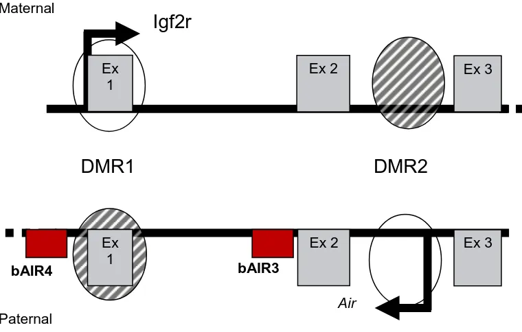
The insulin-like growth factor type 2 receptor (Igf2r) is also known as the cation-independent mannose 6-phosphate receptor (M6PR) and functions as a fetal and placental growth suppressor [169]. Igf2r accomplishes this task primarily by binding Igf2 at the cell surface and targeting it for lysosomal degradation [170]. In mice, Igf2r is an imprinted gene that is maternally expressed and paternally imprinted [171]. The Igf2r gene is part of a 400 kb cluster that contains two other maternally expressed imprinted genes, solute carrier family 22a (Slc22a2 and Slc22a3) , and one paternally expressed antisense non-coding RNA, Air [172, 173]. The promoters for Slc22a2 and Slc22a3 are 190 kb and 260 kb downstream of the Igf2r promoter [174]. Neither Slc22a2 or Slc22a3 are expressed in the embryo, however, both exhibit imprinted expression in the placenta and biallelic expression in adult tissues [173, 175]. In contrast, Igf2r is biallelically expressed in the pre-implantation embryo [176] and maternally expressed in all post-implantation tissues [177] except the brain, where biallelic expression is exhibited [178]. The only paternally expressed gene in the cluster, Air, is transcribed antisense to Igf2r and shares 30 kb of sequence with Igf2r [175]. In addition, the Igfr2/Air cluster contains two differentially methylated regions (DMRs) [175]. DMR1 contains the promoter for Igf2r and is methylated on the paternal allele, but not on the
maternal allele [175]. DMR2 is located within intron2 of Igf2r and contains the promoter for
Imprinting Control in the Igf2r/Air Cluster
Parent-specific expression of murine Igf2r and neighboring genes within the Igf2r/Air
cluster is regulated by DNA methylation, expression of the non-coding RNA, Air, and histone modifications [173, 175, 179-181]. Differentially methylated region 1 (DMR1) contains the promoter for Igf2r and acquires a methylation imprint on the paternal allele during post-implantation development [182, 183]. This acquired methylation is apparently a result of imprinting and not a cause [182]. Differentially methylated region 2 (DMR2) is located within intron 2 of murine Igf2r and is thought to be the imprint control region (ICR) for the cluster [184]. In addition, DMR2 contains the promoter for the antisense non-coding RNA, Air [172]. Methylation of DMR2 occurs during oogenesis on the maternal allele [182] and is maintained during embryogenesis through the period of genome-wide demethylation and remethylation that occurs during cleavage development [185]. A 113 bp imprinting box is thought to establish the methylation imprint at DMR2 by a de novo methylation signal (DNS) and a allele discriminating signal (ADS) within the imprinting box of the maternal allele [186]. During pre-implantation development, murine Igf2r is biallelically expressed and the maternal methylation imprint at DMR2 is already in place [182, 185]. This indicates that the presence of methylation on DMR2 is not enough to induce imprinted expression [176, 177]. Furthermore, loss of methylation at both DMR1 and DMR2 results in a complete lack of Igf2r expression in mice, indicating that the absence of methylation on the Ig2r
expression of Air ncRNA from its promoter in the unmethylated DMR2 of the paternal allele is known to induce silencing of Igf2r, Slc22a2 and Slc22a3 [187]. Therefore, it could be argued that the loss methylation at DMR2 allowed for biallelic expression of Air ncRNA resulting in induced silence of Igf2r expression [175]. In the mouse loss of Air expression from the paternal allele by deletion of DMR2 results in biallelic expression of Igf2r, Slc22a2
and Slc22a3 [173, 179, 180]. Interestingly, imprinting control in the cluster is also lost when
Air ncRNA is truncated to 3 kb even though the methylation imprint at DMR2 is still intact [187]. Therefore, expression of full length Air ncRNA is necessary for imprinting control of the cluster. However, the 30 kb transcriptional overlap between Igf2r and Air is not
necessary to silence the other genes within the cluster [188]. Imprinted expression of Igf2r,
Slc22a2 and Slc22a3 was maintained in mice with a deleted Igf2r promoter and lacked any transcriptional overlap with Air [188].
DNA methylation at DMR1 and DMR2 are not consistently indicative of imprinted expression at the Igf2r loci. Therefore, several studies have examined histone modifications at the DMRs in different tissues in order to more precisely predict imprinted expression of
associated with open chromatin whereas methylation of lysine 9 on histone H3 (H3K9me) is associated with closed chromatin or repressed transcription [191].
In murine fibroblasts the active promoters for Igf2r and Air exhibit histone modifications that are associated with transcriptionally active chromatin [189]. These modifications include tri-methylation of lysine 4 on histone 3 (H3K4me3), di-methylation of lysine 4 on histone 3 (H3K4me2), and acetylation of lysine 9 on histone 3 (H3K9Ac) [189]. The silenced promoters of Igf2r and Air exhibit repressive histone modifications that include tri-methylation of lysine 9 on histone 3 (H3K9me3) and tri-methylation of lysine 20 on histone 4 (H4K20me3) [189]. In murine liver, allele-specific histone modifications in both DMR1 and DMR2 of Igf2r included acetylation of histones H3 and H4, as well as di-methylation of lysine 9 on histone 3 (H3K9me2) [183, 190]. In contrast, in murine neurons
Igf2r is biallelically expressed and Air is not [178]. Interestingly, no allelic differences in histone acetylation and di-methylation at DMR1 are exhibited in these cells [178]. However, in murine glial cells and fibroblasts, Igf2r is imprinted and Air is expressed [178]. In this case histone acetylation and di-methylation of lysine 4 on histone 3 (H3K4me2) were only found on the maternal DMR1 [178]. Thus, histone modifications reliably mark the
promoters of the active and repressed alleles of Igf2r in the mouse [191]. Acquisition of Gametic and Somatic Imprints
when epigenetic reprogramming occurs [182]. Acquisition of methylation at DMR2 during oogenesis signifies the first step to imprinted expression in the Igf2r/Air cluster [182]. During pre-implantation development, Igf2r is biallelically expressed and it is assumed that
Air ncRNA is not expressed [176, 177]. Additionally, it is assumed that Air ncRNA begins to be expressed around the time implantation as the paternal Igf2r allele begins to be silenced [176, 177]. It is unclear when Slc22a2 and Slc22a3 begin to exhibit imprinted expression in the placenta [175]. However, Slc22a2 is imprinted in placenta between 11.5 days post coitum (dpc) to 15.5 dpc [173]. Similarly, Slc22a3 is observed to have imprinted expression 11.5 dpc but does not at 15.5 dpc [173]. Interestingly, neither Slc22a2 or Slc22a3 are
imprinted in the embryo or adult tissues [175]. Following implantation, a somatic
methylation imprint is acquired at DMR1 on the paternal allele [182]. Acquisition of DNA methylation and repressive chromatin modifications at DMR1 maintain transcriptional silence of paternal Igf2r [189, 190]. The somatic imprint at DMR1 is not completed until after birth [182]. It is thought that this imprint does not directly cause paternal silencing of
Igf2r, but rather, this imprint is the result of silencing induced by the expression of Air
ncRNA [192].
Antisense to Igf2r (Air)
Antisense to the Igf2 receptor (Air) is an antisense non-coding (nc) RNA found in the mouse that regulates imprinted expression of three protein coding genes in cis [184].
ncRNA induces silence in cis on the Igf2r/Air cluster. Several models have been proposed to explain how Air mediates gene silencing in the Igf2r/Air cluster. In the expression
competition model it is proposed that Igf2r, Slc22a2, Slc22a3 and Air are all competing for common factors required for promoter and enhancer activation [117]. Transcription of one gene in the cluster may modulate expression of the other genes in the cluster by reducing their access to the necessary common factors [117]. However, this model is likely not correct because the truncated version of Air can still be expressed in cis with the other genes in the cluster [187].
In the RNAi model it is proposed that the transcriptional overlap between Air and
Igf2r could result in silencing of the Igf2r promoter by RNAi-mediated processes [181]. The silent chromatin state induced at the Igf2r promoter could then be spread to Slc22a2 and
Slc22a3 by recruitment of unknown factors that act to suppress expression [181]. In opposition to this model, it has been demonstrated that the transcriptional overlap between
Air and Igf2r is not necessary for imprinted expression of the Igf2r/Air cluster [188]. Similar to X-inactivation, in the RNA-directed targeting model it is proposed that the
In the transcriptional interference model it is proposed that Air induces silence of
Igf2r, Slc22a2 and Slc22a3 by transcription through a domain regulatory element. Based on this model, transcription of Air would prevent binding of RNA polymerase II (RNAPII) to the promoter or binding of a domain regulator to a cis-acting enhancer resulting in silenced expression from the paternal chromosome [181]. The instability of Air ncRNA supports the transcriptional interference model because instability is associated with a lack of splicing [181]. Therefore, the absence of splicing may trap the Air ncRNA close to the site of transcription on the paternal allele and prevent it from acting in trans on the maternal allele [181].
DNA methylation of the Air promoter is required to silence Air expression on the maternal allele [181]. In murine embryos, loss of DNA methylation results in greatly reduced levels of Igf2r mRNA, as well as, a doubling of Air ncRNA expression [64, 181]. Decreased DNA methylation allowed biallelic expression of Air and subsequent silencing of
Igf2r on the maternal and paternal alleles [181]. In contrast, aging mice exhibit de novo methylation on DMR2 of the parental alleles without any change to DMR1 [194]. De novo methylation of DMR2 increased with the age of the mice and was associated with a decrease in Air expression, whereas Igf2r expression was unaffected [194]. Together these studies illustrate that Igf2r and Air expression can be very sensitive to changes in methylation patterns as well as of their corresponding levels. Similarly, Igf2r and Air expression are sensitive to changes in histone modifications [195]. The expressed alleles of Igf2r and Air
acetylation of histones and decreased methylation of DNA demonstrating that histone acetylation and DNA methylation are interdependent [195]. TSA relaxed imprinting of Igf2r
but stimulated the relaxation of Air imprinting to a greater degree [195]. Therefore, factors apart from DNA methylation and histone acetylation may be involved in imprinting of Igf2r
and Air [195].
EVOLUTION OF IGF2R IMPRINTING
Parent-of-origin specific expression of the insulin-like growth factor type 2 receptor (IGF2R) is thought to have first appeared in the mammalian lineage between 180 and 210 million years ago (MYA) [196]. The appearance of imprinted IGF2R expression is
coincident with the divergence of the monotremes from the therian lineage 210 MYA and the departure of the marsupials from eutherians around 180 MYA [196, 197]. Imprinted
expression of Igf2r/IGF2R has been demonstrated in most mammals including mice [171], rats [198] sheep [53], cows [199], pigs [191], dogs [200] and opossums [201]. Animals that are more ancestral to marsupials exhibit biallelic expression of IGF2R and include
monotremes [202] and aves [203]. Interestingly, all mammals in the Euarchonta clade also exhibit biallelic expression of IGF2R [204]. Theseinclude the tree shrew, flying lemur, ringtail lemur, and humans [204].
Evolutionary Pressure for Imprinted Expression of Igf2r/IGF2R
secrete milk from their abdomens [196]. Concurrent with the divergence of marsupials and eutherians from monotremes is the appearance of viviparity and imprinted expression of
Igf2r/IGF2R and Igf2/IGF2 [196]. Both IGF2R and IGF2 are imprinted in marsupials [202, 206, 207]. These species have a non-invasive choriovitelline placenta [208], short gestation period and give birth to altricial young [202]. Similarly, imprinting of Igf2r/IGF2R and
Igf2/IGF2 is exhibited by eutherians [53, 209] which are true placental mammals. However, loss of imprinted IGF2R expression occurred in primates around 75 MYA [191] while imprinted expression of IGF2 is maintained [205]. Interestingly, IGF2R imprinting in humans appears to be polymorphic in a small subset of the population [210, 211]. IGF2R
serves as a tumor suppressor gene and loss of heterozygosity or mutations of IGF2R are frequently found in early stage tumors indicating that monoallelic expression of IGF2R may be an early mechanism for initiating cancer growth [191, 212-214]. The occurrence of polymorphic IGF2R imprinting in humans may be the result of ancestral imprinted alleles still in the population or the re-emergence of IGF2R imprinting [204].
Currently, there are several theories that have been proposed to explain the
evolutionary pressure that may have stimulated creation of imprinted gene expression [76, 191, 196]. Based on the ovarian time bomb hypothesis, it is proposed that an allele favoring imprinted expression would suppress malignant trophoblastic disease resulting from
hypothesis fails to account for genes regulating post-natal development [76]. The kinship theory, also known as the conflict hypothesis, arose from the observation that in the mouse Igf2r, a growth suppressor, was maternally expressed and Igf2, a growth promoter, was paternally expressed [216]. The theory states that the investment made in offspring is
different between males and females resulting in different selective pressures on the parental alleles [217]. An intra-genomic conflict arises within the offspring between the maternal and paternal sets of alleles over potential resources supplied to the offspring by the mother [196]. Based on the conflict hypothesis, a ancestor of mammals may have evolved imprinted
expression of Igf2r and Igf2 as a result of conflict between the parental genomes [196]. Therefore, it would be advantageous for the paternal genome to increase fetal size at the expense of the mother by favoring expression of Igf2. In contrast, would be advantageous for the maternal genome to minimize fetal growth by favoring expression of Igf2r. The conflict theory predicts that the degree to which offspring develop in utero, gestation length and type of postnatal care are all selective pressures influencing imprinted gene expression [202].
Species Differences in Requirements for Imprinted Expression of Igf2r/IGF2R
nc(RNA), Air. In addition, an imprinting box in DMR2 was identified that contains a de novo methylation signal (DNS) and an allele discriminating signal (ADS) [186]. However, an imprinting box has not yet been identified in any other species [191]. DMRs are
composed of cytosine guanine repeats (CpGs) that are differentially methylated between the parental alleles. In the mouse, CpG1 corresponds to DMR1 and CpG2 corresponds to DMR2. In contrast to the mouse, the opossum, which is a marsupial, does not have a CpG2 island comparable to the CpG2 island in DMR2 of the mouse [201]. However, although the opossum does have a CpG1 island orthologous to the CpG1 island in DMR1 of the mouse [201], it is not differentially methylated [201]. In addition, Air ncRNA is not detected in the opossum [201]. Therefore, none of the known requirements to imprint Igf2r in the mouse exist in the opossum [201]. Notably, IGF2R in the opossum binds IGF2 with far less affinity than that observed in the mouse [218]. Consistent with the conflict hypothesis is the idea that because altricial offspring of marsupials are only exposed for a limited time to a non-invasive intrauterine environment there is less selective pressure to exploit maternal resources [196]. Therefore, this would potentially result in less pressure for a strong imprinting response from the maternal allele.
The Artiodactyla clade contains sheep, cows and pigs all of which exhibit imprinted expression of IGF2R and IGF2 [53, 209, 219]. The ruminants both display differential methylation on the CpG islands of IGF2R consistent with DMR1 and DMR2 of the murine
Recently, it was determined that the canine IGF2R gene is imprinted [200]. Dogs belong to the superordinal group called Laurasiatheria, which is a sister group to the
superordinal group that contains both rodents and primates [200]. The canine IGF2R appears to be similar to murine Igf2r gene in that it does have a CpG2 island that exhibits differential methylation [200]. However, in contrast to the mouse, the promoter of IGF2R on the canine paternal allele is not methylated and maternal expression of IGF2R is not accompanied by paternal expression of AIR ncRNA [200].
The human IGF2R gene has been extensively studied [178, 211, 220-222]. IGF2R in the human exhibits similar elements to the mouse for regulating imprinted expression of
Igf2r. IGF2R/Igf2r in both humans and mice is differentially methylated at the CpG2 island of DMR2 in intron 2 of IGF2R/Igf2r [191, 211]. Additionally, IGF2R/Igf2r in both humans and mice have a CpG1 island that contains the promoter of IGF2R/Igf2r [191]. However, the CpG1 island of human IGF2R does not display differential methylation [191]. In contrast to all other eutherian mammals and despite similar elements necessary for imprinted expression of Igf2r in the mouse, human IGF2R is biallelically expressed in most of the population [211, 221]. Additionally, it has also been shown that human AIR ncRNA does not exist [211, 223]. Therefore, methylation at DMR2 is not sufficient to induce imprinted expression of IGF2R.
alleles corresponds to the biallelic expression of IGF2R in humans [191]. Furthermore, differential patterns of acetylation and methylation between the parental alleles do not occur at the DMR2 of human IGF2R [178, 189].
Histone modifications and DNA methylation may work together in marking specific promoters for expression or silencing [191]. Expression may result from histone acetylation and H3K4me on a promoter in combination with the loss of H3K9me3 and DNA methylation [191]. Losing H3K9me3 and DNA methylation from the promoter without addition of
histone acetylation and H3K4me may only result in an unrepressed chromatin state but may not facilitate transcription [191]. For example, human IGF2R contains an unmethylated DMR2 on the paternal allele, which lacks expression of AIR ncRNA [191]. Therefore, the human DMR2 may only lack histone modifications necessary to promote active transcription of AIR ncRNA [191].
there are imprinted alleles still circulating in the population [191, 210]. An ancestor of primates may have had a selective advantage of biallelically expressing IGF2R over those that were monoallelically expressing IGF2R. IGF2R is a regulator of fetal growth, a
suppressor of cell proliferation and is involved in T-cell mediated apoptosis [204]. A primate ancestor that inherited an imprinted IGF2R allele would potentially be subjected to a greater risk of fetal overgrowth and carcinogenesis due to haploinsufficiency [204]. Thus, the original evolutionary pressures that created imprinted IGF2R expression in the ancestor of marsupials and placental mammals may have been over come by another selective force favoring biallelic expression of Igf2r/IGF2R.
genes. DNA methylation and histone modifications are very dynamic during pre-implantation development and function to modulate chromatin structure and regulate transcription [143]. The pre-implantation embryo is highly sensitive to the external environment and exposure to culture or in vitro manipulation alters the pattern of DNA methylation and histone modifications of imprinted alleles, resulting in aberrant mRNA expression [230].
Disruption of DNA Methylation
Expression of imprinted genes is dependent upon DNA methylation to mark the parental alleles in a sex-specific manner [230]. During pre-implantation development, methylation imprints are maintained as the parental genomes first undergo global
demethylation which is then followed by remethylation [230]. Embryo culture and in vitro manipulation can affect imprinted gene expression by disrupting DNA methylation imprints [120, 219, 227, 231, 232]. Therefore, some of the phenotypes associated with AOS may be attributed to aberrant expression of imprinted genes [37, 54].
Epigenetic reprogramming errors may occur during erasure, acquisition or
imprinted genes have also been observed following SCNT [62, 228, 233, 234]. Furthermore, cloned mouse, sheep and bovine embryos and offspring exhibit imprinting-related
abnormalities that are thought to be derived from incomplete epigenetic reprogramming [53, 62, 229, 235-237].
Aberrant expression of imprinted genes associated with abnormalities in the placenta have also been described following the transfer of IVP [238] or SCNT embryos [62, 226, 239-241]. Abnormal placentation has been suggested to be the major cause of failed NT pregnancies [242]. In pre-implantation SCNT-derived embryos, aberrant methylation patterns have been mostly found in the trophectoderm, indicating that dysregulation of the extraembryonic lineages may be a major contributor to the inefficiency of SCNT [243]. Perturbed Histone Modifications
hypomethylated DNA, an open chromatin state, and increased gene transcription [96]. In contrast, methylation of lysine residue 9 on histone H3 (H3K9me) and deacetylated core histone tails are associated with hypermethylated DNA, a closed chromatin state, and repressed gene transcription [96]. Acetylation and deacetylation of histone tail residues occurs by the actions of two groups of histone modifying enzymes, the histone
acetyltransferases (HATs) and the histone deacetylases (HDACs) [248]. Methylation of histone tail residues occurs by the actions of histone methyltransferases (HMTs) [96]. It has been demonstrated that before and after embryonic genome activation, in vitro culture environments and cloning procedures aberrantly affect the patterns of mRNA expression for histone modifying enzymes resulting in altered histone modifications [248, 249]. In murine clones, the pattern and level of histone acetylation varies according to the type of donor cells used [250]. Similarly, perturbed histone acetylation has been observed in SCNT-derived bovine embryos [234]. Additionally, bovine SCNT embryos exhibited altered histone acetylation and methylation patterns in conjunction with delayed and decreased DNA methylation [251, 252]. Thus, the effects of both in vitro culture environments and SCNT procedures can aberrantly affect patterns of histone modifications and potentially contribute to altered gene expression ultimately resulting in AOS phenotypes.
Contribution of Imprinted Expression of IGF2R to AOS
growth, such as the insulin-like growth factor family [153, 157, 164, 253]. Transgenic mice that overexpress Igf1 were approximately 21% larger at birth than in vivo controls and exhibit organomegaly [253]. Similarly, murine fetuses that overexpress Igf2 exhibit
organomegaly, as well as fetal and placental overgrowth [254]. In addition, murine fetuses inheriting a nonfunctioning insulin-like growth factor type 2 receptor (Igf2r) gene died around the time of birth and exhibited major cardiac abnormalities, elevated circulating Igf2 levels, and were 25 to 30% larger than normal siblings [164]. Patterns of mRNA expression for members of the IGF family are also altered in bovine fetuses, placentas and offspring that are derived from the transfer of IVP or SCNT embryos [10, 54, 239, 255-257]. However, only aberrant expression of IGF2R in sheep has been shown to be directly correlated with AOS phenotypes [53]. Altered expression of the IGF2R gene has been demonstrated in embryos, fetuses, placentas and offspring in cattle [199, 226, 255, 257-261] following the transfer of IVP or SCNT embryos. Thus, aberrant expression of IGF2R may contribute to some of the phenotypes associated with AOS; for example, fetal and placental overgrowth.
In cattle, the IGF2R gene is imprinted and is expressed from the maternal allele [199]. Expression from this allele is mediated by two differentially methylated regions (DMRs) [199]. Alterations to the methylation patterns of these DMRs were observed following the transfer of SCNT-derived embryos and resulted in altered IGF2R expression [199]. In that study, 3 of 5 cloned fetuses were reconstructed using granulosa cells and 2 of them died near term and exhibited organomegaly, fetal overgrowth, and respiratory failure [199]. In
the heart to 99% in the brain, indicating that methylation patterns at DMR2 were regulated in a tissue-specific manner and may be mediated by other regulatory elements as previously reported in the mouse [183, 187, 195, 199].
In the mouse, imprinted expression of Igf2r is regulated by DNA methylation at two DMRs, expression of the antisense non-coding RNA, Air, and covalent histone modiciations [178, 187, 195]. Placental overgrowth, a common characteristic of AOS, was observed in embryonic Day 9.5 (E9.5) SCNT murine clones that exhibited significantly reduced Igf2r expression [240]. In a separate study, murine clones were created using an ES cell line known to produce fetal and placental overgrowth [262]. The cloned fetuses exhibited a 30% increase in weight over controls at Day 17.5 of gestation [262]. This was attributed to the elevated expression of Igf2 in the cloned fetuses at Day 9.5 and Day 12.5 of gestation [262]. In contrast, Igf2r expression was not different between clones and controls at Day 9.5, Day 12.5 and Day 17.5 [262]. Interestingly, expression of Air ncRNA was significantly higher in clones at Day 17.5 [262]. Therefore, a failure to increase Igf2r expression in response to elevated Igf2 expression may result from the silencing effects of Air on Igf2r and may ultimately result in fetal and placental overgrowth late in gestation. Sheep also share similar methylation imprints at DMR1 and DMR2 as those identified in mice and cattle [199]. IVP-derived ovine fetuses exhibiting AOS showed a reduction of 30-60% in IGF2R mRNA expression relative to the controls [53]. Methylation at DMR2 in 9 of the 12 AOS fetuses was completely lost [53]. Similarly, in Dnmt1 null mice DMR2 is unmethylated and Igf2r
to be hypomethylated at the DMR2 of IGF2R in liver, brain and heart, whereas DMR2 in the lung was hypermethylated [199]. If loss methylation at DMR2 is associated with a reduction of IGF2R, then organomegaly observed in AOS fetuses may result from loss of methylation at DMR2 of IGF2R in those organs. Interestingly, when murine fibroblasts were cultured in the presence of trichostatin A (TSA), a deacetylase inhibitor, Air expression was dramatically increased whereas Igf2r expression was only slightly increased [195]. Increased histone acetylation at DMR1 should have allowed for an increase in Igf2r expression; however, it resulted in hypomethylation of DMR2 and a dramatic increase in Air expression [195]. This may have inhibited the expression of Igf2r despite the open chromatin state [195].
Interestingly, when a bovine kidney cell line was cultured with TSA, IGF2R expression was decreased [199]. These observations potentially indicate that relaxation of IGF2R imprinting may have been inhibited by a relaxation of imprinted expression of bovine AIR.
The level of IGF2R expression has consistently been shown not to differ between in vivo, IVP and SCNT derived bovine blastocysts [67, 226, 244, 263-266]. Recently, the level of IGF2R expression was shown to be increased in the placentas of gestational day 25 bovine concepti derived from SCNT and IVP embryos compared to in vivo controls [226]. The increase in IGF2R expression observed in the placenta at Day 25 may be in response to the high expression levels of IGF2 observed in both SCNT derived placentas and fetuses [226]. Alterations in expression of IGF2R within the placenta may contribute to abnormal
development and function of the placenta, which has been proposed as one of the major
Imprinted expression of Igf2r/IGF2R is altered in fetuses and placentas derived from the transfer of IVP and SCNT of murine [240, 262, 268], ovine [53, 219, 269] and bovine [199, 226, 257] embryos. In the mouse, manipulation of regulatory elements that control
Igf2r expression, including DNA methylation at DMR2 and expression of Air, result in aberrant expression of Igf2r [181, 195, 262]. In sheep and cattle, it is unknown if AIR
ncRNA is involved in the regulatory mechanism mediating imprinted expression of IGF2R
[53, 199]. However, DMR1 and DMR2 are present and hypo- or hyper-methylation of these two sites results in aberrant expression of IGF2R in sheep and cattle [53, 199]. Improper expression of Igf2r resulting in AOS-like phenotypes is associated with mutations or deletions of Igf2r in the mouse [64, 153]. In addition, repressed IGF2R expression was demonstrated in AOS ovine fetuses [53]. Thus, some phenotypes of AOS displayed by bovine fetuses and placentas may result from aberrant expression of IGF2R.
STATEMENT OF THE PROBLEM