Diagnosis of Brain Tumor Through MRI
Image Processing using Clustering with
Optimization Technique
Ruchita A. Banchpalliwar 1, Dr. Suresh S. Salankar 2
Dept. of Electronics and Telecommunication, G.H. Raisoni College of Engineering, Nagpur, India 1
Assistant Professor, Dept. of Electronics and Telecommunication, G.H. Raisoni College of Engineering, Nagpur, India2
ABSTRACT: To Analysis and Diagnosis of tumor in MRI Brain images segmentation technique is used. The paper
presents standard Fuzzy C Means with Particle swarm optimization technique for the effectiveness of fuzzy C means clustering used to mark the position of brain tumor all the way through MRI. This methodology is tested on 15 MRI images. Fuzzy C means and Fuzzy C means with PSO is compared. Due to the limitation in initialization the FCM algorithm is not efficient. FCM with PSO has the advantage of increasing accuracy over FCM. It has accuracy improvement in segmentation of 26.89 % over FCM and FCM with PSO. And it also detect the stage of MRI brain tumour
.
KEYWORDS: Magnetic Resonance Image (MRI), Brain Tumor Segmentation, Fuzzy C Means Algorithm (FCM) ,
Particle Swarm Optimization (PSO).
I. INTRODUCTION
Analysis of medical images decreases the doctor’s workload. It measures the brain tumor variation. But the automatic segmentation of brain tumor is still a tough problem due to various tumors types with difference structure, shapes and size in morphological.
For MRI segmentation numerous techniques have been developed. The most important four classes are region based, model based, threshold-based techniques and pixel classification. In region based segmentation techniques pixels are grouped into homogeneous regions and it Segments the region of homogeneous properties and the connected region are generated. This region growing technique is analogous to an algorithm which is called as split-and merge, but the seed point are not needed. Region growing can also be induced to separate the regions, graceful to noise. This all problems which are coming in region growing techniques can be removed by using homotopic region-growing algorithm.The disadvantage is that it is Expensive in computational time and memory. In model-based segmentation, advanced knowledge of the object like structure, position and situation is used to build a particular anatomic shape. Connected model are also constructs in a continuous manner. These are costly and tough to initialize. The threshold based techniques is one of the older method of segmentation. In this the object of the intensities in the image are differentiate with one or many thresholds intensity and are classified. These threshold techniques are global and local. These methods of segmentation cannot utilize the MRI information. Supervised and Unsupervised are used in pixel classification. It is used as a classifier to cluster pixels in the quality space. Bayes classifiers and artificial neural networks (ANN) are comes under the supervised techniques and K-means and Fuzzy clustering techniques (FCM) comes under unsupervised techniques.
gives us the details of Standard fuzzy C means Clustering algorithm. PSO Algorithm are described in section IV. Modified the proposed FCM with PSO is described in section V. Experiments and results are obtained in section VI and section VII concludes the paper.
II. PREVIOUS WORK AND ITS DISADVANTAGES
Previously the paper referred have introduced regarding how the brain tumour segmentation is extremely tedious job. The brain consists of cells and mixture of tissues which can also been seen in the brain tumour image of MRI, so it’s the most critical part to recognize the brain tumour exactly and efficiently. The Previous work was skilful to diagnose the tumour but the accuracy and efficiency level of segmentation was not up to the mark and for correctly diagnose the tumour the processing time was very high. There was a possibility of a group of parts of tissues and the inner swelling to be categorized as a tumours region. Hence for brain tumour detection and segmentation in MRI images there is a need of computer aided techniques. There are many segmentation techniques which are used for MRI detection. But from this techniques most suitable technique for detection is FCM. And to improve this we will combined FCM technique with the optimization technique PSO.
III. STANDARD FUZZY C MEANS CLUSTERING ALGORITHM
During the segmentation Standard FCM clustering algorithm scan only local information. The variable values enclose from 0 to 1. To each and every data point equivalent to center cluster depending on the distance between the cluster center and data point membership is allowed. After every iteration the membership and cluster centers are updated.
Algorithm steps for FCM:
Let the set of data points X ={x1, x2, x3 ..., xx} and the set of centers V = {v1, v2, v3 ..., vv}.
Step 1: Select c cluster centers randomly.
Step 2: Function µij of fuzzy membership is determined as:
(3.1)
Step 3: Function vj fuzzy centers are calculated as:
(3.2)
Step 4: Until the minimum ’J’ value is accomplished repeat 2 & 3 step or ||Uk+1−Uk|| < β where,
k = Step of iteration.
Β = Termination criterion between [0, 1].
U = The fuzzy membership matrix is (µij) + c
J= Objective function.
IV. PARTICLE SWARM OPTIMIZATION ALGORITHM
Particle swarm optimization is the population based algorithm. It is a basic of swarm intelligence technique which grouping the behaviour in which the food is explore between the source origin. The bird searches the area space for the solution which is used in particle swarm algorithm. Each and every particle have their individual fitness value which are assess in the form of fitness functions and also the data is in the form of velocity which give the results to the location. The particles which are proceeding are in the form of the majority useful purpose in given trouble space.
The main algorithm which starts while grouping the particles generating the particular solutions having the optimal value is the survey with the help of the orderly iterations. Then every particles are updated iteratively according to the most superior values. The values which are found by the maximum particles are called as “pbest”. And the second value is determined with the population in the particles value is called as “gbest”.
This technique is in the form of numeric based optimization nature. One proposed on the other hand that while in segmenting the image this cluster forming can be applied. The centre of the cluster is defined by using the population searching techniques.
Algorithm steps for PSO:
Step 1. Initialize the population, velocity and location. Step 2. The fitness of the individual particle evaluate (Pbest). Step 3. Track to keep the individual highest fitness (Gbest). Step 4. Pbest and Gbest location modify the velocity. Step 5. The particle position updated.
Step 6. Close if condition is met else go to step 3.
The first two steps are fairly trivial. To the objective function by providing the candidate solution Fitness analysis is conducted. Individual, global best fitness value and positions are corrected and updated by differentiating the newly evaluated fitness against the previous individual and global best fitness value, and exchanging the best fitness and positions as necessary. The velocity and position update step is important for the optimization capacity of the PSO algorithm.
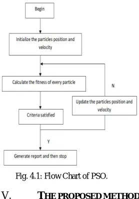
Fig. 4.1: Flow Chart of PSO.
V. THE PROPOSED METHOD
Step 1: Processing of morphological operation Step 2: Changing image data into fuzzy domain data Step 3: Membership moderation
Step 4: Defuzzification
Step 5: The enhanced image displayed
Fig. 5.1: The Proposed Methodology for Brain MRI Segmentation
Using the Fuzzy C Means the enhanced image is segmented with particle swarm optimization technique.
VI. EXPERIMENTAL RESULTS
The segmentation is performed of brain tumour by using Matlab 13. The MRI data base of Brain Tumor has collected from Datta Meghe Institute of Medical Science, Sawangi, Wardha and also from opendata source
http://www.cancerimagearchive.net/display/public/collections .
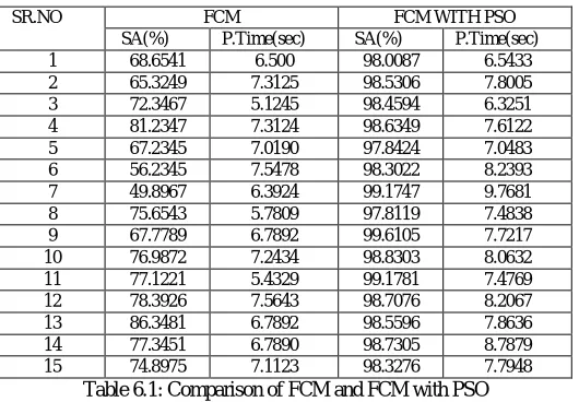
For the segmentation purpose 15 MRI brain images are considered. These all images are with the default size 0f 512 x 512.
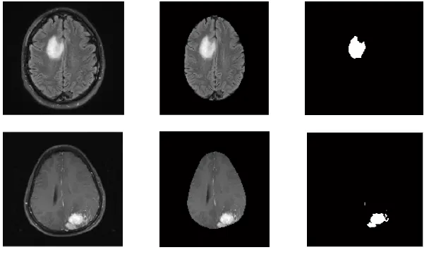
1)Output of FCM with PSO on MRI Brain Image: (a) Original Image (b) Skull Removed Image (d) Standard FCM with PSO
(a) Original Image (b) Skull Removed Image (d) Standard FCM with PSO
(a) Original Image (b) Skull Removed Image (d) Standard FCM with PSO
(a) Original Image (b) Skull Removed Image (d) Standard FCM with PSO
Fig 6.1:Segmentation Results of FCM with PSO 2) Performance Analysis
SR.NO FCM FCM WITH PSO
SA(%) P.Time(sec) SA(%) P.Time(sec)
1 68.6541 6.500 98.0087 6.5433
2 65.3249 7.3125 98.5306 7.8005
3 72.3467 5.1245 98.4594 6.3251
4 81.2347 7.3124 98.6349 7.6122
5 67.2345 7.0190 97.8424 7.0483
6 56.2345 7.5478 98.3022 8.2393
7 49.8967 6.3924 99.1747 9.7681
8 75.6543 5.7809 97.8119 7.4838
9 67.7789 6.7892 99.6105 7.7217
10 76.9872 7.2434 98.8303 8.0632
11 77.1221 5.4329 99.1781 7.4769
12 78.3926 7.5643 98.7076 8.2067
13 86.3481 6.7892 98.5596 7.8636
14 77.3451 6.7890 98.7305 8.7879
15 74.8975 7.1123 98.3276 7.7948
Table 6.1: Comparison of FCM and FCM with PSO
We have also calculated the PSNR value, iterations and also detect the stages for FCM with PSO technique as shown in the following table:
3) Parameters for FCM with PSO
SR.NO PSNR (dB)
In the previous work they are able to detect the tumors but the segmentation accuracy not upto the mark and hence the processing time for the computation process while correctly determining the tumor was very high .
The table shows that the average processing time is reduced with improvement in segmentation accuracy.
FCM FCM WITH ACO
FCM WITH PSO Time
(sec)
6.7139 32.03 7.7823
Accuracy (%)
71.69 97.57 98.58
Table 6.3: Comparison between FCM , FCM with ACO and FCM with PSO
VII. CONCLUSION
The segmentation accuracy of FCM is less due to the fact that it is practical to initialization. Thus to increase the accuracy of segmentation of FCM, Particle swarm optimization is used. The centre cluster is initialized to proper ideal value which gives the results in increased accuracy segmentation. The proposed algorithm results for 15 MRI images are shown. Hence with the help of FCM it is fast for cluster formation and with the help of particle swarm optimization we have found the rise in the time along with the segmentation accuracy.
REFERENCES
1. Rastgarpour M., And Shanbehzadeh J., Application Of Ai Techniques In Medical Image Segmentation And Novel Categorization Of Available Methods And Tools, Proceedings Of The International Multiconference Of Engineers And Computer Scientists 2011 Vol I, Imecs 2011, March 16-18, 2011, Hongkong.
2. M.H. Ahmed, S.M. Yamany, N. Mohamed, A. Farag, T. Moriarty, A modified fuzzy c-means algorithm for bias field estimation and segmentation of MRI data, IEEE Transactions on Medical Imaging 21 (3) (2002) 193–199.
3. K.S. Chuang, et al., Fuzzyc-means clustering with spatial information for image segmentation, Comput. Med. Imaging Graph. 30 (2006) 9–15. 4. Zhang, Y. J, An Overview Of Image And Video Segmentation In The Last 40 Years, Proceedings Of The 6th International Symposium On
Signal Processing And Its Applications, Pp. 144-151, 2001.
5. Wahba Marian, An Automated Modified Region Growing Technique For Prostate Segmentation In Trans-Rectal Ultrasound Images, Master’s Thesis, Department Of Electrical And Computer Engineering, University Of Waterloo, Waterloo, Ontario, Canada, 2008.
6. K.Selvanayaki, Dr.P.Kalugasalam Intelligent Brain Tumor Tissue Segmentation From Magnetic Resonance Image (Mri) Using Meta Heuristic Algorithms Journal Of Global Research In Computer Science Volume 4, No. 2, February 2013
7. Kshitij Bhagwat, Dhanshri More, Sayali Shinde, Akshay Daga, Assistant Prof. Rupali Tornekar, “ Comparative Study Of Brain Tumor Detection Using K Means ,Fuzzy C Means And Hierarchical Clustering Algorithms ” International Journal Of Scientific & Engineering Research , Volume 2,Issue 6,June 2013,Pp 626-632.
8. S.Roy And S.K.Bandoyopadhyay, “Detection And Qualification Of Brain Tumor From Mri Of Brain And Symmetric Analysis”, International Journal Of Information And Communication Technology Research, Volume 2 No.6, June 2012, Pp584-588
9. M. Karnan, T. Logheshwari, "Improved implementation of brain MRI image segmentation using ant colony system", IEEE International Conference on Computational Intelligence and Computing Research (ICCIC), pp. 1-4, 2010.
10. Shen S, Sandham W, Granat M, Sterr A. MRI fuzzy segmentation of brain tissue using neighborhood attraction with neural-network optimization. IEEE Transaction on Information Technology in Biomedicine 2005; 9(3):459–67.
11. S Luo, R Li, S Ourselin, A new deformable model using dynamic gradient vector flow and adaptive balloon forces, in APRS Workshop on Digital Image Comp(APRS, Brisbane,(2003), pp. 9–14.
12. Tahir Sag Mehmet Cunkas, “Development Of Image Segmantation Techniques Using Swarm Intelligence”,Iccit 2012
13. Mohammed Sabbih Hamoud Al-Tamimi Ghazali Sulong, “Tumor Brain Detection Through Mr Images: A Review Of Literature”, Journal Of Theoretical And Applied Information Technology 20th April 2014. Vol. 62 No.2
14. Sayali D. Gahukar et al Int. Journal of Engineering Research and Applications Vol. 4, Issue 4( Version 5), April 2014, pp.107-111 15. Ganesh S. Raghtate, Suresh S. Salankar, “Modified Fuzzy C Means With Optimized Ant Colony Algorithm For Image