University of Windsor University of Windsor
Scholarship at UWindsor
Scholarship at UWindsor
Electronic Theses and Dissertations Theses, Dissertations, and Major Papers
2010
Characterization of cellular stress systems using biological mass
Characterization of cellular stress systems using biological mass
spectrometry
spectrometry
Anna Kozarova
University of Windsor
Follow this and additional works at: https://scholar.uwindsor.ca/etd
Recommended Citation Recommended Citation
Kozarova, Anna, "Characterization of cellular stress systems using biological mass spectrometry" (2010). Electronic Theses and Dissertations. 8114.
https://scholar.uwindsor.ca/etd/8114
This online database contains the full-text of PhD dissertations and Masters’ theses of University of Windsor students from 1954 forward. These documents are made available for personal study and research purposes only, in accordance with the Canadian Copyright Act and the Creative Commons license—CC BY-NC-ND (Attribution, Non-Commercial, No Derivative Works). Under this license, works must always be attributed to the copyright holder (original author), cannot be used for any commercial purposes, and may not be altered. Any other use would require the permission of the copyright holder. Students may inquire about withdrawing their dissertation and/or thesis from this database. For additional inquiries, please contact the repository administrator via email
Characterization of cellular stress systems using
biological mass spectrometry
by Anna Kozarova
A Dissertation
Submitted to the Faculty of Graduate Studies through Chemistry and Biochemistry in Partial Fulfillment of the Requirements for
the Degree of Doctor of Philosophy at the University of Windsor
Windsor, Ontario, Canada 2010
1*1
Library and Archives CanadaPublished Heritage Branch
395 Wellington Street OttawaONK1A0N4 Canada
Bibliotheque et Archives Canada
Direction du
Patrimoine de I'edition
395, rue Wellington Ottawa ON K1A 0N4 Canada
Your file Votre reference ISBN: 978-0-494-80248-9 Our file Notre reference ISBN: 978-0-494-80248-9
NOTICE: AVIS:
The author has granted a
non-exclusive license allowing Library and Archives Canada to reproduce, publish, archive, preserve, conserve, communicate to the public by
telecommunication or on the Internet, loan, distribute and sell theses
worldwide, for commercial or non-commercial purposes, in microform, paper, electronic and/or any other formats.
L'auteur a accorde une licence non exclusive permettant a la Bibliotheque et Archives Canada de reproduire, publier, archiver, sauvegarder, conserver, transmettre au public par telecommunication ou par Plnternet, preter, distribuer et vendre des theses partout dans le monde, a des fins commerciales ou autres, sur support microforme, papier, electronique et/ou autres formats.
The author retains copyright ownership and moral rights in this thesis. Neither the thesis nor substantial extracts from it may be printed or otherwise reproduced without the author's permission.
L'auteur conserve la propriete du droit d'auteur et des droits moraux qui protege cette these. Ni la these ni des extraits substantiels de celle-ci ne doivent etre imprimes ou autrement
reproduits sans son autorisation.
In compliance with the Canadian Privacy Act some supporting forms may have been removed from this thesis.
Conformement a la loi canadienne sur la protection de la vie privee, quelques formulaires secondaires ont ete enleves de cette these.
While these forms may be included in the document page count, their removal does not represent any loss of content from the thesis.
Bien que ces formulaires aient inclus dans la pagination, il n'y aura aucun contenu manquant.
• • I
DECLARATION OF CO-AUTHORSHIP / PREVIOUS PUBLICATION
I. Co-Authorship Declaration
I hereby declare that this thesis incorporates material that is the result of joint research, as
follows:
This thesis incorporates the outcome of joint research undertaken in collaboration
with Michelle A. Sharon under the supervision of Dr. Alden H. Warner, Department of
Biological Sciences. The collaboration is covered in Chapter 2 of the thesis. In all cases,
the key ideas, primary contributions, experimental designs, data analysis and
interpretation, were performed by the authors; Michelle purified a 21 kDa protein from
Artemia franciscana, PCR amplified cDNA from an embryonic library corresponding to
its gene and performed the initial characterization of this protein. My contribution to this
manuscript was the de novo identification of this protein using mass spectrometry (MS).
Since the genome of this organism has not yet been sequenced, this included in silico
analysis for the identification. Based on the peptide sequence corresponding to a
conserved motif, the 21 kDa protein was identified as a member of group 1 Late
embryogenesis abundant (LEA) family. Once Michelle completed the gene amplification,
I was able to match the peptide sequences generated by MS and thus further confirm the
identity of the 21 kDa protein.
This thesis also incorporates the outcome of joint research undertaken in
collaboration with Inga Sliskovic under the supervision of Dr. Bulent Mutus, Department
of Chemistry and Biochemistry. The collaboration is covered in Chapter 3 of the thesis.
In all cases, the key ideas, primary contributions, experimental designs, data analysis and
interpretation, were performed by the authors, and the contribution of co-authors was
Inga prepared the PDI protein sample in a reduced and in an auto-oxidized form. Dr. Eric
Simon and Dr. Philip Andrews at the Michigan Proteome Consortium, University of
Michigan, provided knowledge and instrumentation for the peptide separation and
MALDI TOF/TOF analysis. My contribution to this manuscript was application of the
isotope-coded affinity tag (ICAT) technology and identification of the cysteine redox
state on PDI using mass spectrometry (MS). I performed all experimental steps involved
in the ICAT labeling protocols, followed with acquisition and interpretation of the MS
spectra.
This thesis also incorporates the outcome of joint research undertaken in
collaboration with Dr. John W. Hudson, Department of Biological Sciences. The
collaboration is covered in Chapter 4 of the thesis. In all cases, the key ideas, primary
contributions, experimental designs, data analysis and interpretation, were performed by
the authors, and the contribution of co-authors was through the provision of experimental
help, analysis and data interpretation with flow cytometry based approach. My
contribution to this manuscript in preparation was identification of phosphorylation sites
on Flag-hYVHl expressed and immunoprecipitated from mammalian cells; generation of
phosphomutants, localization experiments, RNAi experiments and characterization of the
phenotype, as well as sample processing for flow cytometry experiments.
I am aware of the University of Windsor Senate Policy on Authorship and I certify
that I have properly acknowledged the contribution of other researchers to my thesis, and
have obtained written permission from each of the co-author(s) to include the above
I certify that, with the above qualification, this thesis, and the research to which it
refers, is the product of my own work.
II. Declaration of Previous Publication
This thesis includes 2 original papers that have been previously published/submitted for
publication in peer reviewed journals, and 1 manuscript in preparation as follows:
Thesis Chapter Chapter 2
Chapter 3
Chapter 4
Publication title/full citation
Sharon, M.A., Kozarova, A., Clegg, J.S., Vacratsis, P.O., and Warner, A.H. (2009). Characterization of a group 1 late embryogenesis abundant protein in encysted embryos of the brine shrimp Artemia franciscana. Biochem Cell Biol 87, 415-430 Kozarova, A., Sliskovic, I., Mutus, B., Simon, E.S., Andrews, P.C., and Vacratsis, P.O. (2007). Identification of redox sensitive thiols of protein disulfide isomerase using isotope coded affinity technology and mass spectrometry. J Am Soc Mass Spectrom 18, 260-269
Kozarova, A., Hudson, J.W., Vacratsis, P.O.
Publication status*
Published
Published
To be submitted
I certify that I have obtained written permission from the copyright owner(s) to
include the above published material(s) in my thesis. I certify that the above material
describes work completed during my registration as graduate student at the University of
Windsor.
I declare that, to the best of my knowledge, my thesis does not infringe upon
quotations, or any other material from the work of other people included in my thesis,
published or otherwise, are fully acknowledged in accordance with the standard
referencing practices. Furthermore, to the extent that I have included copyrighted material
that surpasses the bounds of fair dealing within the meaning of the Canada Copyright Act,
I certify that I have obtained a written permission from the copyright owner(s) to include
such material(s) in my thesis.
I declare that this is a true copy of my thesis, including any final revisions, as
approved by my thesis committee and the Graduate Studies office, and that this thesis has
ABSTRACT
In recent years mass spectrometry has become an invaluable tool to address an
array of biological questions. The versatility of this experimental approach does not only
allow assignment of protein identity and identification of sequence specific modifications,
but with the help of particular derivatization techniques facilitates the determination of
peptide quantity. Each of these approaches were applied to the following biological
projects:
The 21 kDa heat stable protein purified from the encysted embryo of Artemia
franciscana was characterized by time-of-flight mass spectrometry. De novo sequencing
of peptides identified this protein as a group 1 Late embryogenesis abundant (LEA)
protein. The amino acid sequence assignment to these peptides allowed amplification of
the entire gene sequence from an embryonic cDNA library. This was deposited into the
NCBI database (EF656614). The expression of group 1 LEA protein is consistent with
and supports a role in desiccation tolerance. In addition, this is a first report describing
identification of a group 1 LEA protein in an animal species.
A MS-based quantitative analysis was performed in order to analyze relative
changes in the dynamic thiol and disulfide states of the redox sensitive protein disulfide
isomerase, PDI. PDI cysteine residues were derivatized with an isotope-coded affinity tag
(ICAT), thus allowing a direct comparison between the reduced and auto-oxidized forms.
Quantitation was based on relative ratios between light and heavy isotopically labeled
cysteine containing peptides. The application of the ICAT-labeling approach to PDI
related studies, allowed direct assignment of individual cysteine residues and their
information regarding the average number of modified cysteine residues within PDI, not
their exact identity.
A combination of a phosphopeptide enrichment step and a MS-based approach
was utilized to identify three phosphorylation sites on hYVHl, an atypical dual
specificity phosphatase that functions as a cell survival factor. With the help of
phosphomimetic and non-phosphorylable mutants, we were able to decipher their effect
on localization and progression through the cell cycle.
Collectively, these studies manifest the power of MS-generated data to influence
DEDICATION
ACKNOWLEDGMENT
First, and most importantly, I would like to thank my supervisor, Dr. Otis
Vacratsis, for giving me the opportunity to work with him and for sharing his personal
"tricks of the trade" related to mass spectrometry. I would also like to thank him for the
hours long scientific discussions, help and patience, and for the words of encouragement
whenever experiments went sour. Overall, thank you Otis for providing a great
environment for discovery in the complicated world of science.
I would also like to thank the members of my committee; Drs. Andrew
Hubberstey, Lana Lee and Siyaram Pandey for their valuable discussions. I would like to
thank my collaborators, Drs. Bulent Mutus and Alden Warner, for the ability to work
together on their projects while acquiring extraordinary technical experience. I would also
like to thank Dr. John Hudson, for all his help during my tenure as a graduate student.
Additionally, I would like to express words of appreciation to all members of the
Chemistry and Biochemistry Department who made their equipment available to me. A
special thank you is in place for Dr. Michael Crawford from the Department of Biological
Sciences, for letting me use his microscope. Additionally, I thank Drs. Eric Simon and
Philip Andrews at the Michigan Proteome Consortium, University of Michigan, for
providing knowledge and access to high end mass spec instruments. Last, but not least, I
would like to thank all members of the Vacratsis lab, present and past, for a good working
relationship and friendship.
The members of my family deserve an award for their support and understanding
during my time in the graduate school and writing of this thesis. I am very grateful to
TABLE OF CONTENTS
Declaration of co-authorship/previous publication iii
Abstract vii Dedication viii
Acknowledgment x List of Tables xiii
List of Figures xiv List of Appendices xv List of Abbreviations xvi
Chapter 1 1 Literature Review 2
ABC's of mass spectrometry 2 Mass spectrometry and biological questions 6
Biological mass spectrometry and sample complexity 9
Mass spectrometry as a quantitative tool 12 Mass spectrometry and isotopic in vivo labelling 13
Mass spectrometry and non-isotopic quantitation 17 Mass spectrometry and isotopic in vitro labelling 18
Mass spectrometry and post-translational modifications 23 Mass spectrometry and identification of phosphorylation sites 24
Mass spectrometry and dual specificity phosphatases 28
Objectives of this Thesis 31 References 32
Chapter 2 40 Identification of group 1 LEA protein from A. franciscana by de novo MS
Introduction 41 Materials and Methods 43
De novo protein sequencing by MALDI-PSD 43 De novo protein sequencing by MALDI-MS/MS 44
Results 45 Discussion 52
Acknowledgments 60
References 61
Chapter 3 63 Identification of PDI redox sensitive thiols by ICAT-based MS
Introduction 64 Materials and Methods 66
Purification of Protein Disulfide Isomerase 66 PDI reduction and spontaneous auto-oxidation 66 ICAT labelling of reduced and auto-oxidized PDI 66 HPLC separation of ICAT labelled peptides 67
Mass spectrometry analysis 68
Tryptophan oxidation adjacent to the active site cysteine 74 Dynamic changes in the redox state of PDI active site 77 Redox-induced changes of b' domain cysteines 79
Acknowledgment 81 References 82
Chapter 4 86 Identification of hYVHl phosphorylation sites by MS and their function
Introduction 87 Materials and methods 89
DNAplasmids 89 Tissue culture, transfections and treatments 90
Immunoprecipitation experiments 91 Preparation of phosphopeptides and MS analysis 91
siRNA experiments 92 Immunofluorescence analysis 93
Flow cytometry analysis 94 Senescence (3-galactosidase assay 94
Results 95 hYVHl is regulated by phosphorylation 95
Serl4 phosphorylation affects hYVHl localization and cell cycle
progression 97 Ser335 phosphorylation affects cellular DNA content 101
The zinc binding domain mediates the hYVHl induced cell cycle
profile 108 Knockdown of hYVHl expression blocks cells in Go/Gi and
induces cellular senescence 109
Discussion 112 Acknowledgements 115
References 116
Chapter 5 120 Conclusions and Future Directions 121
References 127
Appendices 129 Press license for LEA publication 129
Press license for ICAT publication 131
LIST OF TABLES
Chapter 3
LIST OF FIGURES
Chapter 1
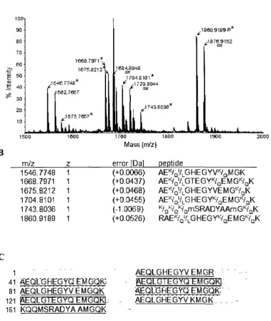
1. A simplified version of a Mass spectrometry (MS) experiment 3 2. Basics of peptide ionization and time-of-flight instruments 4
3. The ABC's of peptide sequencing 7 4. Alternative approaches to decrease sample complexity 11
5. Quantitative mass spectrometry 15
Chapter 2
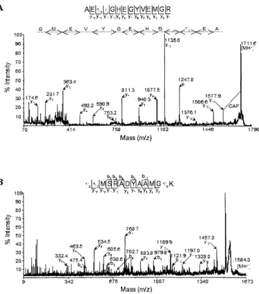
1. Analysis of the 21 kDa protein by matrix-assisted laser desorption-ionization
(MALDI)-tandem mass spectrometry and de novo sequencing 46 2. Analysis of trypsin generated peptides from the 21 kDa protein by
matrix-assisted laser desorption-ionization (MALDI)-tandem mass spectrometry 48
continued D-H 49-51 3. Analysis of GluC generated peptides from the 21 kDa protein by
matrix-assisted laser desorption-ionization (MALDI)-tandem mass spectrometry 53
continued D-J 54-57
Chapter 3
1. Schematics of ICAT strategy 70
2. Reduced PDI labelled with light 12C-ICAT reagent 71
3. Separation of the modified 12C-ICAT-labelled peptides 75
4. MALDI-TOF MS/MS analysis of the a domain active site labelled with light
12C-ICAT reagent displaying tryptophan oxidation 76
5. Quantitation of reduced (light 12C-ICAT-labelled) and auto-oxidized (heavy
13C-ICAT-labelled) PDI following reverse-phase nano-LC separation 78
6. Redox regulation of Cys-295 within the b' domain 80
Chapter 4
1. Phosphorylation of hYVHl 96 2. Analysis of hYVHl N-terminal phosphorylation site 98-99
3. Analysis of hYVHl C-terminal phosphorylation site 102 4. Analysis of hYVHl TP phosphorylation site 104-105 5. Analysis of hYVHl double and triple phosphorylation site mutants 106-107
6. Effect of domain specific constructs on progression through cell cycle 110
LIST OF APPENDICES
Appendix 1
LIST OF ABBREVATIONS ACN ATM ATR BSA CAF CalA CDK CG CID CIP CsA DiFMUP DNA-PK DTT DUSP EGTA ERK ESI HPLC HRP Hsp70 ICAT IgG IMAC iTRAQ JNK LEA m/z MALDI MALDI-TOI MAPK MOAC MS MudPIT NEM OA PAI PBS PCR PDI PSD PTP RNAi SCX SDS-PAGE acetonitrile
ataxia telangiectasia mutated ATM- and Rad3 related bovine serum albumin
chemically assisted fragmentation calyculin A
Cyclin-dependent kinase cytosine-guanine
collision-induced dissociation calf intestinal alkaline phosphatase cyclosporin A
6,8-diFluoro-4-methylumbelliferyl phosphate DNA-dependent protein kinase
dithiotreitol
dual specificity phosphatase
bis(aminoethyl)glycolether-N,N,N',N'-tetraacetic acid Extracellular signal-regulating kinase
electrospray ionization
high pressure liquid chromatography horseradish peroxidase
heat shock protein 70 isotope-coded affinity tags immunoglobulin G
immobilized metal affinity chromatography
isobaric tags for relative and absolute quantification c-Jun NH2 terminal kinase
Late embryogenesis abundant mass-to-charge ratio
matrix assisted laser desorption/ionization
7 matrix assisted laser desorption/ionization time-of-flight Mitogen activated protein kinase
metal oxide affinity chromatography Mass Spectrometry
multidimensional chromatography N-ethylmaleimide
okadaic acid
protein abundance index phosphate-buffered saline polymerase chain reaction protein disulfide isomerase po st- source-decay
protein tyrosine phosphatase RNA interference
strong cation exchange chromatography
siRNA small interfering RNA TAP tandem affinity purification
TBST tris-buffered saline containing 0.1% Tween-20 TOF time-of-flight
CHAPTER 1
Recent advances in mass spectrometry have brought this technique to the forefront
of discovery based system biology research. Since a major theme of this dissertation is
the application of different mass spectrometry based approaches to answer significant
biological questions, an overview of several different mass spectrometry based
approaches will be discussed, including methods employed during this dissertation.
ABC's of mass spectrometry
Mass spectrometry (MS) in recent years has become a fundamental experimental
approach in biochemistry. Mass spectrometers by definition measure mass-to-charge ratio
(m/z) of ionized analytes (Figure 1) [1]. A mass spectrometer is characterized by three
major components, an ion source, a mass analyzer and an ion detector (Figure 2A).
Originally, MS was restricted to measurement and analysis of small and
thermostable molecules, because large organic molecules (such as peptides and proteins)
would undergo extensive fragmentation and decomposition upon vaporization into the gas
phase [2]. This was overcome by the development of "soft" ionization methods (where
ions are created with low internal energy and thus undergo limited fragmentation), such
as electrospray ionization (ESI) [2] and matrix assisted laser desorption/ionization
(MALDI) [3]. These two ionization methods are fundamentally different. In the case of
ESI, a liquid sample is injected through a needle and the surface of the emerging liquid is
charged by an electric field, thus forcing it into a fine spray of charged droplets [2],
thereby producing a continuous beam of ions (Figure 2B) [4]. In the case of MALDI, the
gas phase ions are produced from a dry co-crystallized liquid sample with an organic
matrix [3]. The energy from the laser is deposited into the matrix causing rapid thermal
Biological ^
peptide fingerprint
Sample Proteolytic Peptide Mass preparationAJWL, digestion ^ 0 \ N ionization spectrometry
question ^s^b *^- MS MS
Protein Peptide sample sample
Figure 1. A simplified version of a Mass spectrometry (MS) experiment
A
Ion source
Mass analyzer
Detector
B
LaserSpray needle
Peptide ions Peptide ions
D
Laser
Of
_r
Sample plate
TOF
Reflector
E
Laser
"X
Br
-CD-Sample T 0 Fl
plate Collision
cell
TOF,
Reflector
Figure 2. Basics of peptide ionization and time-of-flight instruments
producing packets of ions (Figure 2C) [4]. Another common distinction for peptide
analysis is that mainly singly charged ions are generated upon MALDI, whereas ESI
generates multiply charged species [5].
There are currently four basic types of mass analyzers for the study of peptides
and proteins; time-of-flight (TOF), ion trap, quadrupole and Fourier transform
ion-cyclotron (FTICR) [1, 6, 7]. The separation of ions in these mass analyzers is based on
flight time (TOF instruments), stability (quadrupoles) and resonance frequency (ion traps,
orbitraps, FTICR instruments) [5]. Each analyzer has a characteristic behaviour with
regards to resolving power, sensitivity and dynamic range. In a quest to make the
analyzers better and more versatile, a new generation of 'hybrid' instruments have been
introduced. These instruments containing a parallel configuration of more than one
analyzer, can be either identical, as in case of TOF-TOF, or different as for examples in
quadrupole TOF, linear ion trap-orbitrap, etc. [1, 5, 6, 8]. The detector, the last but not the
least important part of the mass spectrometer, monitors voltage pulses and converts these
into a signal. This signal represents an ion intensity for a particular m/z and is displayed
as mass spectra [7]. As the work described in this dissertation was performed on
MALDI-TOF and MALDI-MALDI-TOF/MALDI-TOF mass spectrometers, only these instruments will be
described in further details.
In the TOF instrument, the m/z is determined from flight times of peptides
through a tube of specified length under vacuum. This measures the mass of intact
peptides, which have been generated from intact proteins by enzymatic proteolysis prior
to analysis. The major difference between the TOF (Figure 2D) and
MALDI-TOF/TOF instrument (Figure 2E) is in how the information regarding the amino acid
primary sequence analysis can be obtained by a process called post-source-decay (PSD),
which is based on metastable fragmentation. PSD can be observed when the reflecting
electrical field (reflectron) is placed at the end of the flight tube [9]. Upon selection of the
parental ion [MH]+ for analysis, a MALDI-PSD spectrum is obtained as a stitch of
manually acquired spectra corresponding to different portions of the peptide under
investigation (from the parental ion to the immonium ions) in a form of windows. Each
window (correlates to a part of the spectra) corresponds to gradual changes in reflectron
voltages [9]. A more common method to induce fragmentation for MS/MS analysis is
collision-induced dissociation (CID), which is based on multiple collisions between a
peptide under study with inert gas atoms [10]. The MALDI TOF-TOF instrument
contains a collision cell between two TOF sections (Figure 2E). Upon selection of the
parental ion [MH]+ the entire MS/MS spectrum is automatically acquired.
The MS/MS spectrum of a peptide precursor ion allows the assignment of an
amino acid sequence. As the amide bond is the lowest energy bond in peptides, this is
where most of the fragmentations will occur, thus generating y-ions (C-terminal fragment
ions) and b-ions (N-terminal fragment ions) (Figure 3A) [7]. Additionally, internal
fragments due to double cleavage of peptide bond may also be observed [7].
Mass spectrometry and biological questions
Biological mass spectrometry mainly focuses on the complex world of proteins.
This diversity is not only due to the fact that a particular gene can give rise to multiple
distinct proteins as a result of alternative splicing or that some proteins can differ in a few
amino acids due to sequence polymorphisms, but it is also a reflection of the vast array of
A
H2N
' 3 - i O!
R,
ni
' 4 - 1
R<i
N H
• y5
o
H N
' 5 n
o!
R, 1 ^N•-!H be.^J
iff
H^
o;
\ >
Rj
i
N H
- y3
RB
OH
O
0 o
g^-R^
H 0" 6 o
-HN
f
T
Sulfonated N-tenniims (+136 amu)
ol
• N '
H
R, l 1
i
o\
HQ!
1 iN-R
3jH
R4 ! H
\r
o|
o|
^ R5 1 y2 RB OH OFigure 3. The ABC's of peptide sequencing
glycosylation and ubiquitination just to mention a few. The changes in post-translational
modifications can be either analyzed conceptually for the entire proteome (proteomics) or
for a single protein. This will be described later.
With the advancement of the instrumentation and analysis software, one could
now answer questions like what proteins are present within the yeast 80S ribosome [11],
what dynamic changes in the complex composition does the pre-60S ribosome undergo
[12] and what proteins are residents in which mammalian organelle [13]? These are a just
few examples of the many proteomics studies reported in the last ten years. One
limitation of this technology is the under-representation of low abundance proteins.
Furthermore, as MS-based identifications rely on the detection of peptides, additional
factors must be considered including that some peptides might be too small or too large to
be detected, not all peptides are ionized efficiently and some might saturate the detector
[14]. In order to lessen experimental bias due to the presence and ionization of high
abundance peptides, a selected reaction monitoring (SRM)-based MS has been recently
implemented [15-17]. In this experimental approach, the parameters for MS/MS data
acquisition are pre-set, so that the instrument will only record three to five unique
proteotypic peptides for each protein, if these correspond to calculated y-ion series under
two main charge states. Furthermore, in a multiple reaction monitoring (MRM)-based
MS, multiple peptides can be targeted in a single experiment by repeatedly cycling
through a list of SRM-ion pairs associated with a set of specific retention times [18].
One of the major forces enabling proteomic analyses was the completion of entire
genome sequences for numerous organisms. These allow comparison between acquired
MS and MS/MS spectra with theoretically calculated fragment sizes from the in-silico
commonly used to digest proteins into peptide are sequence specific, for example trypsin
only cleaves C-terminal to lysine and arginine residues.
However, to date, not all of the organisms have their genomes sequenced and not
all of the proteins sequences have been deposited into databases. As the annotations of
MS/MS spectra containing y- and b-ions without prior sequence information are
challenging, simplifications of the spectra can be achieved by chemical derivatization (so
called chemically assisted fragmentation, CAF) [19, 20]. This method allowing de novo
sequencing is based on attachment of sulfonic acid to the N-terminus of each peptide
(+136 Da), which promotes an efficient charge-site initiated cleavage of peptide amide
bonds, and thus enables selective detection of a single series of fragment ions, the
C-terminal y-ions (Figure 3B) [19, 20]. A step to protect lysine side chains from sulfo nation
includes conversion to homoarginine (+42 Da) [20].
One of the organisms for which the genome has not been sequenced yet is the
brine shrimp Artemia franciscana. In collaboration with Dr. A. Warner, Department of
Biological Sciences, University of Windsor we employed a MS-based approach to
identify a novel protein from this species. A heat-stable protein was purified from
encysted A. franciscana embryos and identified from peptide fragments by CAF
derivatization combined with de novo sequencing. This study will be discussed in detail
in Chapter 2 of this thesis.
Biological mass spectrometry and sample complexity
The complexity of peptide samples can be decreased by an array of separation
techniques. Peptides generated from proteins digested in solution (the "bottom-up"
where the first separation is based on charge (strong cation exchanger) and the second on
hydrophobicity (reverse-phase) prior to direct injection into the electrospray based
MS-instruments (Figure 4A) [11,21].
Alternatively, instead of decreasing complexity of peptide samples by
chromatographic separation, proteins can be first separated on one-dimensional (ID) or
two-dimensional (2D) SDS-PAGE prior to enzymatic digestion into peptides. This allows
identification by either MALDI or ESI-MS (Figure 4B).
An elegant protocol based on the use of tandem affinity purification (TAP) tags
allows co-purification of interacting partners at substoichiometric levels [22]. Originally
this method included an SDS-PAGE separation of the protein components of the
immunoprecipitated complex and the collection of unique protein bands allowing separate
proteolytic digests to be performed. Overall, this decreased the complexity of analyzed
peptide samples [22]. The classical route for identifying interacting partners involves
over-expression systems, that usually employ genetically engineered fusion proteins
containing one tag, for example Flag-tag, Myc-tag, etc. These tags allow affinity
purification and/or detection, when a suitable antibody directed against endogenous
protein is not available, or not suitable for these applications. However, the ability to
detect fusion proteins at low concentrations in a complex mixture in these one-tag
over-expression systems is limited. This problem was circumvented by the introduction of two
different tags (the TAP-tag system) separated by a protease recognition sequence (tev) in
frame with the protein for which the goal is to identify interacting partners (Figure 4C).
After expression of this fusion protein, the first purification step is based on the affinity
between IgG -binding domain (the first tag) and the IgG beads. Upon removal of
A
Proteolytic
1 digestion ^^\
Protein sample
Peptide sample
2D chromatographic separation of peptide
• * I sex I-HRP k = > Spray needle
Mass spectrometry
I Pepbde ions
B
Protein sample
Proteolytic
digestion „., ^
Peptide sample
->• Mass spearomefry
SDS-PAGE
C D i d s *
Target Protein A
protean i XEV protease cleavage sfte
C akno dulin binding peptide
Contaminating ^ proteins • ^ ^ ^ f
4S ^ »
i
TEA proteas< cleai,Ta^e j |
* ^ ^ S l * *
i .
. Associated ^ proteins
*:'
• 1st affinity
§|f§§ Column
IjlilgG
HHill beads
2U affinity
j . ' .
Column (Ca:~)
Calmodulin beads
I
EDTAFigure 4. Alternative approaches to decrease sample complexity
cleavage with tev protease. Thus the IgG binding domain remains attached to the beads.
The second purification step is based on affinity of a calmodulin binding peptide (the
second tag) to calmodulin beads in a presence of calcium. Upon removal of any
carry-over non-specific proteins and tev protease by washing steps, the carry-over-expressed protein
and its interacting partners are eluted from the beads in the presence of chelating agents,
such as EGTA, and then separated by SDS-PAGE (Figure 4D). The protocols described
so far allow for the identification of proteins as cellular components and members of
protein-protein interaction networks. However, if the biological questions are rephrased to
monitor differences in protein abundance or their post-translational modifications under
various conditions, labelling protocols that allow quantification and direct comparison of
two different states in the same analysis are required.
Mass spectrometry as a quantitative tool
Mass spectrometry is not inherently a quantitative technique due to the fact that
different molecular species (e.g. different peptide sequences) will have diverse ionization
efficiencies. Several quantitation methods for MS-based approaches have been devised
and can be divided into two types, absolute and relative [14]. The general principle
behind these methods takes advantage of the fact that chemical and physical properties of
isotopically labelled compounds are identical to their naturally occurring counterparts,
with the exception of their mass [23, 24]. Thus stable isotopic nuclei (carbon-13,
nitrogen-15, oxygen-18 and deuterium) will not alter ionization efficiency and can be
either incorporated into peptides and function as relative references, or included into
The relative labelling can be either accomplished by metabolic incorporation (i.e. stable
isotope labelling by amino acids in cell culture, SILAC [23]), by post-harvest chemical
derivatization method (i.e. isotope-coded affinity tag, ICAT, [24], isobaric tags for
relative and absolute quantification, iTRAQ, [26]), or by incubation with H2180 during
enzymatic digestion [27].
Absolute quantification is based on 'spiking' known quantities of chemically
synthesized stable isotope labelled peptide as internal standards (absolute quantitation,
termed AQUA, [28]). The major limitation of the AQUA method is the requirement for
chemical synthesis of isotope containing peptide equivalents to proteolytic digest of all
generated peptides, and thus is restricted to a small number of pre-selected proteins [14].
With regards to the relative quantification strategies, the least commonly utilized
approach is based on proteolytic labelling with oxygen-18 during proteolytic digestion.
As this can result in a 2 or 4 Da shift, due to incorporation of either one or two oxygen-18
atoms into the carboxyl terminus of each generated peptide, this may complicate the
quantification. Also, if only one oxygen is replaced it might cause an overlap of isotopic
envelopes [14, 29], as each MS-detected signal corresponding to a peptide consists of
isotope cluster of peaks [7]. These peaks are separated by 1 Da, caused by the 1%
probability in nature of each carbon-12 atom to be a carbon-13 isotope [7]. In addition,
further complications exist in that variability is added as a result of slow or incomplete
incorporation of the oxygen-18 for highly acidic peptides [30].
Mass spectrometry and isotopic in vivo labelling
Biologists often ask whether a certain protein is present within a particular
to address the quantitative part of their questions, so called "metabolic" or "in vivo"
labelling methods were developed, which are based on the incorporation of stable
isotopes containing essential amino acids into each synthesized protein. The original
proof-of-principle SILAC report used incorporation of an isotopically labelled deuterated
form of leucine (2H3-Leu) in sample A ('heavy') which was compared to sample B, in
which cells were grown in a media containing normal leucine (H-Leu, 'light') [23]. After
experimental treatments (in this case myoblast differentiation), the protein populations
were harvested and mixed together, separated on SDS-PAGE and proteolytically digested
(Figure 5A). As the exact ratio between labelled and unlabelled proteins/peptides is not
affected, quantitation (based on peak intensity) can be performed either on the peptide
mass or fragment spectrum as the fragmentation patterns are identical and no
complications are introduced due to label (3 Da difference). The advantages of this
labelling technique include the ability to label many proteins in a cell (as more than half
of tryptic peptides contain leucine), there is no difference in labelling efficiencies
between samples (cells are exposed to isotope containing amino acids for a prolonged
period of time, five days in culture, prior to treatment), and the labelling is consistent
among numerous peptides from the same protein and is sequence specific [23]. However,
problems were encountered upon reverse phase separation of peptides due to both, the
deuterium label and the fact that not all peptides generated by trypsin digest were
labelled. In the second generation of labelled amino acids, the stable isotopes were
carbon-13 and nitrogen-15, and the label was incorporated into lysine and arginine,
instead of leucine. As trypsin (peptide bond cleavage at arginine and lysine residues) is
the most commonly used protease in MS-based experiments, when isotopicaly labelled
B
Sample A Sample BSample A
:heaw"
Sample B 'light'
2HrLeu H-Leu
(!3C653NrLvs) ( " C g ^ j - L v s )
(!3C6!5Nj-Arg) (^C^NYArg)
X , \ . / / Protein purification (optional)
1
Combine, prior to protease digest
1
Peptide fractionation (optional)I
Quantification by MS
\
'I
Separately denature and reduce proteins
1" 1
Chemical Modification Chemical Modification by i3C-ICAT reagent by 32C-ICAT reagent
'heavy Iliajht:
s y
Combine prior to protease digest
I
Cation exchange column
1
Avidin affinity column
1"
Quantification by MS
Figure 5. Quantitative mass spectrometry
(A) A schematic presentation of the SILAC method, the "metabolic" or "in vivo" labelling
are labelled with an 8 Da mass shift difference for Lys and a 10 Da for Arg containing
peptides [31].
An alternative approach to the TAP-tag system for identification of
protein-protein interactions involves the use of untagged endogenous protein-proteins, at their normal
cellular levels, in a quantitative immunoprecipitation combined with a knockdown screen
[32]. This method, QUICK is a hybrid of SILAC, RNA interference (RNAi) and
coimmunoprecipitation [32]. As RNAi allows specific depletion of a protein, its
interacting partners can be identified upon immunoprecipitation with endogenous
antibody, elution, digestion and MS-based comparison of peptide ratios between 'light'
cells, where the protein under study has been down-regulated and non-treated 'heavy'
cells. After the immunoprecipitation and digestion, MS-analysis detects peptides at a
higher intensity corresponding to the protein under study and its interacting partners in
the 'heavy' labelled sample (non-treated), as these are decreased or absent from RNAi
treated cells; whereas peptides corresponding to contaminating proteins are observed at
similar levels [32].
Another promising adaptation of SILAC is in a screen for identification of novel
DNA-protein interactions [33]. Protein levels are often a reflection of the regulation of
transcription as the case of the presence/absence of specific transcription factors
recognition sites within promoter regions (upstream of transcription start site). DNA
methylation also affects transcript levels. For example, in mammals in approximately
80% of all genes the promoter regions contain stretches of cytosine-guanine (CG)
nucleotides, so called CpG islands [34]. The cytosine within CpG dinucleotides is
susceptible to methylation, an event that potentially may disrupt the ability of a
promoter regions of numerous genes have also been shown to display tissue specific
patterns as well as a correlation with many malignancies [34]. In order to identify
signalling networks mis-regulated due to methylation of a specific CpG island,
biotinylated double stranded synthetic oligonucleotide containing either fully methylated
cytosine residues within a CpG island or a corresponding non-methylated sequence were
separately immobilized onto streptavidin beads [33]. Affinity chromatography was
performed separately for DNA containing the methylated CpG island and 'heavy' (2
H4-Lys) labelled nuclear extract and for DNA containing the non-methylated CpG island and
'light' labelled (H-Lys) nuclear extract. Afterwards samples are combined and
DNA-protein complexes were eluted by restriction enzyme digestion, as the nucleotides were
engineered to contain endonuclease recognition site. Thereafter protein complexes are
either immediately digested with a protease in-solution or proteins can be first separated
on SDS-PAGE and then in-gel digested. Lysine containing tryptic peptides originating
from DNA methylation specific interacting proteins were detected at a higher peak
intensity for the 2H4-form (mass shift difference of 4 Da), while DNA affinity purified
non-specific proteins displayed 1:1 ratio between isotopic forms [33].
Mass spectrometry and non-isotopic quantitation
In the case where samples have not been or cannot be isotopically labelled,
approaches for label-free quantitative proteomic have been developed. These methods
allow for extraction of quantitative data from mass spectra, and require implementation of
computational algorithms. One of the label-free quantitative methods is based on
normalization of the number of observed peptides to the number of observable peptides
was later modified to exponentially modified PAI (emPIA) method, as it was observed,
that the number of peptides observed has a logarithmic relationship to the amount of
protein within that sample [37]. Furthermore, this method was recently further enhanced
by the inclusion of correction factors that allow the measurement of absolute protein
concentration per cell from the proportionality between protein abundance and the
number of observed peptides [38]. The second label-free quantitative method is only
applicable in highly reproducible liquid-chromatography interfaced MS-instruments. This
method is based on extracted ion current, which is the area under the curve, when signal
intensity as peptide elutes from chromatographic column, is plotted versus time [39]. The
extracted ion current is linearly related to the amount of particular peptide (extracted ion
currents for different peptides of the same protein are very different) under the same
experimental conditions [14]. Samples under comparative analysis are processed and
analyzed separately, one after another. It is noted that slight changes in sample processing
or the presence of contaminants, such detergents, interfere with quantification [14].
Applications of this label-free quantitative analysis include, but are not limited to, protein
correlation profiling, an adaptation to provide protein abundance profiles upon
fractionation procedures [40] and detection of changes in the cell surface proteome [41].
Mass spectrometry and isotopic in vitro labelling
A second category of isotopic labelling strategies is based on chemically targeting
reactive groups on side chains of amino acids or peptide termini. Some of these labelling
approaches are performed at the peptide level and some at the protein level, depending
upon the experimental questions. As the above-described in vivo labelling techniques are
incorporation of labels allowing quantitative proteome analysis can be performed on a
wide variety of samples, including patient specimens.
The most popular of the isobaric mass tags, iTRAQ allows concurrent analysis of
up to eight different samples and consists of an amine-specific reactive group, a reporter
mass group (variable mass of 114-117 Da (for 4-plex assays) or 113-121 Da (for 8-plex
assays)) and a balancer group (variable mass of 191-188 Da or 192-184 Da) [26]. The
iTRAQ label introduces a highly basic group at the N-terminus of peptides and lysine side
chains, and during MS/MS the reporter groups are released as singly charged ions of
masses at 114, 115, 116 and 117 m/z (in a 4-plex experiment) thus allowing
quantification based on their ratios.
Recently introduced (by Applied Biosystems) the non-isobaric, amine based
isotope-coded reagent, mTRAQ, allows concurrent quantification of three samples, as
addition of 13C and 15N containing tags as precursors that result in a mass shift of 0, 4 and
8 Da for arginine and 0, 8 and 16 Da for lysine tryptic peptides [29].
Another class of labelling reagents has specificity towards sulfhydryl groups, a
reduced form of the side-chain in cysteine residues. These isotope-coded affinity tags
(ICAT) contain a thiol-specific reactive group (iodoacetamide) and a biotin affinity tag,
separated by an isotopically coded linker [24]. This quantitative labelling technique was
designed in order to simplify the complex peptide pool prior to MS-analysis, while
maintaining high proteome coverage. Cysteine was chosen because it is a rare, but
functionally essential amino acid, with most proteins containing at least one cysteine.
magnitude (Figure 5B) [24]. The major caveat of this method is that the representation of
proteins that lack cysteine residue is compromised. Based on current estimates this
represents approximately 5% of human proteins [42]. In the first generation of ICAT
reagents the difference between 'light' and 'heavy' was 8 Da, due to incorporation of
eight deuterium atoms into the 'heavy' tag [24]. There were a few shortcomings related to
the design of these labelling tags, such as a shift in the elution of corresponding peptides
from reverse phase chromatography due to deuterium modification and ambiguities in
assigning peptides with more than one cysteine. Note, a difference of 16 Da may
represent the presence of either two labelled cysteine residues or oxidation of methionine.
An additional difficulty was the possibility of undesired fragmentation at the biotin tag,
instead of fragmentation at the peptide backbone during MS/MS. This led to development
of a second generation of ICAT reagents [43], which are now commercially available.
These ICAT reagents include an acid-cleavable linker group between the biotin moiety
and sulfhydryl reactive isotope tag, while the 'heavy' reagent contains nine 13C atoms
instead of eight deuterium atoms [43, 44]. These reagents allow the comparison of the
content and relative abundance between two samples, by first reducing the side chains of
cysteinyl residues in a sample representing one cell state followed by derivatization with
Tight' ICAT reagent. In the second sample, equivalent groups are first reduced and then
derivatized with 'heavy' ICAT reagent [24]. After labelling, the two samples were
combined and proteolytically digested; thereafter the resulting peptides were separated by
multidimensional chromatography, including isolation of tagged peptides by
avidin-affinity. Quantification is based on comparison of the relative signal intensities (ratios)
between peptide ions pairs of identical sequence, with a mass difference due to
allowing an accurate measure of the relative abundance of the peptide (hence the protein)
[24]. The application of ICAT-labelling to study changes in protein abundance at the
entire proteome level ranges from analysis of differences in microsomal fractions
between naive and differentiated human myeloid leukemia cells [45], in tracheal tissue of
cystic fibrosis patients [43], and serum of hepatocellular carcinoma patients [46], just to
mention a few from a long list.
The thiol group of cysteine residue is very susceptible to oxidation by reactive
oxygen species or reactive nitrogen species [47]. Furthermore, thiol groups (-SH) are
essential for the function of many proteins, in that oxidation (in a form of disulfide, -S-S)
may increase or decrease a protein's activity, thus affecting their function. ICAT reagents
have provided an invaluable tool to identify which cysteines are susceptible to oxidation
(for example after exposure to hydrogen peroxide) and for the quantification of the
amount of oxidized cysteine residues [48, 49]. This procedure is based on the premise that
oxidized cysteine residues are not susceptible to ICAT derivatization, when samples are
labelled under non-reducing conditions. Thus normal sample would be labelled with
Tight' ICAT reagent, and the experimental sample exposed to oxidant stress, would be
labelled with 'heavy' ICAT reagent, thus the decrease in specific labelling of
oxidant-sensitive cysteine thiols can be quantified from the ratio of ICAT reagents.
One of key redox modulators that contain a CysXXCys motif within two active
sites is protein disulfide isomerase (PDI). In collaboration with Dr. B. Mutus, Department
of Chemistry and Biochemistry, University of Windsor, ICAT labelling was utilized to
identify and quantitate redox sensitive cysteines on PDI after auto-oxidation. This study is
In addition to identification of redox-sensitive thiols, the ICAT in combination
with labelling has been adapted to trap differential thiols, in a method called OxICAT
[50]. The OxICAT method is based on sequential labelling of the same sample; first free
thiols are labelled with Tight' ICAT reagent after protein(s) denaturation and afterwards
disulfides are disrupted by strong reducing agent, thus allowing for these thiols to be
labelled with 'heavy' ICAT reagent. Cysteines that were originally present as thiols, will
be detected only as Tight' ICAT labelled; whereas cysteines which were originally
oxidized, will be identified by the 'heavy' ICAT modification. This will result in a 9 Da
shift if the peptide contains one cysteine and if more cysteines, multiples thereof. If
cysteines are at redox equilibrium, then both forms (Tight' and 'heavy' ICAT labelled)
for the same peptide would be detected. This approach also provides a novel method to
monitor the kinetics of thiol oxidation by trapping oxidation intermediates within a
purified protein (for example upon oxidative stress, thus activating or inactivating the
protein) [50].
Thiols can also be modified by either S-nitrosylation or S-glutathiolation [51]. The
MS-based approach to identify Cys-SNO modifications, SNO-Site Identification method
SNOSID, is essentially an adaptation of biotin-switch assay [52, 53]. First, all free thiols
in the protein mixture are methylated, then Cys-SNO are selectively reduced (by
ascorbate), thus generating a new unmodified thiols, at which position biotin will be
attached through a disulfide bond (upon addition of
N-[6-(biotinamido)hexyl]-3'-(2'-pyridyldithio)propionamide, biotin-HPDP). At this time, proteins are proteolytically
digested, biotin-tagged peptides are isolated by avidin-affinity, and subsequently released
ICAT technology, it serves as an example that alternative methods are available to study
cysteine modifications.
Mass spectrometry andpost-translational modifications
So far, MS-based approaches designed to identify novel proteins and quantitative
differences among samples have been described. Nevertheless, biological functions of
most proteins are regulated through post-translational modifications. The ability to
unambiguously identify these post-secondary modifications (and their location), further
highlights the power of MS. Identification is achieved from MS/MS spectra analysis, as
the observed fragmentation pattern is altered due to the mass shift introduced by the
modification. The acquired spectra can be compared to a predicted fragmentation pattern
(generated by open-source software) including defined modifications introduced at a
specific amino acid. For example, acetylation on lysine residue introduces a mass shift
difference of +42 Da of the parent ion [MH]+ compared to unmodified peptide in MS
spectra (prior to fragmentation), while the position of modified lysine can be assigned by
+42 Da shift detected in MS/MS spectra at the position of this residue. In the case of
methylation, the extent of mass shifts introduces depends on the number of methyl groups
present, i.e. +14 Da for methylation, +28 Da for dimethylation and +42 Da for
trimethylation. Ubiquitination plays an important role in protein stability, as ubiquitinated
proteins are targeted for degradation [54]. The MS-identification of lysine residues
originally modified by ubiquitination is based on detection of ubiquitin remnants, in a
form of a covalently attached glycine-glycine dipeptide, the signature peptide (mass shift
difference at the lysine residue of 114.1 Da), after trypsin proteolysis; sustained by
lysines) [55]. Protein modifications by carbohydrates (O-linked and N-linked
glycosylation) have been increasingly recognized as biochemical alterations associated
with malignant transformations and tumorigenesis [56]. Due to the incredible variability
in the glycan structures, and complexity of these modifications, methodologies applied to
their identification and characterization are beyond the scope of this overview.
Mass spectrometry and identification of phosphorylation sites
Last, but not least, phosphorylation is one of the most important regulatory
modifications, affecting a broad range of biological functions and processes.
Phosphorylation occurs most commonly on serine residues (86.4%), followed by
threonine residues (11.8%) and rarely on tyrosine residues (1.8%) (values are based on in
vivo phosphoproteome analysis) [57]. Other amino acids that can be phosphorylated are
histidine and glutamic acid [42]. Phosphorylation is detected as a modification of the
peptide by a +80 Da shift in the MS spectra in comparison to the non-phosphorylated
peptide and typical dominant -98 Da and -80 Da fragments detected in the MS/MS
spectra due to neutral losses (the mass of H3PO4, and HPO3 respectively, derived from
phosphoserine and phoshothreonine). In the case of phosphotyrosine, a specific
immonium ion (an internal fragment containing just a single side chain of an amino acid)
is detected at 216.02 m/z [58].
Due to the dynamic nature of phosphorylation, phosphorylated proteins are often
present at low levels, thus the fraction of phosphorylated peptides will be at a much lower
concentration compared to their non-phosphorylated counter-parts (high abundance
peptides cause ion suppression during ionization process) [59]. In addition to low
problems can be circumvented by techniques that promote enrichment of
phosphopeptides.
Originally, three methods were described for the identification of phosphopeptides
from a complex protein mixture. The first one included replacement of phosphate groups
by ethanedithiol, followed by the addition of a biotin tag, the latter allowing
phosphopeptide affinity purification [60]. A limitation of this method includes
complications that arise during the derivatization process and the inability to detect
phosphotyrosine residues, as only phosphoserine and phosphothreonine undergo
(3-elimination [61]. The second method was based on a chemistry intensive six-step
derivatization/purification protocol for tryptic peptides, where the capture of
phosphopeptides was accomplished through a reversible covalent linkage to a solid
support [62]. The third method employs phosphopeptide enrichment from whole cell
lysates by immobilized metal affinity chromatography (IMAC) on peptides derivatized by
esterification of carboxylate groups [63]. IMAC has become one of the most commonly
used strategies for phosphopeptide enrichment [5] and will be described in further details
later.
In addition to IMAC, numerous techniques have recently been developed to enrich
for phosphopeptides. For example, strong cation exchange chromatography (SCX-based
strategy), which separates peptides according to ionic charge, allows enrichment of
tryptic phosphopeptides from unmodified peptides (singly charged species elute before
multiply charges ones) [64]. At low pH (2.7) only lysine, arginine, histidine and the
amino terminus are charged with a net charge state of+2 on non-phosphorylated peptide,
phosphopeptide [64]. The major caveats of this enrichment technique include the fact that
it is limited to studying trypsin generated proteolytic fragments as well as having an
inability to detect phosphopeptides containing a single histidine residue [65]. The
presence of histidine within a phosphopeptide alters the net charge to +2, the same as
non-phosphopeptides, thus effecting the elution properties of this phosphopeptide from
the SCX-column and hindering detection [65]. Nevertheless, this phospho-enrichment
protocol is powerful and has allowed the phosphoproteomic analysis of the developing
mouse brain [65]. Furthermore, in combination with other phosphopeptide enrichment
methods, SCX-based strategies have been implemented as the first fractionation step in
numerous studies, including the effect of epidermal growth factor induced simulation in
HeLa cells [57], profiling of mitotic phosphorylation [66] and analyses of the
phosphoproteome in mouse liver [67], and Drosophila melanogaster embryos [68].
A motif-specific phosphopeptide enrichment method has also been described that
is useful for characterizing specific kinase targets. The method is based on the
precipitation of phosphopeptides with Ba2+ in the presence of acetone under varying pH
[69]. For example, Ba2+-based precipitation in combination with pH dependent
fractionation and MudPIT separation of peptides was used to analyze nuclear extracts
from HeLa cells. When the identified phosphopeptides were analyzed in a pH dependent
manner, at pH 3.5, 86% of the phosphopeptides were unique and the sequence adjacent to
the phosphorylated residue contained acidic amino acids; at pH 4.6 70% and at pH 8.0
56% of the phosphopeptides were unique. Ba2+-based precipitation at pH 4.6 and 8.0
displayed an increased preference for proline residues C-terminal to the phosphorylation
site. Overall, the pattern of Ba2+ precipitation of phosphopeptides at each pH coincided
As phosphorylation at tyrosine residue ranges between 0.05%> (as determined by
phosphoamino acid analysis [70]) to 1.8% (as determined by phosphoproteome analysis
[57]) of the total cellular phosphorylation, selective enrichment for phosphotyrosines
containing phosphopeptides is necessary when the quest is to identify tyrosine
phosphorylation sites. The method of choice to selectively enrich for these
phosphopeptides is based on peptide affinity purification with an anti-phosphotyrosine
antibody. The immuno-affinity phosphopeptide strategy was first applied to profile
analysis of oncogenic Src (tyrosine kinase) down-stream targets and comparative analysis
of two cancer cell lines derived from anaplastic large cell lymphomas [71]. The same
experimental approach was utilized to study the phosphotyrosine proteome in mouse
brain [72].
In addition to IMAC, a metal oxide affinity chromatography (MOAC) based
method, which utilizes Zr02 and Ti02, has been shown to selectively bind
phosphopeptides [73-75]. The limitation of this enrichment method is similar to IMAC
(discussed below), i.e. the possibility of retention of acidic non-phosphopeptides [74, 75].
When a phosphopeptide standard mixture was isotopically labelled with iTRAQ (to allow
direct comparison) and phosphopeptides were enriched by either Ti02 or Fe3+-based
affinity and then mixed together prior to MS-analysis, comparable performance between
these two technique was observed [58]. Furthermore, when phosphopeptides from
biological sample were SILAC labelled (to quantitate changes in phosphorylation after
EGF stimulation), phosphotyrosine containing proteins were immuno-precipitated with an
anti-phosphotyrosine antibody, protein complexes were separated on a gel and after in-gel
Immobilized metal ions, such as Fe and Al bind phosphoproteins and
phosphopeptides with high specificity [76, 77]. Furthermore, after enrichment of
phosphopeptides on either Fe3+ and Ga3+ loaded agarose, phosphorylated residues can be
identified by MS [78]. This experimental protocol is the basis of commercially available
PHOS-Select resin (from Sigma-Aldrich). The major limitations of this enrichment
technique include binding of non-specific peptides and variability in the retention of
phosphopeptides [63, 79]. IMAC enrichment of phosphopeptides has been employed to
identify human mitotic spindle phosphoproteins [80], in the phosphoproteome analysis of
a-factor arrested Saccharomyces cerevisiae [81] and phosphoproteins in
Schizosaccharomyces pombe [82].
Mass spectrometry and dual specificity phosphatases
Dual specificity phosphatases (DUSPs) are able to remove a phosphate group
from phosphoserine, phosphothreonine and phosphotyrosine residues [83, 84]. This very
heterogenous sub-group is a subfamily of the protein tyrosine phosphatase (PTP)
superfamily [83]. All PTP family members (including DUSPs) contain the invariant
catalytic sequence C(X)sR and the hydrolysis of phosphorylated substrate involves
formation of a stable phosphoryl-intermediate [85, 86]. The DUSPs are the most diverse
group in terms of substrate specificity, and include the following families; MKPs
(mitogen-activated protein kinase phosphatases), Slingshots, PRLs (phosphatase of
regenerating liver), Cdcl4s, PTENs (phosphatase and tensin homologue deleted on
The atypical DUSPs, a group of 16 members, are generally small, under 250
amino acids, and lack specific mitogen activated protein kinase (MAPK) targeting motifs
even through a few family members can dephosphorylate at least one of the MAPKs
(either Erk, Jnk or p38). These includes DUSP3 (VHR) [87], DUSP14 [88], DUSP18
[89], DUSP19 [90], DUSP22 [91], DUSP23 [92] and DUSP26 [93]. Other members
display a broad range of substrate specificities. For example, laforin dephosphorylates
complex carbohydrates [94] and DUSP11 interacts with RNA [95]. If all of this
heterogeneity and variability within this sub-class of DUSPs is taken into consideration,
we can expect that hYVHl (also known as DUSP 12), the DUSP of interest in the
Vacratsis lab and the only atypical DUSP that contains a zinc-binding domain, to have a
unique role.
YVH1 orthologues are extremely conserved throughout evolution; present in
species as diverse as Plasmodium falciparum [96] to humans signifying YVH1
orthologues as regulators of conserved cellular functions. Interestingly, YVH1
orthologues are present as single gene copies even in advanced mammals such as humans,
suggesting that there has been evolutionary pressure against yvhl gene duplication or
deletion.
Recent projects from our laboratory have used several MS-based approaches to
discover novel hYVHl functions. A MS-based approach was utilized to identify the
cytoprotective heat shock protein 70 (Hsp70) as the first known hYVHl interacting
protein [97]. Phosphatase assays showed that hYVHl is a heat stable enzyme whose
activity during oxidative conditions can be modestly increased in the presence of Hsp70.
phosphatase and can prevent both thermal and oxidative stress induced cell death. This
cytoprotective property requires catalytic competence and the zinc binding domain [97].
Our laboratory also used differential thiol labelling and MS to reveal that the
cysteines in the zinc binding domain serve a redox buffer role, allowing hYVHl to
function under oxidative conditions known to inactivate cysteine based phosphatases
[98]. In the present study, outlined in Chapter 4, a MS-based approach was also utilized to
identify phosphorylation sites within hYVHl. Not only are these the first phosphorylation
sites reported for any YVH1 orthologues, but this result led us to discover that hYVHl
Objectives of this Thesis
Recent developments in the mass spectrometry field have facilitated the
applicability of this experimental tool to study a variety of biological questions. These
include, but are not limited to, identification of proteins or post-translational
modifications, and quantitative analysis of proteins after modification of peptides with
isotopically labelled tags. This dissertation is comprised of three research chapters that
examine various applications of MS-based methods to study biological questions.
Chapter 2 investigates the identity of a 21 kDa protein from encysted embryos of
brine shrimp Artemia franciscana as a group 1 Late embryogenesis abundant protein by a
MS-based de novo sequencing approach.
Chapter 3 examines the redox status of cysteine residues within recombinant
human protein disulfide isomerase after auto-oxidation conditions employing the
isotope-coded affinity tag (ICAT) technology and mass spectrometry.
Chapter 4 identifies three in vivo phosphorylation sites on hYVHl using a
combination of a phosphopeptide enrichment protocol and mass spectrometry. This study
also examines the effect of overexpressing hYVHl and phosphospecific mutants on
hYVHl localization and progression through the cell cycle. In addition, phenotypes


![Figure 4. Alternative approaches to decrease sample complexity (A) Peptide fractionation by the MudPIT, multidimensional chromatography method [11, 21]](https://thumb-us.123doks.com/thumbv2/123dok_us/1452876.1177992/29.601.105.475.88.503/alternative-approaches-decrease-complexity-peptide-fractionation-multidimensional-chromatography.webp)