DOI: 10.1534/genetics.109.107508
Centromere Replication Timing Determines Different Forms of
Genomic Instability in
Saccharomyces cerevisiae
Checkpoint
Mutants During Replication Stress
Wenyi Feng,* Jeff Bachant,
†David Collingwood,
‡M. K. Raghuraman* and Bonita J. Brewer*
,1*Department of Genome Sciences and‡Department of Mathematics, University of Washington, Seattle, Washington 98195 and†Department of Cell Biology and Neuroscience, University of California, Riverside, California 92521
Manuscript received July 17, 2009 Accepted for publication September 23, 2009
ABSTRACT
Yeast replication checkpoint mutants lose viability following transient exposure to hydroxyurea, a replication-impeding drug. In an effort to understand the basis for this lethality, we discovered that different events are responsible for inviability in checkpoint-deficient cells harboring mutations in the mec1andrad53genes. By monitoring genomewide replication dynamics of cells exposed to hydroxyurea, we show that cells with a checkpoint deficient allele ofRAD53,rad53K227A, fail to duplicate centromeres. Following removal of the drug, however,rad53K227Acells recover substantial DNA replication, including replication through centromeres. Despite this recovery, the rad53K227A mutant fails to achieve bio-rientation of sister centromeres during recovery from hydroxyurea, leading to secondary activation of the spindle assembly checkpoint (SAC), aneuploidy, and lethal chromosome segregation errors. We dem-onstrate that cell lethality from this segregation defect could be partially remedied by reinforcing bipolar attachment. In contrast, cells with themec1-1 sml1-1mutations suffer from severely impaired replication resumption upon removal of hydroxyurea.mec1-1 sml1-1cells can, however, duplicate at least some of their centromeres and achieve bipolar attachment, leading to abortive segregation and fragmentation of in-completely replicated chromosomes. Our results highlight the importance of replicating yeast centromeres early and reveal different mechanisms of cell death due to differences in replication fork progression.
C
ENTROMERES have long been known to be one of the earliest regions of the budding yeast genome to replicate during S phase (McCarrolland Fangman 1988). However, the biological significance of early replication of centromeres remains speculative, partly owing to the lack of mutants showing altered or delayed timing of centromere replication. During our investi-gation of chromosome replication dynamics during nucleotide shortage brought upon by the treatment with hydroxyurea (HU), we discovered that problems with centromere replication can lead to fundamentally different forms of genome instability.The two mutations that exhibit interesting centro-mere replication phenotypes are in the genes encoding two essential protein kinases, Mec1 and Rad53, which play pivotal roles in the cellular response to DNA damaging agents as well as in cell cycle arrest in response to HU (Branzei and Foiani 2006; Tourriere and Pasero 2007). Mutations in the kinase domains of Mec1 and Rad53 render the proteins checkpoint
de-ficient and cause the cells carrying such mutations to be hypersensitive to HU. Because Mec1 is an upstream effector of Rad53 in the replication checkpoint pathway, checkpoint-deficient alleles of the two genes are thought to lead to similar phenotypes in response to replication impediments. Whenrad53cells encounter HU during S phase, they fail to slow the temporal program of origin firing, expose large regions of single-stranded DNA (ssDNA) at effectively all origins, and elongate their spindles, a phenotype that indicates that the cells are at-tempting premature chromosome partitioning (Allen et al. 1994; Weinert et al. 1994; Desany et al. 1998; Santocanaleand Diffley1998; Sogoet al.2002; Feng et al.2006). Similarly,mec1cells have also been shown to initiate precocious segregation of unreplicated chromo-somes upon exposure to HU (Weinert et al. 1994; Sanchezet al.1996). Even after the removal of HU, both mec1 and rad53 cells show considerable reduction in their ability to produce progeny. However, the reason for inviability after HU exposure is ill defined. We reasoned that understanding the molecular basis of cell death would help elucidate the role of checkpoint control in DNA replication and cell cycle regulation and in the maintenance of genome integrity.
Previous reports indicated that following transient exposure to HU, rad53 checkpoint-deficient cells are Supporting information is available online athttp://www.genetics.org/
cgi/content/full/genetics.109.107508/DC1.
1Corresponding author:Department of Genome Sciences, University of Washington, Box 355065 Foege Bldg., Room S041, 1705 NE Pacific St., Seattle, WA 98195. E-mail: [email protected]
unable to complete DNA replication, which in turn was thought to constitute the primary reason for the loss of viability (Desany et al. 1998; Lopes et al. 2001). This conclusion was corroborated by the observation that delaying premature chromosome segregation by inhib-iting microtubule assembly (via nocodazole treatment) following HU exposure was unable to improve the viability ofrad53cells (Desanyet al. 1998). Together, these studies suggested that incomplete replication rather than premature chromosome segregationper se was the major reason for loss of viability in bothrad53 and mec1 cells, following treatment with HU. By flow cytometric measurements it appeared thatrad53 cells were able to replicate slowly a significant amount of genomic DNA following transient (30–60 min) expo-sure to HU, but that these chromosomes did not enter pulse field gels (Desany et al. 1998). The authors proposed that the DNA synthesized byrad53cells must contain gaps, branches, or other structures that retard the mobility of chromosomal DNA in the pulse field gels. The presence of putative abnormal replication intermediates was later revealed by two-dimensional gel electrophoresis and by electron microscopy (Lopeset al. 2001; Sogoet al.2002).
Intrigued by the results of Desany et al. (1998), we decided to ask where in the genome DNA replication occurred during recovery from HU—in particular, whether forks established during HU treatment were able to resume or whether replication was occurring from unfired origins. By addressing this question, we hoped to identify what precisely was the defect, if any, in DNA replication during the recovery phase from HU in cells lacking the checkpoint function.
In this study, we examine the cellular responses of two checkpoint-deficient mutants,mec1-1 sml1-1and rad53K227A(Weinertet al.1994; Sogoet al.2002), upon exposure to HU during the initiation of S phase and during recovery after HU is removed. We chose themec1-1 sml1-1 strain for our studies because it is in an isogenic background (A364a) as therad53K227Amutant that we have previously studied. The mec1-1allele is phenotypi-cally identical to amec1null allele and it requires the sml1-1mutation for viability (Zhaoet al.1998; Basraiet al. 1999). In contrast, therad53K227Amutation (Fenget al. 2006) is not lethal and the strain harboring this mutation does not contain the sml1-1 allele (data not shown). Through the usage of these specific mutations in the checkpoint pathway, we observed that themec1-1 sml1-1 andrad53K227Acells, upon exposure to HU, lose viability through distinct mechanisms that arise from differences in centromere replication in the two mutants. We further demonstrate that the extent to which the two mutants recover DNA synthesis upon the removal of HU also differs. Our study reveals the importance of early replica-tion of centromeres and underscores the involvement of the replication checkpoint pathway in the establishment of chromosome biorientation.
MATERIALS AND METHODS
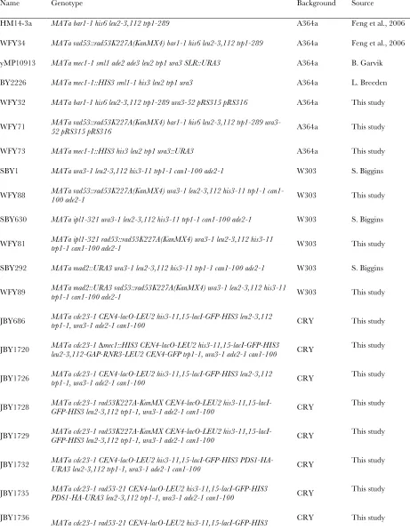
Yeast strains and media:Yeast strains used in this study are listed insupporting information,Table S2. Cells were grown at 30°in synthetic complete medium unless otherwise indicated.
a-Factor was used at 200 nmforbar1strains and 3mmforBAR1 strains. Pronase was used at 25mg/ml and 300mg/ml forbar1 and BAR1strains, respectively, to remove a-factor from the culture medium. HU was added at 200 mmand nocodazole was used at 15mg/ml.
Measurement of cell viability by colony formation assay:
Cells synchronized in G1 bya-factor treatment were released into medium containing 200 mmHU or in combination with 15mg/ml nocodazole as detailed in the main text. Aliquots of 100ml were removed for serial dilutions in ice-cold minimal medium lacking a nitrogen source. The cell suspension of the appropriate concentration was sonicated briefly before plat-ing aliquots in triplicate on solid medium. The plates were incubated at 30° for 2–3 days before colonies were counted and analyzed.
Contour-clamped homogeneous electric field gel electro-phoresis and Southern blotting: Contour-clamped homoge-neous electric field (CHEF) gel analysis was performed as described previously (vanBrabantet al.2001). Electropho-resis was conducted at 14°for 25 hr with a switch time ramped from 60 to 120 sec at 200 V. The Chr IX probe was amplified from genomic DNA with primers of the following sequences: forward, 59-CTATGACGAGGGCGAAGAAG-39; reverse, 59-ATT TCACAGGGCCAGACACG-39. Southern blotting was performed according to standard procedures.
Flow cytometry:Cells were collected and mixed with 0.1% NaN3, followed by fixing with 70% ethanol. Flow cytometry was performed using standard procedures after staining the cells with Sytox Green (Molecular Probes) and the data were analyzed with CellQuest software (Becton-Dickinson).
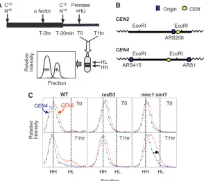
Chromosome biorientation assay:Cells carrying the CEN4-GFP tag and thecdc23-1mutation were blocked in G1 with
a-factor and released into YPD medium at 35°(to block cells at the metaphase/anaphase transition by inactivating the Cdc23 protein) in the presence or absence of 200 mmHU. After 1 hr, cells were allowed to recover in fresh medium lacking HU at 35°. At the indicated times, samples were evaluated for the percentage of cells with two separated CEN4-GFP foci, in-dicative of successful biorientation at thecdc23block.
Indirect end labeling: Yeast chromosomes embedded in agarose gels were prepared as previously described (van Brabantet al.2001). A detailed protocol for in-gel restriction digestion can be found at http://fangman-brewer.genetics. washington.edu/fork-D.html. The digested agarose plugs were then placed in wells of a 0.4% agarose gel (without ethidium bromide) and electrophoresed at 1 V/cm for 26 hr at room temperature. Standard Southern blotting techniques were used. The primer sequences for the DSF2 probe are: DSF2-F, 59-TTTCATTACCTCCAACGCCA-39; DSF2-R, 59-TTTCGGACCTTGTTTCATGT-39. The TRP1 probe was iso-lated as a HindIII fragment from plasmid pTA-DIR (M. K. Raghuraman, unpublished results.).
Genomic ssDNA mapping: ssDNA analysis on rad53 and mec1 sml1cells was performed as previously described (Feng et al.2006, 2007).
(forrad53andmec1 sml1cells) or 2 [for wild-type (WT) cells] hr, cells were filtered to remove HU and allowed to recover in fresh medium without HU. Samples were collected at the times indicated in the figure legends and genomic DNA was extracted followed by fractionation after ultracentrifu-gation in a CsCl density gradient. An aliquot of each gradi-ent fraction was slot blotted, followed by hybridization with centromere DNA probes (CEN2andCEN4) or a whole--genomic DNA probe to identify the unreplicated (HH) DNA and replicated (HL) DNA. TheCEN2fragment was amplified from genomic DNA with the following primers: forward, 59-TAGTCTATCAGCCTCCGAAG-39; reverse, 59-GTA GGT GCCAGTTGAATAGC-39. TheCEN4fragment was excised as a XhoI-SpeI restriction fragment from the plasmid YCpG. EDamtd (M. K. Raghuraman, unpublished results). For microarray analysis, those fractions containing HH and HL DNA identified by slot blotting and hybridization with a genomic DNA probe were pooled, respectively. The HH and HL DNA for each timed sample were differentially labeled with cyanine (Cy3 or Cy5)-conjugated dUTP (Perkin Elmer) as described by the Brown laboratory (http://cmgm.stanford.edu/ pbrown/protocols/4_genomic.html) followed by purification through a Sephadex G-50 column and ethanol precipitation for microarray hybridization.
Microarray hybridization and analysis:SeeFile S2.
RESULTS
Extent of replication fork progression in HU inferred from ssDNA profiles: Treatment of check-point-deficientrad53K227A cells (henceforth referred to asrad53cells) with HU during a synchronized S phase generates regions of ssDNA restricted to the immediate vicinity of virtually all replication origins in the yeast genome (Fenget al.2006, 2007). The colocalization of ssDNA with origins suggests that replication forks are unable to progress very far into adjacent regions before they become arrested. To assess whether the accumula-tion of ssDNA at origins is unique to therad53mutation, we analyzed amec1-1 sml1-1 (henceforth referred to as mec1 sml1) checkpoint deficient mutant under similar conditions.
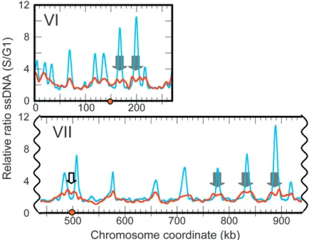
When we releasedmec1 sml1cells from a G1 arrest into HU for 1 hr, ssDNA also appeared at virtually all rep-lication origins in the genome. However the peaks of ssDNA in mec1 sml1 cells, though significantly above background, were lower, broader, and often ‘‘split’’ in comparison to those in rad53K227A cells (data from Fenget al.2006; Figure 1 andFigure S1), suggesting that replication forks inmec1 sml1cells were able to proceed further than were forks inrad53cells. While the majority of centromeres in rad53 cells are located in troughs between ssDNA peaks, inmec1 sml1cells, ssDNA extended into some centromeric regions (such as Chr VII, Figure 1, open block arrow). This observation prompted us to investigate whether centromere duplication during HU treatment differs between these two mutants.
Centromere replication in HU: We examined the extent of centromere replication inrad53andmec1 sml1 cells during exposure to HU using a density transfer
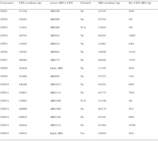
nearest ARSs (using a compiled origin list by Niedus-zynski, http://www.oridb.org) only two centromeres (CEN3 andCEN12) are closer to an ARS thanCEN2is (Table S1). While we have not done an exhaustive test to ask whether any of the other centromeres was repli-cated inrad53cells in HU, we note that we have never been able to detect any shift toward hybrid DNA for the genome as a whole or even for efficient, early firing origins such asARS305andARS306(data not shown). We conclude that a very small percentage ofrad53cells are capable of replicating even a single centromere whereas, under the same conditions,mec1 sml1cells can potentially duplicate at least three centromeres (CEN2, -3, and -12).
Two concurrent studies showed that chromosomes remained monopolarly tethered to one of the spindle pole bodies (SPBs) during premature spindle extension in HU-treated checkpoint mutants (Krishnan et al. 2004; Bachant et al.2005). It was proposed that, for rad53cells, it was due to the lack of centromere rep-lication in HU that the chromosomes fail to biorient and the cells undergo precocious spindle extension (Bachantet al.2005). This hypothesis is consistent with our finding thatrad53 cells failed to replicate centro-meres during exposure to HU.
Replication resumption after HU exposure: To ask whether the difference in the extent of replication by rad53andmec1 sml1cells in the presence of HU has an impact on how cells recover once HU is removed, we measured the progress of replication after removal of HU. Density transfers (Figure 3A) were identical to those described earlier with the exception that after cells were exposed to HU for 1 hr, they were filtered and
allowed to recover in fresh light medium without HU for up to 2 hr. The extent of genomic DNA replication was measured by hybridization with total genomic DNA (Figure 3B). We found that rad53 cells were able to recover from HU and replicate the bulk of the genome (Figure 3B), including the centromeres (Figure 3C;
Figure S3), by 120 min. In contrast, mec1 sml1 cells accumulated far less HL DNA (,50%) during the same recovery period (Figure 3, B and C). These observations suggest that mec1 sml1 cells were less capable of re-suming synthesis after exposure to HU thanrad53cells. We hybridized HH and HL DNA from these recovery samples to microarrays to investigate which parts of the genome were able to resume replication. By comparing samples that had recovered for 45 min after a 1-hr HU exposure to a sample of WT control cells that were also given a 45-min recovery period following a 2-hr expo-sure to HU, we observed that the temporal order of replication in the rad53 and mec1 sml1 mutants was altered: WT cells simply resumed a normal pattern of chromosome replication; however, for both mec1 sml1 andrad53cells accumulation of HL DNA in normally early replicating regions was delayed relative to later replicating regions, resulting in rather flat replication profiles (Figure 3C, Figure S3). These results are consistent with the notion that in the mutants, replica-tion forks become ‘‘damaged’’ and are less likely to resume replication after HU exposure. Therefore, the cells would need to rely on initiation of new forks from any unused late/dormant origins to complete genome replication.
High level of chromosome breakage after HU exposure inmec1 sml1but notrad53 cells:As budding Figure2.—Differences in centromere replica-tion in mec1 sml1 (WFY73) andrad53 (WFY34) cells in HU. HM14-3a cells were used as a WT con-trol. (A) Experimental scheme for density transfer and subsequent analyses. Cells were propagated in isotopically dense (13C and 15N) medium for at
least eight generations prior to G1 arrest. At 30 min prior to release into S phase (time ¼ T-30 min), cells were switched to light medium (12C and14N) to allow nucleotide pools to
yeast cells assemble intranuclear mitotic spindles during S phase, it has been suggested that early replication of centromeres might be crucial for ensuring bipolar attachment of the chromosomes during S phase and thus their proper segregation later in mitosis (Tanaka et al. 2005). Ifmec1 sml1 but notrad53cells were able to duplicate at least some of their centromeres during HU exposure, thenmec1 sml1cells would have a much higher probability of at least a few chromosomes achieving bipolar attachment during HU treatment and thereby reducing overall spindle extension. How-ever, becausemec1 sml1cells have difficulty in complet-ing replication after HU is removed, we would predict that they might be prone to chromosome breakage during recovery from HU.
We used CHEF gel electrophoresis to examine the chromosomes in cells following transient exposure to HU. Cells that had been exposed to HU for 1 hr were washed and resuspended in fresh medium without HU and samples were collected every hour for up to 3 hr. Chromosome breakage was assayed by Southern blot-ting using probes located near the ends of specific chromosomes. For all three strains (WT,rad53, andmec1 sml1) chromosomes remained mostly intact for up to 3 hr in HU, withmec1 sml1cells experiencing a slightly elevated level of chromosome IX breakage (Figure 4A). However, upon the removal of HU at 1 hr,mec1 sml1cells showed substantial chromosome breakage, whilerad53
cells were comparable to WT cells (Figure 4A). Similar results were obtained with a chromosome V probe (data not shown).
If the chromosome breakage in mec1 sml1 cells resulted from tension exerted by the spindle on the partially replicated chromosomes, we would expect that blocking spindle extension via nocodazole treatment would prevent or reduce chromosome breakage dur-ing recovery from HU. To test this hypothesis, we released mec1 sml1 cells from the G1 block into me-dium containing HU (‘‘HU’’) or HU and nocodazole (‘‘HU1Noc’’) and, after a 1-hr exposure, allowed them to recover in just fresh medium (‘‘Noc’’) or in the continued presence of nocodazole (‘‘1Noc’’) (Figure 4B). As expected, for the samples recovering in the absence of nocodazole (‘‘Noc’’) most of the genomic DNA was reduced to fragments of a median size of 200 kb (Figure 4B). In contrast, fragmentation was greatly reduced in cells that recovered in the presence of nocodazole (‘‘1Noc’’) leaving chromosomes mostly intact. This result supports our hypothesis that the incompletely replicated chromosomes inmec1 sml1cells break directly or indirectly as a consequence of tension exerted by the spindle. The residual breakage occurring in the presence of nocodazole could be attributed to either incomplete blockage of spindle extension by nocodazole or spontaneous breakage that occurred independently of tension.
Chromosome breakage near the centromere inmec1 cells:If spindle tension on the bioriented centromeres of partially duplicated chromosomes is responsible for some of the breaks in themec1 sml1cells recovering from HU treatment, then breaks should be detected near centromeres such asCEN2but notCEN4becauseCEN2 has a higher probability of being duplicated during HU treatment than CEN4. We employed an indirect end-labeling method to ask whetherin vivobreakage occurs nearCEN2andCEN4.mec1 sml1cells at different stages of HU treatment and recovery was embedded in agarose plugs and genomic DNA was digested in-gel withPstI, which generates 20-kb and 24.6-kb fragments contain-ingCEN2andCEN4, respectively (Figure 4C). Hybrid-izing the Southern blot with probes located near the end of each fragment (DSF2for Chr II andTRP1for Chr IV, Figure 4C) allowed us to detect and map the potential sites of chromosome breakage on each fragment.
We detected two breakage sites in the pericentric region ofCEN2, labeled B1 and B2, located2 kb and 10 kb to the right ofCEN2, respectively (Figure 4, C and D). Breakage at B1 is observed only during recovery; breakage at B2 is detected during HU treatment but increases in frequency during recovery (Figure 4D). The relative percentages of breakage at these sites at 2 hr during recovery are 3.8 and 4.9% for B1 and B2, respectively. These values are consistent with the notion that chromosome breakage occurs at a low rate at any given locus but collectively contribute to the overall level of chromosome breaks. Neither breakage is detected in
a-factor arrested cells, suggesting that replication is required to generate both breaks (Figure 4D). In contrast, no significant sites of breakage nearCEN4were observed using aTRP1probe on the right end of thePstI fragment on Chr IV (Figure 4D). These observations are consistent with our hypothesis that incompletely replicated chro-mosomes in HU-treated mec1 sml1 cells are subject to Figure4.—mec1 sml1but not rad53 cells show chro-mosome breakage during recovery from transient (1 hr) exposure to HU in S phase. (A) Top: CHEF gel and Southern blot (probed with a telomeric-proximal Chr IX probe) of WT (HM14-3a), mec1 sml1 (WFY73), and rad53 (WFY34) cells exposed to HU continuously for up to 3 hr (HU: 1, 2, and 3 hr) or, after 1-hr exposure to HU, allowed to recover for another 3 hr (recovery: 1, 2, and 3 hr). M, yeast chro-mosome markers; 0, a
breakage, and that at least some of this breakage occurs as a direct consequence of bipolar spindle force.
Partial improvement of rad53 cell viability by eliminating reorientation of kinetochore-to-SPB con-nections:Ifrad53cells can resume replication after HU removal rapidly enough to escape chromosome frag-mentation, why do they die after exposure to HU? We propose that at least one type of lethal event inrad53 cells in HU is the precocious segregation of chromo-somes with an unreplicated centromere that can only experience monopolar attachments. Furthermore, if the spindle assembly checkpoint (SAC) is active during the HU-challenged S phase, then attachments to the old spindle will be repeatedly broken as the error correction mechanisms attempt to biorient chromosomes by dis-tributing attachments between the two spindle poles. With randomly oriented, unreplicated centromeres, the genome would undergo a reductional segregation to-ward the two spindle poles.
This proposed scenario is reminiscent of the pheno-type of acdc6mutant that is defective in the initiation of DNA synthesis—cdc6cells undergo reductional mitosis to randomly segregate their intact unreplicated chro-mosomes (Hartwell1976; Buenoand Russell1992; Lianget al.1995; Piattiet al.1995). However, in acdc6 ipl1double mutant (IPL1encodes the yeast homolog of the mammalian Aurora B kinase) (Chanand Botstein 1993; Biggins et al.1999), the chromosomes predom-inantly remain attached to the old spindle pole (Tanaka et al.2002). Consequently, all the chromosomes segre-gate to the daughter cell (Tanakaet al. 2002).
There-fore, we hypothesized that inactivating the Ipl1 kinase to avert the reductional mitosis might improverad53cell viability in HU by increasing the chance that one of the cells would inherit the entire genome and proceed with replication once HU is removed.
A rad53 ipl1 culture was split upon release from
a-factor arrest, with one half incubated at 30° (the permissive temperature for theipl1-321mutation), and the other half at 37°(the nonpermissive temperature). Cells incubated at 37°showed significantly higher levels of viability than did the cells incubated at 30°(Figure 5A). This observation held true even after we accounted for the difference in cell cycle entry kinetics at the two temperatures (Figure 5B). The viability of either single mutant was unaffected by temperature (Figure 5A). These results support our predictions that (1) the inactivation of the Ipl1 kinase would allow at least some of the rad53 cells to inherit a full genome in the presence of HU, and (2) during recovery from HU, rad53cells would be able to finish replicating the bulk of their chromosomes and retain viability. Among the rad53 ipl1 cells that had survived HU exposure, we would expect to find some cells with greater than one genome equivalent of DNA content. We screened 14 colonies ofrad53 ipl1 HU survivors by flow cytometry. Most of these colonies gave rise to cultures that were heterogeneous in DNA content, ranging from one to two genome equivalents, indicative of aneuploidy ( Fig-ure S4).
Because theipl1mutation is defective in the tension-sensing branch of the SAC, it is formally possible that it Figure 5.—Lethality in rad53 cells in HU can be partially rescued by inactivating the Ipl1 ki-nase (A and B) or by the introduc-tion of CEN-ARS plasmids (C and D). The percentage of colony forming units was calculated for each sample by normalizing to the control T0 sample. Error bars represent standard deviations. (A) Effect of theipl1-321 tempera-ture sensitive mutation on viability ofrad53cells released into HU at 30° or 37°. ipl1 (SBY630), rad53 (WFY88), andipl1 rad53(WFY80) cells are shown. (B) The cell viabil-ity ofipl1 rad53cells at 30°and 37°
is the inactivation of the SAC rather than the elimina-tion of the kinetochore-to-pole reorientaelimina-tion that res-cuesrad53lethality in HU. If this postulate were true, then one would expect that the inactivation of the SAC via a mutation in the MAD2 gene would also rescue rad53lethality in HU. We found that the double mutant rad53 mad2Ddid not have elevated viability in HU com-pared to therad53 single mutant (Figure S5). There-fore, we conclude thatipl1mutation rescuesrad53HU sensitivity by forcing all chromosomes to cosegregate with the same pole, thereby preventing reductional segregation of the genome.
Partial restoration of rad53 cell viability in HU by CEN-ARS plasmids: We have estimated from themec1 sml1cells that a minimum of three duplicated centro-meres is sufficient to block precocious spindle exten-sion and random segregation of the genome. Previously, it was demonstrated that the introduction of multiple CEN-ARS plasmids in which a centromere was placed immediately adjacent to an ARS to increase the number of replicated centromeres was able to reduce the level of spindle extension inrad53cells in HU (Bachantet al. 2005). We wanted to ask whether reinforcing bipolar attachment would actually ameliorate the loss of viabil-ity inrad53cells in HU. We tested both mec1 sml1and rad53cells bearing two CEN-ARS plasmids (pRS314 and pRS316) for their ability to form colonies after HU treatment.
Cells grown under selection for the plasmid markers were released from a G1 arrest into selective medium containing HU. Samples were collected during S phase and plated on nonselective medium lacking HU. At the time of transfer the culture was quite heterogeneous with only 30% of the population containing both plas-mids. Therefore we chosenotto select for the presence of the plasmids after cells were plated because we wanted to examine specifically whether the subset of cells that actually contained plasmids were able to improve cell viability during HU exposure, not whether the ensuing plasmid replication and segregation were successful. After 3 days of incubation, colonies were counted to calculate the relative viability of cells over time in HU.
The culture ofrad53cells transformed with the two CEN-ARS plasmids had improved viability (10% at 3 hr in HU) when compared to the control cells without the plasmids (,1% at 3 hr in HU) (Figure 5C). To ensure that the improved survival was not simply due to slowed S phase entry of the transformed cells, we also compared cell viability at times when the two cell cultures had reached similar levels of budding or cell cycle entry. To make this comparison, we plotted the cell viability as a function of the budding indices (Figure 5D). This plot confirms that the viability of cells that contained plasmids was higher than that of control cells at the same cell cycle stage. Moreover, rescue was specific to therad53mutant, as we did not observe any increase in viability ofmec1 sml1cells with the plasmids (Figure 5C).
Thus, we conclude that increasing the number of cen-tromeres inrad53cells that could potentially be replicated in the presence of HU, and thus become bioriented, stabilizes the mitotic spindle and prevents the precocious random segregation of chromosomes with only mono-polar attachment.
rad53 mutants are unable to biorient or separate chromosomes after HU treatment: Our observations thus far are consistent with the hypothesis that rad53 cells suffer from reductional chromosome segregation during HU treatment, but can substantially replicate centromeres following removal of HU. To determine if these replicated centromeres can establish bipolar connections to the spindle, we compared the ability of rad53K227A, rad53-21, and mec1D GAP-RNR3 cells to recover chromosome biorientation following a 1-hr HU treatment;rad53-21is a well-characterized S phase checkpoint defective allele (Allen et al. 1994) and mec1D GAP-RNR3 (where the essential function of MEC1is compensated by the overproduction ofRNR3 under the glyceraldehyde 3-phosphate dehydrogenase promoter, similar to the sml1mutation; Desany et al. 1998) is phenotypically identical tomec1-1 sml1. Acdc23-1 mutation was introduced to block anaphase entry and allow ample time for chromosome biorientation. The centromere on Chr IV was tagged with GFP (CEN4-GFP) to assay biorientation (Goshimaand Yanagida2000); bipolar attachment normally causes sister CENs to split into two distinct GFP foci. Following a 1-hr exposure to HU, we observed that WT and the mec1D GAP-RNR3 mutant exhibited fairly similar kinetics of biorientation. Bothrad53mutants, however, largely failed to achieve biorientation (Figure 6).
Defective biorientation in rad53 cells would be expected to activate the SAC, leading to a Mad2-dependent delay in sister chromatid separation and progression through the metaphase-to-anaphase transi-tion. Indeed, we found the anaphase inhibitor Pds1/ securin (Yamamotoet al.1996) was stabilized inrad53 cells for up to 5 hr after a 1-hr HU exposure. This stabilization was largely alleviated in a rad53 mad2D
DISCUSSION
Why replication-checkpoint-deficient yeast cells die when challenged with a replication impediment is a long-standing question. Do cells die because they cannot complete some aspect of genome replication after the impediment is removed, or is it because, in the absence of the checkpoint, they have executed some critical cell cycle events out of order? Characterizing replication and segregation phenotypes of mutants in two checkpoint kinases, we have been able to distinguish how failure of each of these two processes contributes to distinct types of genome instability (Figure 7).
We found that whenrad53cells encounter HU in S phase they are unable to duplicate their centromeres
and therefore do not achieve the bipolar attachment that normally occurs early in S phase to generate tension within the spindle. As a consequence of premature spindle elongation, unreplicated chromosomes become randomly partitioned to the two spindle poles. Though rad53 cells are capable of nearly completing genomic DNA replication after the removal of HU, we propose that by the time that replication restarts, the window of opportunity to establish bioriented chromosomes has passed and/or that rad53 cells suffer from specific defects that preclude chromosome biorientation (such as structural defects of the centromeres and/or kinet-ochores) or sister chromatid disjunction during re-covery from HU. Thus, rad53 cells are destined for Figure6.—Lack of biori-entation of chromosomes in rad53K227A cells after removal of HU. (A) cdc23 ( JBY686) andcdc23 mec1D
GAP-RNR3 ( JBY1720; two experiments) cells carrying CEN4-GFP were released froma-factor arrest into S phase in YPD media at 35°
random chromosome segregation by both precocious spindle extension during HU treatment and lethal defects in chromosome attachment/separation after HU removal. In contrast, mec1 sml1 cells are largely proficient for bipolar attachment but experience lethal chromosome breaks due to nondisjunction of incom-pletely replicated chromosomes. Although the force exerted by a spindle microtubule (Nicklas 1983; Bloom 2008) is thought to be insufficient to make a dsDNA break (Bensimon et al. 1995), the presence of ssDNA brings spindle force-induced breakage into the realm of possibility. Alternatively, spindle force may induce chromatin unraveling, making regions more accessible to nucleases—an idea consistent with our observation of discrete breakage sites (Figure 4D; see
File S1for a fuller discussion).
While we refer to these differences in replication and cell cycle execution as resulting from the loss of Rad53 or Mec1 checkpoint function, we do not mean to imply that we have necessarily detected distinct functions for Rad53 and Mec1 kinases in the DNA replication checkpoint pathway. It may be the case that Rad53 and Mec1 kinases do have distinct phosphorylation targets, and that the two mutations reflect these differences. However, there are at least two other explanations for the phenotypic differences that we have observed in the two mutants. (1) The different point mutations in the rad53K227Aandmec1-1 sml1alleles may retain different residual levels of checkpoint activation and the specific phenotypes may reflect this difference. (2) Themec1 sml1 strain may have higher level of dNTPs thanrad53due to thesml1mutation. However, we were informed that by chromatin immuoprecipitation of bromodeoxyuridine-labeled DNA coupled with microarray analysis, it was observed thatmec1mutants (mec1-1andmec1-100) consis-tently showed longer tracks or more extensive fork pro-gression than arad53mutant (rad53-11) in the presence
of HU (L. Crabbe, P. Pasero and A. Lengronne, per-sonal communication). Because neither themec1-100nor therad53-11allele requires thesml1mutation for survival, this observation suggests that the difference betweenmec1 sml1and rad53mutants may not be simply attributed to different levels of dNTPs in the cell. Regardless of which explanation is correct, the specific mutations we have used have allowed us to identify an important step in the chromosome duplication cycle that is regulated by the DNA replication checkpoint. Interestingly, another check-point mutant mrc1, which similarly exhibits precocious spindle elongation upon HU treatment, is able to maintain substantial viability (Alcasabas et al. 2001). However, because a detailed study of replication dynamics and chromosome segregation of mrc1 cells under replication stress has not been performed, it is possible thatmrc1cells can replicate some centromeres in HU asmec1 sml1cells do and recover replication asrad53 cells do, thus averting both precocious chromosome segregation and chromosome breakage. Our study underscores the importance for yeast cells to execute linked steps of the cell cycle in their correct order and specifically the importance of replicating centromeres early in S phase to ensure proper spindle assembly and chromosome segregation.
nocodazole affects not only microtubule dynamics but also the behavior of spindle poles. Indeed, we note that even treatingRAD53cells simultaneously with HU and nocodazole during S phase for just half an hour led to a reduction of viability by50% (data not shown). Consistent with this finding is the observation that nocodazole not only prevents spindle pole body (SPB) separation (Yoderet al.2003) but also abolishes cen-tromere clustering near the SPBs during G1, causing them to drift away from the SPBs (Tanakaet al.2002). Recent studies that followed kinetochore recapture after nocodazole treatment demonstrated that yeast mitotic chromosomes have difficulty attaching and biorienting on the spindle when the SPBs are in the unseparated (side by side) configuration, and require the functional Sgo1 protein for correction (Indjeian et al. 2005; Indjeian and Murray 2007). If the un-duplicated kinetochores in rad53 cells in HU were released from the SPBs to which they were first attached, subsequent duplication and recapture by both SPBs might be an inefficient and error-prone event. Therefore, nocodazole’s potential ability to rescuerad53cell lethality in HU may be masked by its negative effect on SPB segregation and subsequent capture and biorientation of chromosomes.
We note that in the population of cells transformed with the CEN-ARS plasmids we achieved only modest increase in viability (from 3% without CEN-ARS plasmids to 12% with two CEN-ARS plasmids at 60 min in HU). While we do not know for certain how many replicated centromeres are required to restrain the spindle in WT cells, we can make estimates on the basis of our replication studies ofrad53 cells in HU. As we were unable to detect any replication ofCEN2, which is500 bp from its closest origin of replication, it would leave only two centromeres as candidates, and one of these is adjacent to an ARS that is rarely used under normal circumstances (ARS308). If we were to assume a success rate for replication of either of these two centromeres of 20% (an estimate based on the sensitivity of our density transfer assay to detect even partial replication of fragments contain-ing the efficient origins ARS305and ARS306), and if two replicated centromeres were sufficient to restrain the spindle to ensure viability upon recovery from HU, then we could expect a survival rate of 4%. Introducing plasmids with similarly spaced centro-meres and ARSs would theoretically improve viability to 20%. The fact that only 30% of the cells in the culture actually had both plasmids reduces the expectation of survival to be ,20%. We predict that introducing origins of replication closer to additional chromosomal centromeres might demonstrate even better rescue of HU sensitivity inrad53 cells than the CEN-ARS plasmids. Likewise, the rescue ofrad53HU sensitivity by ipl1 mutation was only partial presum-ably because many of the cells that survived are
aneuploid (Figure S4) and thus suffer a growth disadvantage (Torreset al.2007).
Although we have shown that rad53K227A cells suffer from reductional mitosis, it is difficult to ascertain how much cell death of rad53 cells can be attributed to segregation defects as opposed to other causes. In fact, we believe that rad53 cells also suffer substantially from replication defects during recovery from HU, despite near completion of DNA synthesis. It is noteworthy that the observation of the substantial ability of rad53K227A cells to replicate DNA, follow-ing transient (1 hr) exposure to HU, is somewhat at odds with a previous study where it was reported that rad53K227A cells are virtually incapable of DNA synthesis after exposure to HU (Lopes et al. 2001). We believe the difference between the two studies lies in the fact that Lopes et al. employed a much more extensive (3 hr) exposure of rad53 cells to HU. We have observed that the ability ofrad53cells to recover from HU decreased as the duration of exposure to HU increased and that the replication occurring during recovery relied even more on new initiation events in cells exposed to HU for longer period than shorter ones, suggesting that replication forks are destabilized in a time-dependent fashion in HU (data not shown). The genomic replication profiles during recovery from HU also showed that the normal replication timing pattern was somewhat reversed in both mutants, suggesting that many replication forks established during HU exposure were defective in resuming synthesis and that some portion of the observed replication during recovery may have orig-inated from new initiation events from previously unfired origins. We are currently investigating the extent to which replication resumption in rad53 and mec1 sml1cells relies on new initiationsvs.resumption from preexisting forks and whether ssDNA gaps are filled or become sites of chromosomal breakage.
assembly. Engaging the checkpoint accomplishes two important replication functions: (1) stabilizing ongoing forks so that they can continue at a slow rate to incorporate nucleotides during HU treatment and can efficiently resume once HU is removed and (2) delaying activation of unfired origins so that the cell can concentrate the few available nucleotides to sites of the earliest activated replication forks—among which are those near a few centromeres.
We thank L. Breeden, B. Garvik, and S. Biggins for providing yeast strains and U. Surana for personal communications at early stages of this work. We are grateful to T. Davis, S. Biggins, B. Byers, J. Sidorova, and the Brewer/Raghuraman lab members for insightful discussions and to the anonymous reviewers for helpful suggestions. We also thank P. Pasero for personal communication of unpublished data. We extend our gratitude to the staff at the Center for Array Technologies in Seattle for microarray hybridization and scanning. This work was supported by the National Institute of General Medical Sciences (NIGMS) grant 18926 (to B.J.B. and M.K.R.). W.F. was supported by a Pathway to Independence award (1K99GM081378-01) from the National Institutes of Health.
LITERATURE CITED
Alcasabas, A. A., A. J. Osborn, J. Bachant, F. Hu, P. J. Werleret al.,
2001 Mrc1 transduces signals of DNA replication stress to acti-vate Rad53. Nat. Cell. Biol.3:958–965.
Allen, J. B., Z. Zhou, W. Siede, E. C. Friedbergand S. J. Elledge,
1994 TheSAD1/RAD53protein kinase controls multiple check-points and DNA damage-induced transcription in yeast. Genes Dev.8:2401–2415.
Bachant, J., S. R. Jessen, S. E. Kavanaugh and C. S. Fielding,
2005 The yeast S phase checkpoint enables replicating chromo-somes to bi-orient and restrain spindle extension during S phase distress. J. Cell Biol.168:999–1012.
Basrai, M. A., V. E. Velculescu, K. W. Kinzler and P. Hieter,
1999 NORF5/HUG1 is a component of the MEC1-mediated checkpoint response to DNA damage and replication arrest in
Saccharomyces cerevisiae.Mol. Cell. Biol.19:7041–7049.
Bensimon, D., A. J. Simon, V. V. Croquette and A. Bensimon,
1995 Stretching DNA with a receding meniscus: experiments and models. Phys. Rev. Lett.74:4754–4757.
Biggins, S., F. F. Severin, N. Bhalla, I. Sassoon, A. A. Hymanet al.,
1999 The conserved protein kinase Ipl1 regulates microtubule binding to kinetochores in budding yeast. Genes Dev.13:532– 544.
Bloom, K. S., 2008 Beyond the code: the mechanical properties of
DNA as they relate to mitosis. Chromosoma117:103–110. Branzei, D., and M. Foiani, 2006 The Rad53 signal transduction
pathway: replication fork stabilization, DNA repair, and adapta-tion. Exp. Cell Res.312:2654–2659.
Bueno, A., and P. Russell, 1992 Dual functions ofCDC6: a yeast
protein required for DNA replication also inhibits nuclear divi-sion. EMBO J.11:2167–2176.
Chan, C. S., and D. Botstein, 1993 Isolation and characterization
of chromosome-gain and increase-in-ploidy mutants in yeast. Genetics135:677–691.
Desany, B. A., A. A. Alcasabas, J. B. Bachantand S. J. Elledge,
1998 Recovery from DNA replicational stress is the essential function of the S-phase checkpoint pathway. Genes Dev. 12: 2956–2970.
Feng, W., D. Collingwood, M. E. Boeck, L. A. Fox, G. M. Alvino
et al., 2006 Genomic mapping of single-stranded DNA in hy-droxyurea-challenged yeasts identifies origins of replication. Nat. Cell Biol.8:148–155.
Feng, W., M. K. Raghuramanand B. J. Brewer, 2007 Mapping
yeast origins of replication via single-stranded DNA detection. Methods41:151–157.
Goshima, G., and M. Yanagida, 2000 Establishing biorientation
oc-curs with precocious separation of the sister kinetochores, but not the arms, in the early spindle of budding yeast. Cell100:619–633. Hartwell, L. H., 1976 Sequential function of gene products
rela-tive to DNA synthesis in the yeast cell cycle. J. Mol. Biol.104: 803–817.
Indjeian, V. B., and A. W. Murray, 2007 Budding yeast mitotic
chromosomes have an intrinsic bias to biorient on the spindle. Curr. Biol.17:1837–1846.
Indjeian, V. B., B. M. Sternand A. W. Murray, 2005 The
centro-meric protein Sgo1 is required to sense lack of tension on mitotic chromosomes. Science307:130–133.
Krishnan, V., S. Nirantar, K. Crasta, A. Y. Chengand U. Surana,
2004 DNA replication checkpoint prevents precocious chromo-some segregation by regulating spindle behavior. Mol. Cell16: 687–700.
Liang, C., M. Weinreichand B. Stillman, 1995 ORC and Cdc6p
interact and determine the frequency of initiation of DNA repli-cation in the genome. Cell81:667–676.
Lopes, M., C. Cotta-Ramusino, A. Pellicioli, G. Liberi, P. Plevani
et al., 2001 The DNA replication checkpoint response stabilizes stalled replication forks. Nature412:557–561.
McCarroll, R. M., and W. L. Fangman, 1988 Time of replication of
yeast centromeres and telomeres. Cell54:505–513.
Nicklas, R. B., 1983 Measurements of the force produced by the
mitotic spindle in anaphase. J. Cell Biol.97:542–548.
Piatti, S., C. Lengauerand K. Nasmyth, 1995 Cdc6 is an unstable
protein whose de novo synthesis in G1 is important for the onset of S phase and for preventing a ‘reductional’ anaphase in the bud-ding yeastSaccharomyces cerevisiae.EMBO J.14:3788–3799. Raghuraman, M. K., E. A. Winzeler, D. Collingwood, S. Hunt, L.
Wodickaet al., 2001 Replication dynamics of the yeast genome.
Science294:115–121.
Sanchez, Y., B. A. Desany, W. J. Jones, Q. Liu, B. Wang et al.,
1996 Regulation ofRAD53by the ATM-like kinasesMEC1and
TEL1in yeast cell cycle checkpoint pathways. Science271:357–360. Santocanale, C., and J. F. Diffley, 1998 A Mec1- and
Rad53-dependent checkpoint controls late-firing origins of DNA repli-cation. Nature395:615–618.
Sogo, J. M., M. Lopesand M. Foiani, 2002 Fork reversal and ssDNA
accumulation at stalled replication forks owing to checkpoint de-fects. Science297:599–602.
Tanaka, K., N. Mukae, H. Dewar, M.vanBreugel, E. K. Jameset al.,
2005 Molecular mechanisms of kinetochore capture by spindle microtubules. Nature434:987–994.
Tanaka, T. U., N. Rachidi, C. Janke, G. Pereira, M. Galovaet al.,
2002 Evidence that the Ipl1-Sli15 (Aurora kinase-INCENP) complex promotes chromosome bi-orientation by altering kinet-ochore-spindle pole connections. Cell108:317–329.
Torres, E. M., T. Sokolsky, C. M. Tucker, L. Y. Chan, M. Boselli
et al., 2007 Effects of aneuploidy on cellular physiology and cell division in haploid yeast. Science317:916–924.
Tourriere, H., and P. Pasero, 2007 Maintenance of fork integrity
at damaged DNA and natural pause sites. DNA Repair (Amst)6: 900–913.
van Brabant, A. J., C. D. Buchanan, E. Charboneau, W. L.
Fangmanand B. J. Brewer, 2001 An origin-deficient yeast
ar-tificial chromosome triggers a cell cycle checkpoint. Mol. Cell7: 705–713.
Weinert, T. A., G. L. Kiserand L. H. Hartwell, 1994 Mitotic
checkpoint genes in budding yeast and the dependence of mitosis on DNA replication and repair. Genes Dev.8:652–665. Yamamoto, A., V. Guacciand D. Koshland, 1996 Pds1p, an
inhib-itor of anaphase in budding yeast, plays a critical role in the APC and checkpoint pathway(s). J. Cell Biol.133:99–110.
Yoder, T. J., C. G. Pearson, K. Bloomand T. N. Davis, 2003 The
Saccharomyces cerevisiaespindle pole body is a dynamic structure. Mol. Biol. Cell14:3494–3505.
Zhao, X., E. G. Mullerand R. Rothstein, 1998 A suppressor of
two essential checkpoint genes identifies a novel protein that negatively affects dNTP pools. Mol. Cell2:329–340.
Supporting Information
http://www.genetics.org/cgi/content/full/genetics.109.107508/DC1
Centromere Replication Timing Determines Different Forms
of Genomic Instability in
Saccharomyces cerevisiae
Checkpoint
Mutants During Replication Stress
Wenyi Feng, Jeff Bachant, David Collingwood, Mosur K. Raghuraman
and Bonita J. Brewer
W. Feng et al. 2 SI
W. Feng et al. 3 SI
B
W. Feng et al. 4 SI
FIGURE S2.–Differences in centromere replication in mec1 and rad53 cells in HU after in vitro fill-in synthesis of ssDNA. (A)
W. Feng et al. 5 SI
W. Feng et al. 6 SI
B
W. Feng et al. 7 SI
W. Feng et al. 8 SI
W. Feng et al. 9 SI
FIGURE S5.—Mitotic progression in rad53 and rad53mad2 mutants during HU recovery. A. WT (JBY649), ∆mad2 (JBY1703), rad53-21 (JBY1701) and rad53-21∆mad2 (JBY1705) strains harboring Pds1-Myc were released from G1 into YPD containing 200 mM HU at 30oC for 1 hr. Cells were then washed into fresh YPD pH 3.9 media lacking HU. Mating pheromone was added to restore G1 arrest following completion of mitosis. Protein samples were prepared at the indicated times and processed for Pds1-Myc immunoblotting. Ponceau staining was used to evaluate equivalency of protein load. B. Densitometry analysis of Pds1-Pds1-Myc immunoblots shown in A. C. WT (JBY1707), ∆mad2 (JBY1714), rad53-21 (JBY444), rad53-21∆mad2 (JBY1718) strains harboring
W. Feng et al.
10 SI
File S1
Supplemental Discussion
The unique response of the rad53 mutant to HU provides us with the opportunity to consider the following questions.
First, if centromeres are not replicated in rad53 cells during HU treatment (resulting in the lack of tension on the spindles), why is
the spindle checkpoint not activated to prevent chromosome partition? It has been shown that cdc6 cells proceed with a
reductional mitosis despite the complete absence of replication (Piatti et al., 1995). Similarly, certain alleles of cdc7 and dbf4
mutants--encoding the catalytic and regulatory subunits, respectively, of the DDK (Dbf4-dependent kinase), an S phase
promoting kinase important for the initiation of DNA replication--were also shown to execute the division of their chromatin
when the initiation of DNA replication is blocked (Toyn et al., 1995). These results suggest that the signal that elicits the
checkpoint response to inhibit nuclear division may require the establishment of replication forks (Piatti et al., 1995). However, in
a population of rad53 cells in HU, most if not all origins have established a fork (as evidenced by the accumulation of stable
stretches of ssDNA). Why, then, can’t rad53 cells activate the spindle checkpoint to inhibit anaphase entry in the presence of HU?
Two recent studies have provided clues to this question. Bachant et al. produced compelling evidence that the premature spindle
extension and nuclear division in rad53 cells upon exposure to HU is not through the execution of a true anaphase entry: many of
the events associated with anaphase initiation were not observed in rad53 cells in HU (Bachant et al., 2005). This result was
corroborated by a concurrent study where mec1 cells were also found to initiate chromosome segregation without anaphase entry
in the presence of HU (Krishnan et al., 2004). Therefore, chromosome partitioning in mec1 and rad53 cells is not a true anaphase
and the spindle checkpoint inhibition may not be able to intercede. It is also possible that the activation of a spindle checkpoint
may require the localization of Ipl1 protein to the freshly duplicated kinetochores. Therefore, when centromere and kinetochore
duplication is absent, Ipl1 may not be recruited to activate the spindle checkpoint.
Second, if rad53 cells can resume replication after HU removal, why are they unable to achieve bipolar attachment and
produce viable offspring? We propose that there exists a small window of time in early S phase for chromosomes to replicate
their centromeres and achieve bipolar attachment. Moreover, in rad53 cells, the lack of early centromere replication in HU also
precludes subsequent bipolar attachment of the chromosomes after HU is removed and centromeres replicated, possibly due to
structural defects associated with the centromeres/kinetochores or sister chromatid disjunction. Nevertheless, we hypothesize
that if premature spindle elongation takes place before centromere duplication, bipolar attachment is inefficient. This hypothesis
is consistent with a previously proposed model in which kinetochore attachment to spindle poles requires dynamic searching by
the microtubules in three dimensional space before being captured by the kinetchores (Mitchison and Kirschner, 1985). An
alternative possibility is that the microtubule dynamics may be attenuated in rad53 cells, making it difficult for the microtubules to
W. Feng et al. 11 SI
up-regulated in mec1 mutant, leading to the unscheduled spindle elongation in the presence of HU (Krishnan et al., 2004). It
would be interesting to determine whether the same mechanistic alterations in microtubule dynamics also occur in rad53 cells. It
is also formally possible that the spindle in rad53 cells in HU suffers damage to the point of irreversible collapse so that even after
HU is removed it can no longer be reestablished. However, though we have not directly investigated this possibility, previous
studies have shown that the spindles formed in either rad53 cells in HU or in cdc6 cells showed rather normal morphology
(Bachant et al., 2005; Tanaka et al., 2002).
Our investigation of cell death in mec1 cells also led to more questions. First, why do mec1 chromosomes break
predominantly during recovery from HU rather than during exposure to HU? It is possible that chromosome breakage due to
persistent tension on the spindle is a time-dependent event. For example, mec1 cells exposed to HU for a prolonged time (up to 3
hrs) also showed increased level of breakage compared to at 1 hr (Fig. 4A), although it is not until HU was removed that the cells
displayed drastically more chromosome breakage. It could well be that it is the very act of DNA replication (during the recovery)
that facilitates the transition of ssDNA strand breaks into chromosome breakage. The finding of B1 is consistent with our
hypothesis that the pericentric region is fragile due to persistent spindle tension. However, it is intriguing that the breakage at this
site is only apparent during recovery from, but not during exposure to HU (Fig. 4D). This observation could be explained by two
mutually non-exclusive explanations. One, chromosome breakage is facilitated by replication resumption after HU was removed.
Two, the level of tension on the spindle might be higher after HU was removed than when it was present. In contrast, B2 was
observed even during exposure to HU but increased in frequency during recovery, suggesting that it might occur through a
different mechanism from the likely tension-induced break at B1. This observation is consistent with the previous observation
that nocodazole treatment does not eliminate all chromosome breaks and supports the notion that there might be breakage that
arises independently of tension. Incidentally, B2 is located between two of the most efficient origins on Chr II, ARS208 and
ARS209 (Fig. 4C). It is possible that when replication forks from these two origins stall as a result of exposure to HU, the stalling
forks also prevent other incoming forks to traverse through and replicate the intervening region, which then becomes persistently
under-replicated and ultimately leads to chromosome breakage.
Second, is the breakage near the centromere a direct consequence of applied force on the chromosome by the spindle
that generates breakage of the phosphodiester bonds? Based on previous calculations of maximum force on a yeast kinetochore
microtubule—47 pN (piconewton) measured in the grasshopper spermatocytes (Nicklas, 1983) and 10 pN in budding yeast
(Bloom, 2008)—it is unlikely that the force from a pair of microtubules per yeast chromosome (10 pN x 2 microtubules = 20 pN)
is enough to break the chromosome, which requires a calculated force of 480 pN (+/-20%) (Bensimon et al., 1995). However, the
estimated force for chromosome breakage was calculated for the breaking of double-stranded DNA (Jannink et al., 1996). In the
W. Feng et al.
12 SI
the chromosome. Therefore, chromosome breakage in these cells should require, in theory, less than half of the force to break
dsDNA, thus placing the spindle force-induced breakage in the realm of possibility. It warrants further biophysical investigation
into the question whether force on the yeast spindle is able to break chromosomes. Nevertheless, we do not posit that the
chromosome breakage in mec1 cells is a direct consequence of the spindle force. In fact, we favor the hypothesis that the spindle
force serves to unravel the chromatin such that it might allow nucleases to cleave the DNA at the centromeric regions based on
the observation that the two break sites detected near CEN2 in mec1 cells were very specific, appearing as discrete bands in the
agarose gel (Fig. 4D). We have verified these breakage sites using other probes on the PstI fragment (data not shown) and in all
instances these sites proved very specific, suggesting an enzymatic action as opposed to mechanical rupture. But we emphasize
that the action of nucleases is facilitated by the spindle force to present the substrate (the chromosomes). Recently it has been
reported that fission yeast Mus81 cleaves DNA at stalled replication forks after HU treatment in the absence of a replication
checkpoint (Froget et al., 2008). It would be interesting to determine whether the budding yeast Mus81 also participates in such
an action at the stalled replication forks. We also cannot rule out the possibility that the cleavage furrow during cytokinesis causes
the chromosome breakage seen in mec1 cells and we are in the process of testing this hypothesis. However, once again, the
discrete nature of the breakage would argue against such a postulate.
Finally, it is interesting that the more extensive fork progression in HU in mec1 cells was accompanied by more
compromised resumption of fork movement after HU removal. In contrast, rad53 cells, while unable to replicate centromeres in
HU, were more capable of resuming DNA synthesis later. We hypothesize that this difference between mec1 and rad53 cells may
reflect differences in the regulation of replication fork components by these two kinases in response to replication impediment. It
is possible that mec1 cells specifically lack a critical component restraining fork progression that is important for subsequent fork
resumption. We plan to determine whether there exists such a component of the replisome that is specifically subject to
regulation by Mec1 but not or to a lesser degree by Rad53 during exposure to HU. Further investigation of what molecular
events take place at the replication forks during HU treatment in these two mutants would no doubt aid our understanding of
how genome integrity is monitored by the replication checkpoint.
Alvino, G. M., Collingwood, D., Murphy, J. M., Delrow, J., Brewer, B. J., and Raghuraman, M. K. (2007). Replication in hydroxyurea: it's a matter of time. Mol Cell Biol 27, 6396-6406.
Bachant, J., Jessen, S. R., Kavanaugh, S. E., and Fielding, C. S. (2005). The yeast S phase checkpoint enables replicating chromosomes to bi-orient and restrain spindle extension during S phase distress. J Cell Biol 168, 999-1012.
Bensimon, D., Simon, A. J., Croquette, V. V., and Bensimon, A. (1995). Stretching DNA with a receding meniscus: Experiments and models. Phys Rev Lett 74, 4754-4757.
W. Feng et al. 13 SI
Feng, W., Raghuraman, M. K., and Brewer, B. J. (2007). Mapping yeast origins of replication via single-stranded DNA detection. Methods 41, 151-157.
Froget, B., Blaisonneau, J., Lambert, S., and Baldacci, G. (2008). Cleavage of stalled forks by fission yeast Mus81/Eme1 in absence of DNA replication checkpoint. Mol Biol Cell 19, 445-456.
Jannink, G., Duplantier, B., and Sikorav, J. L. (1996). Forces on chromosomal DNA during anaphase. Biophys J 71, 451-465.
Krishnan, V., Nirantar, S., Crasta, K., Cheng, A. Y., and Surana, U. (2004). DNA replication checkpoint prevents precocious chromosome segregation by regulating spindle behavior. Mol Cell 16, 687-700.
Mitchison, T. J., and Kirschner, M. W. (1985). Properties of the kinetochore in vitro. II. Microtubule capture and ATP-dependent translocation. J Cell Biol 101, 766-777.
Nicklas, R. B. (1983). Measurements of the force produced by the mitotic spindle in anaphase. J Cell Biol 97, 542-548.
Piatti, S., Lengauer, C., and Nasmyth, K. (1995). Cdc6 is an unstable protein whose de novo synthesis in G1 is important for the onset of S phase and for preventing a 'reductional' anaphase in the budding yeast Saccharomyces cerevisiae. Embo J 14, 3788-3799.
Raghuraman, M. K., Winzeler, E. A., Collingwood, D., Hunt, S., Wodicka, L., Conway, A., Lockhart, D. J., Davis, R. W., Brewer, B. J., and Fangman, W. L. (2001). Replication dynamics of the yeast genome. Science 294, 115-121.
Tanaka, T. U., Rachidi, N., Janke, C., Pereira, G., Galova, M., Schiebel, E., Stark, M. J., and Nasmyth, K. (2002). Evidence that the Ipl1-Sli15 (Aurora kinase-INCENP) complex promotes chromosome bi-orientation by altering kinetochore-spindle pole connections. Cell 108, 317-329.
W. Feng et al.
14 SI
FILE S2
Supplemental Experimental Procedures
Microarray hybridization and analysis
The HH and HL DNA for each timed sample from the density transfer experiments were mixed for co-hybridization and
likewise, the ssDNA-labeled S phase and G1 control samples from the ssDNA mapping experiments were also mixed for
co-hybridization to DNA microarrays (Agilent G4140A) according to the manufacturer’s recommendations. The algorithms used
for analyzing microarray data have been described previously (Alvino et al., 2007; Feng et al., 2007) except that a Lowess
smoothing algorithm instead of Fourier transformation was applied in the current study to avoid an artifact introduced by the
latter at the telomeric regions of a chromosome. Microarray data were processed and analyzed as follows:
1. The output files following Agilent slide scanning and data extraction are edited to remove data corresponding to array spots
flagged by the software as anomalous. Spots corresponding to known sequence repeats are also eliminated. The edited data are
referred to as “Raw data”.
2. Raw data are normalized using the scheme described previously (Alvino et al., 2007). This routine requires input of either one
or two experimentally determined parameters, depending on the type of experiment (%HL vs. ssDNA).
3. Normalized data are smoothed using a Lowess smoothing algorithm. First, a window size w is specified for the overall
smoothing of the profile. The "coordinate set" for a given chromosome is the collection of probe coordinates on the array that lie
on that chromosome. For each coordinate t in the coordinate set, consider the interval I(t)=(t-w/2, t+w/2). Next, determine
the set of all locations in the coordinate set that lie in this interval I(t) and do a weighted regression on the associated data points
using the weight function:
€
wtfunc[x−t, w]= 1−8* Abs(−t+x)
3
w3
3
on the interval I(t). In this way, for each t in the coordinate set we obtain a value L(t,w). The new data set of pairs (t, L(t,w)) is
the Lowess smoothed profile for the given chromosome.
4. Extrema (local maxima and minima) are determined as in (Raghuraman et al., 2001) for the output of either smoothing
W. Feng et al. 15 SI
TABLE S1
The distance between centromeres and their closest potential origin of replication (ARS)
Centromere CEN coordinate (bp) nearest ARS to CEN Checked? ARS coordinate (bp) Dis. (CEN-ARS) (bp)
CEN1 151526 ARS108 No 147197 4329
CEN2 238267 ARS208 No 237762 505
CEN3 114441 ARS308 N/A 114624 183
CEN4 449763 ARS416 No 462565 12802
CEN5 152045 ARS510 No 145661 6384
CEN6 148565 ARS605 No 136030 12535
CEN7 496983 ARS719 No 485046 11937
CEN8 105640 Likely ARS No 111335 5695
CEN9 355684 ARS920 No 357275 1591
CEN10 436360 ARS1015 No 442453 6093
CEN11 439831 ARS1114 No 447775 7944
CEN12 150887 ARS1208 N/A 151168 281
CEN13 268090 ARS1309 No 263179 4911
CEN14 628819 ARS1426 No 635781 6962
CEN15 326644 ARS1513 No 337404 10760
CEN16 556012 Likely ARS Yes 559633 3621
Centromere coordinates were obtained from the Saccharomyces cerevisiae Genome Database (http://yeastgenome.org) and the mid point of each
centromere (column 2) was calculated. The identities of the nearest potential origin to each centromere (column 3) were obtained from oriDB
(http://www.oridb.org) compiled by Conrad Nieduczynski and the mid point of each ARS (column 4) was calculated. The distance between the
W. Feng et al.
16 SI
TABLE S2
Yeast strains used in this study
Name Genotype Background Source
HM14-3a MATa bar1-1 his6 leu2-3,112 trp1-289 A364a Feng et al., 2006
WFY34 MATa rad53::rad53K227A(KanMX4) bar1-1 his6 leu2-3,112 trp1-289 A364a Feng et al., 2006
yMP10913 MATa mec1-1 sml1 ade2 ade3 leu2 trp1 ura3 SLR::URA3 A364a B. Garvik
BY2226 MATa mec1-1::HIS3 sml1-1 his3 leu2 trp1 ura3 A364a L. Breeden
WFY32 MATa bar1-1 his6 leu2-3,112 trp1-289 ura3-52 pRS315 pRS316 A364a This study
WFY71 MATa rad53::rad53K227A(KanMX4) bar1-1 his6 leu2-3,112 trp1-289 ura3-52 pRS315 pRS316 A364a This study
WFY73 MATa mec1-1::HIS3 his3 leu2 trp1 ura3::URA3 A364a This study
SBY1 MATa ura3-1 leu2-3,112 his3-11 trp1-1 can1-100 ade2-1 W303 S. Biggins
WFY88 MATa rad53::rad53K227A(KanMX4) ura3-1 leu2-3,112 his3-11 trp1-1 can1-100 ade2-1 W303 This study
SBY630 MATa ipl1-321 ura3-1 leu2-3,112 his3-11 trp1-1 can1-100 ade2-1 W303 S. Biggins
WFY81 MATa ipl1-321 rad53::rad53K227A(KanMX4) ura3-1 leu2-3,112 his3-11 trp1-1 can1-100 ade2-1 W303 This study
SBY292 MATa mad2::URA3 ura3-1 leu2-3,112 his3-11 trp1-1 can1-100 ade2-1 W303 S. Biggins
WFY89 MATa mad2::URA3 rad53::rad53K227A(KanMX4) ura3-1 leu2-3,112 his3-11 trp1-1 can1-100 ade2-1 W303 This study
JBY686 MATa cdc23-1 CEN4-lacO-LEU2 his3-11,15-lacI-GFP-HIS3 leu2-3,112 trp1-1, ura3-1 ade2-1 can1-100 CRY This study
JBY1720 MATa cdc23-1 leu2-3,112-GAP-RNR3-LEU2 CEN4-GFP trp1-1, ura3-1 ade2-1 can1-100 ∆mec1::HIS3 CEN4-lacO-LEU2 his3-11,15-lacI-GFP-HIS3 CRY This study
JBY1726 MATa cdc23-1 CEN4-lacO-LEU2 his3-11,15-lacI-GFP-HIS3 leu2-3,112 trp1-1, ura3-1 ade2-1 can1-100 CRY This study
JBY1728 MATa cdc23-1 rad53K227A-KanMX CEN4-lacO-LEU2 his3-11,15-lacI-GFP-HIS3 leu2-3,112 trp1-1, ura3-1 ade2-1 can1-100 CRY This study
JBY1729 MATa cdc23-1 rad53K227A-KanMX CEN4-lacO-LEU2 his3-11,15-lacI-GFP-HIS3 leu2-3,112 trp1-1, ura3-1 ade2-1 can1-100 CRY This study
JBY1732 MATa cdc23-1 CEN4-lacO-LEU2 his3-11,15-lacI-GFP-HIS3 PDS1-HA-URA3 leu2-3,112 trp1-1, ura3-1 ade2-1 can1-100 CRY This study
JBY1735 MATa cdc23-1 rad53-21 CEN4-lacO-LEU2 his3-11,15-lacI-GFP-HIS3 PDS1-HA-URA3 leu2-3,112 trp1-1, ura3-1 ade2-1 can1-100 CRY This study
JBY1736
W. Feng et al. 17 SI
PDS1-HA-URA3 leu2-3,112 trp1-1, ura3-1 ade2-1 can1-100
JBY649 MATa PDS1-Myc-LEU2 his3-11,15 leu2-3,112 trp1-1, ura3-1 ade2-1 can1-100 CRY This study
JBY1703 MATa 3,112 trp1-1, ura3-1 ade2-1 can1-100 ∆mad2::URA3 PDS1-Myc-LEU2 his3-11,15-lacI-GFP-HIS3 leu2- CRY This study
JBY1701 MATa rad53-21 PDS1-Myc-LEU2 his3-11,15-lacI-GFP-HIS3 leu2-3,112 trp1-1, ura3-1 ade2-1 can1-100 CRY This study
JBY1705 MATa rad53-21 leu2-3,112 trp1-1, ura3-1 ade2-1 can1-100 ∆mad2::URA3 PDS1-Myc-LEU2 his3-11,15-lacI-GFP-HIS3 CRY This study
JBY1707 MATa trp1-1-lacO-TRP1-LEU2 his3-11,15-lacI-GFP-HIS3 leu2-3,112
ura3-1 ade2-ura3-1 canura3-1-ura3-100
CRY This study
JBY1714 MATa leu2-3,112 ura3-1 ade2-1 can1-100 ∆mad2::URA3 trp1-1-lacO-TRP1-LEU2 his3-11,15-lacI-GFP-HIS3 CRY This study
JBY444 MATa rad53-21 trp1-1-lacO-TRP1-LEU2 his3-11,15-lacI-GFP-HIS3 leu2-3,112 ura3-1 ade2-1 can1-100 CRY This study
JBY1718 MATa rad53-21 GFP-HIS3 leu2-3,112 ura3-1 ade2-1 can1-100 ∆mad2::URA3 trp1-1-lacO-TRP1-LEU2 his3-11,15-lacI- CRY This study