Review
1
Title: Growth Hormone (GH) and Cardiovascular
2
system
3
Diego Caicedo 1, Oscar Díaz 2, Pablo Devesa 3 and Jesús Devesa 4,*
4
1 Department of Angiology and Vascular Surgery, Complejo Hospitalario Universitario de Pontevedra.
5
Spain. email: [email protected]
6
2 Department of Cardiology, Complejo Hospitalario Universitario de Pontevedra. Spain. email:
7
8
3 Research and Development, The Medical Center Foltra; Teo, Spain. email: [email protected]
9
4 Scientific Direction, The Medical Center Foltra; Teo, Spain. email: [email protected] or
10
11
* Correspondence: [email protected] or [email protected]; Tel.: +34-981-802-928
12
Abstract: This review describes the positive effects of growth hormone on the cardiovascular
13
system. We analyze why the vascular endothelium is a real internal secretion gland, whose
14
inflammation is the first step for developing atherosclerosis, as well as the mechanisms by which
15
GH acts on the vascular endothelium improving its dysfunction. We also report how GH acts on
16
coronary arterial disease and heart failure, and on peripheral arterial disease inducing the
17
generation of new collateral vessels able to bypass a major artery occlusion. We include some
18
preliminary data from a trial in which GH or placebo is given to elder people suffering from critical
19
limb ischemia, showing the effects of the hormone on plasma markers of inflammation, and stating
20
that the administration of GH in short periods of time is safe and effective even in diabetic patients.
21
We also analyze how Klotho may have strong relationships with GH, inducing, after being released
22
from the damaged vascular endothelium, the pituitary secretion of GH to repair the damaged tissue.
23
Lastly, we show how GH induces wound healing by increasing the blood flow to the ischemic tissue.
24
In summary, we postulate that short-time GH administration is useful for treating cardiovascular
25
diseases.
26
Keywords: cardiovascular diseases; atherosclerosis; oxidative stress; angiogenesis and
27
arteriogenesis; endothelial dysfunction; growth hormone; IGF-I; wound healing.
28
29
1. Introduction
30
The hGH gene family is composed by two GH genes (GH-N and GH-V), and three placental
31
genes located in the chromosome 17 [1]. It has been considered that the GH-V gene is expressed only
32
in the placenta, although some studies indicated that this gene, or some other GH gene, still
33
unknown, could also be expressed in the human pituitary gland [2,3]. In the case of GH-N, it is
34
already well known that in addition to its pituitary expression, responsible for the actions of the
35
hormone at the endocrine level, the hormone is also expressed in numerous cells and tissues, where
36
it acts in an auto/paracrine manner [4]. Perhaps the heart is an exception to this peripheral expression
37
of GH, as we will see later.
38
The regulation of GH pituitary expression is very complex, since in the last few years the classical
39
knowledge of a positive regulation by GHRH and negative by somatostatin [5], has been changed
40
after the knowledge of a series of factors that are decisively involved in that regulation [6]. This is the
41
case, for instance, of the orexigenic ghrelin, released by the empty stomach, or the postulated
anti-42
senescence factor Klotho, mainly expressed in the kidney, but also in the brain and in the own
43
somatotroph cells where it would act in an auto/paracrine manner for directly regulating GH
44
secretion [7]; in addition, the growth differentiation factor 15 (GDF15), synthesized and released by
45
cardiomyocytes, inhibits GH-induced hepatic expression of IGF-I, therefore inhibiting the IGF-I effect
46
on hypothalamic somatostatin release and the direct negative effect of IGF-I on pituitary
47
somatotrophs, thus acting as a coordinator between cardiac function and body growth or other
IGF-48
I dependent GH effects on the human body [8].
49
Although the regulation of GH expression is not the aim of this review, perhaps the complexity
50
of its regulation would explain the fact that far beyond of the concept that GH is mainly a metabolic
51
hormone responsible for the longitudinal growth of the organism before puberty ends, the hormone
52
exerts many other actions on practically all the organs and tissues in the human body [4], as
53
schematized in Figure 1.
54
55
Figure 1. GH is a pleiotropic hormone acting on many tissues and organs in the human organism.
56
Blue arrows show some of the most important territories in which the hormone produces positive
57
effects. For a better understanding of this schema, see reference [4]. AGs: Adrenal glands.
58
In this review, we will focus on the effects of GH on the cardiovascular system; but before it we
59
will analyze the role of the vascular endothelium as an internal secretion gland, as well as the main
60
pathologies that affect the cardiovascular system, to subsequently assess the effect that GH can play
61
in its treatment.
62
1.1. The role of the vascular endothelium as an internal secretion gland and the effects of GH on it
63
Histologically, the vascular endothelium is a single unicellular layer that covers the internal
64
surface of blood vessels and forms the wall of capillaries. However, despite its simplicity, this layer
65
is very complex in physiological terms, since its location allows it to be able to detect alterations in
66
the hemodynamic forces acting on the vascular wall (shear stress forces), as well as changes in
67
circulating chemical signals, responding to all this by releasing vasoactive compounds, able to act
68
oppositely depending on the signals received. For instance, at the level of hemostasis, the vascular
69
endothelium can produce both anti-hemostatic factors (protein C, prostacyclin PGl2, tissue
70
plasminogen activator, nitric oxide), or factors that favor hemostasis (von Willebrand factor, tissue
71
Factor III, plasminogen activator inhibitor, thromboxane A2). The same occurs with the vascular tone,
72
since vasodilators such as nitric oxide (NO) or prostacyclin (PGl2), and vasoconstrictors such as
73
angiotensin II, endothelin, thromboxane II and superoxide anion, can be released from the vascular
74
endothelium to contribute to vascular homeostasis. Moreover, the vascular endothelium produces
75
many growth factors, such as vascular endothelial growth factor (VEGF), platelet derived growth
76
factor (PDGF), basic fibroblast growth factor (bFGF), and endothelin; but also inhibitory growth
77
factors, as transforming growth factor-ß (TGF-ß). Even, the vascular endothelium can participate in
78
immunological responses by producing interleukins (Il-1, Il-6 and Il-18), tumor necrosis factor-α
(TNF-α), monocyte chemoattractant protein-1 (MCP-1), vascular cell adhesion molecule-1
(VCAM-80
1), intercellular adhesion molecule-1 (ICAM-1) and selectins E and P.
81
Most of these factors act locally by auto/paracrine mechanisms, so that they allow, as stated
82
above, to regulate the vascular homeostasis.
83
In general, the vascular endothelium decreases the vascular tone, inhibits platelet adhesion and
84
aggregation, decreases the activation of the coagulation system, stimulates fibrinolysis, decreases
85
capillary permeability and inhibits the adhesion and migration of neutrophils and
inflammation-86
generating macrophages. Therefore, endothelial dysfunction, a primary event in the development of
87
atherosclerosis, is associated with increases smooth muscle vascular tone with arterial rigidity and
88
elevated intima-media thickness.
89
Interestingly, while there are clear evidences that the GH-receptor (GHR) is expressed in the
90
vascular endothelium [9,10], the possibility exists that GH itself is expressed in this territory, as in
91
vitro studies reflect [11]. In any case, there are not doubts about the fact that GH plays a very
92
important role on vascular endothelium. This statement is supported by early studies carried out in
93
children with GH-deficiency (GHD), in whom GH replacement therapy recovered existing
94
endothelial dysfunction. This is the case, for instance, of children with renal insufficiency; in them,
95
endothelial dysfunction is quite common finding, but GH therapy reverses it [12]. In addition, an
96
improvement of the arterial response to induced vasodilation were observed in GH-deficiency (GHD)
97
adolescents after GH-replacement therapy [13]; or in obese children, in whom obesity negatively
98
affects the secretion of GH and constitutes a risk of developing atherosclerosis prematurely [14].
99
Similar results have been found in GHD adults (AGHD) after receiving replacement therapy [15],
100
suggesting that GH reduces vascular inflammation, therefore reducing the vascular risk. Another
101
study in AGHD patients showed that GH treatment led to a significant decrease in plasma levels of
102
apolipoprotein B (Apo B) and C-reactive protein (CRP), while no changes were observed in IL-6 or
103
on markers of endothelial function; but in all, GH administration decreased the cardiovascular risk
104
in them [16]. AGHD patients show impaired coronary flow reserve, which is improved after receiving
105
treatment with the hormone, suggesting that GH improves microvascular function and then could
106
reduce cardiovascular morbidity and mortality in these AGHD [17]. A more recent study describes
107
that six months of treatment with GH are enough to decrease cardiovascular risk and improve
108
endothelial dysfunction [18].
109
One of the biomarkers for cardiovascular disease is the loss of circulating CD34+ cells [19]; this
110
has been shown to be corrected after one year of GH treatment in AGHD, since the number of these
111
cells increased and endothelial function improved [20]. This agrees with the fact that GH increases
112
the production and release of endothelial progenitor cells (EPC) in non AGHD subjects, which in the
113
vascular endothelium act as a repair cells [21]. Moreover, GH replacement therapy improves
114
fibrinolysis in AGHD patients, most likely by increasing the release of endothelial tissue plasminogen
115
activator as a response to venous occlusion [22]. On the contrary, other studies found that AGHD
116
patients treated with GH showed increased concentrations of E-selectin, indicative of an uncorrected
117
endothelial dysfunction [23]. These authors conclude that the beneficial effect of GH in these patients
118
may be produced by the effects of the hormone on other mechanisms rather than acting on
119
endothelial dysfunction.
120
The effects of GH administration on E-selectin had not been found in previous studies
121
performed in healthy adults and AGHD patients [24]; instead vascular cell adhesion molecule-1
122
(VCAM-1) significantly increased in AGHD patients during GH treatment. Interestingly, serum from
123
healthy patients treated with GH significantly increased the expression of VCAM-1 in cultured
124
umbilical vein endothelial cells, suggesting that GH might act on VCAM-1 expression by an indirect
125
mechanism, most likely related to the modulation of the expression of other circulating factors [24].
126
This might explain the reported negative effects of the hormone when administered to critically ill
127
patients, since VCAM-1 mediates leukocytes extravasation which can lead to multiple organ failure
128
in sepsis [25], although the increased mortality reported was observed with doses of GH quite higher
129
To our knowledge only one study reported no positive effects of GH replacement therapy on
131
the endothelial dysfunction in AGHD patients [23], as only one report indicates that GH does not
132
recover the endothelial impairment present in GHD children [26]. Perhaps, the small number of
133
subjects, or the methodology used, or the time during which these studies were carried out justifies
134
the contradictory results.
135
1.2. GH, IGF-I, Klotho and the vascular endothelium.
136
Klotho was first described in 1997 as a product of a gene involved in the suppression of several
137
aging phenotypes in mouse. Initially, it was thought that Klotho would be implied in a signaling
138
pathway regulating senescence and the severity of diseases related with the process of aging, such as
139
atherosclerosis [27]. In mice, the gene codifies a membrane protein homologue to β-glucosidases,
140
while in humans the gene has been shown to be composed of five exons and is located on
141
chromosome 13q12. The gene suffers a physiological alternative RNA splicing giving origin to two
142
transcripts, one of them being a membrane protein while the other one is secreted and predominates
143
over the former [28].
144
The possible effects of Klotho on the physiology of the human vascular endothelium were first
145
postulated in 1998, indicating that it protects the cardiovascular system by inducing NO endothelial
146
production [29], although their possible mechanisms of action had not yet been clarified [30]. Further
147
studies in mice of the same group demonstrated that secreted Klotho promoted endothelial increase
148
of NO in aorta and arterioles [31], and that adenovirus-mediated Klotho gene delivery to a typical rat
149
model of multiple atherogenic risk (OLETF rat) improved endothelial dysfunction, increased NO
150
production, reduced increased blood pressure and prevented medial hypertrophy, meaning that
151
Klotho was a clear positive regulator of vascular function [32]. This was confirmed in Klotho mutant
152
mice when observing that in these animals the density of blood capillaries was decreased at the tissue
153
level and angiogenesis was impaired, as it was the release of NO from the vascular endothelium [33].
154
These effects have been related to an action of Klotho on oxidative stress, responsible for inducing
155
apoptosis and senescence in vascular cells [34].
156
While studies in animal models indicate a clear role for Klotho on the vascular endothelium,
157
there are still no clear data on the physiological role that this hormone plays in men on the
158
cardiovascular system [35]. In vitro studies demonstrated that Klotho suppress TNF-α-induced
159
expression of ICAM-1 and VCAM-1 in human umbilical vein endothelial cells, as well as the
160
inhibition of eNOS phosphorylation induced by the administration of TNF-α [36], effects consistent
161
with its previously postulated role in the modulation of endothelial inflammation.
162
Klotho is mainly produced in kidneys; however, it seems that it could be expressed also in the
163
vascular endothelium, with the only exception of endothelial cells from human brain [37]. In any case,
164
Klotho is a circulating protein that increases NO production and protects the vascular endothelium
165
[38,39].
166
To analyze the role of Klotho on the vascular endothelium nor is the aim of this review, however
167
since it has been shown that this protein plays a role on pituitary GH secretion [7], we think important
168
to try to establish a relationship between Klotho and GH, given both are effective factors to prevent
169
damage to the vascular endothelium and repair it if a damage exists.
170
Mice that do not express Klotho die early than normal mice showing many symptoms of aging,
171
most of them typical of GHD [7]. Plasma levels of Klotho are low in GHD subjects, and the pituitary
172
somatotrophs of Klotho-deficient mice are hypotrophic [7], suggesting that Klotho exerts a trophic
173
effect on them. Besides this, Klotho-deficient mice are smaller than normal mice, and their
GH-174
producing cells in the pituitary show lesser secretory granules [40]. In addition, Klotho strongly
175
inhibits the negative effects of IGF-I on GH secretion, and increases GH secretion in cultured human
176
GH-secreting adenomas [40].
177
All these data indicate that Klotho is a positive active regulator of GH secretion, both in animal
178
models and in humans. However, it is still unknown how GH and Klotho interact to repair a damaged
179
vascular endothelium. For instance, in anorexia nervosa patients, in which the existence of an
180
due to malnutrition, plasma levels of Klotho are lower than expected for the age of the patients [41],
182
but they increased significantly after the patients increased their body weight and, concomitantly,
183
IGF-I increased too. This suggests that IGF-I led to the increase of Klotho [41], perhaps for the
184
maintenance of a physiological feedback loop between GH, IGF-I and Klotho. Those supposed
185
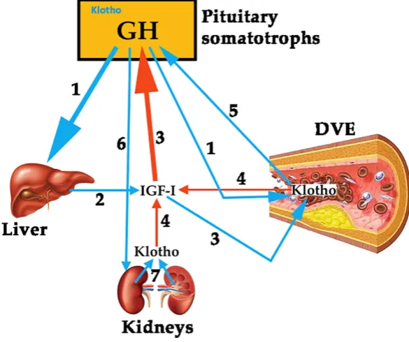
relationships between the three hormones are schematized in Figure 2.
186
187
Figure 2. Schematic representation about the possible relationships between GH, IGF-I and Klotho,
188
and its actions on the vascular endothelium. 1. Pituitary GH induces the hepatic expression of IGF-I
189
(2) and acts on the repair of the damaged vascular endothelium (DVE), although it is also possible
190
that the hormone enhances the production of Klotho by this damaged tissue. 3. Besides its inhibitory
191
effects on pituitary GH release,IGF-I also contributes to repair DVE, and, as in the case of GH, it could
192
enhance Klotho production in DVE. 4. DVE secretes Klotho and it inhibits the negative effect of
IGF-193
I on pituitary GH release, but plasma Klotho may also proceed from kidneys (7), contributing or being
194
responsible for the inhibition of IGF-I effects on GH secretion. 5. The possibility exists that Klotho
195
released from DVE stimulates GH secretion for repairing DVE. 6. GH plays an important role on the
196
physiology of kidneys, being particularly important when there is a chronic kidney disease; since in
197
this pathology there is a state of systemic Klotho deficiency, it is possible that GH tries to correct this
198
problem associated to cardiovascular diseases. Some of these concepts are merely speculative, but
199
existing data lead to think that there is a feedback regulation circuit between GH, IGF-I and Klotho.
200
Blue arrows indicate stimulation and red arrows indicate inhibition.
201
1.3. Cardiovascular disease as an inflammatory condition.
202
Several diseases have been related to inflammation since many years, including atherosclerosis
203
[42–46]. It is considered that inflammation plays a key role in atherogenesis, since it is not only
204
involved in the development and progression of this process [46], but also in the associated symptoms
205
[43]. Circulating monocytes and lymphocytes are present in the vascular wall early in atherogenesis,
206
and both are responsible for the formation and complication of the atherosclerotic plaque [46].
207
A current study has demonstrated the high influence of inflammation in cardiovascular disease
208
(CVD) from a clinical point of view. As known, IL-6 has been previously associated with an increased
209
risk of cardiovascular events, with independence of the cholesterol levels in plasma [42]. IL-6
210
amplifies the inflammatory cascade and is the main circulating cytokine linking systemic
211
smooth muscle cells (SMC) in atherosclerotic plaque [47], and stimulates coagulation by increasing
213
messenger ribonucleic acid transcription of tissue factor and factor VIII [49].
214
IL-1β mediates the IL-6 signaling pathway [42], and canakinumab, a fully human monoclonal
215
antibody targeting IL-1β, leads to a marked reduction of both, plasma levels of IL-6 and CRP without
216
lowering the level of low-density lipoprotein (LDL) in patients with diabetes who were at high
217
vascular risk [50]. This drug led to a significant lower rate of recurrent cardiovascular events than
218
placebo [42].
219
The development of the atheromatous plaque is a multi-factorial process. SMC from the middle
220
layer in the elastic arteries show a differentiated phenotype with a low proliferation and migration
221
rate. Unlike the skeletal and cardiac myocyte, mature SMC may suffer a phenotypic modulation,
222
because of an atherogenic stimulus, with a re-entry in the cellular cycle. These activated state makes
223
them proliferate and migrate to the vascular lumen, and synthesize some extracellular matrix (EM)
224
components and proteases that modify the matrix, contributing to the atheromatous plaque [51].
225
The key aspect of the plaque formation is the endothelial dysfunction secondary to some
226
atherogenic stimuli, such as hypercholesterolemia, hypertension, diabetes, tobacco, etc. The
227
consequence of this endothelial dysfunction is the appearance of an inflammatory response.
228
SMC are essential in the stability of these plaques. When there is a scarcity of these cells into the
229
plaque, the atheroma will be highly vulnerable to rupture [52]. Plaque rupture and subsequent
230
thrombus formation can lead to an acute event [53–55], although in the lower extremities this event
231
can be better tolerated because of the numerous and large collateral network.
232
It is well known the role of LDL in this setting. Oxidized LDL (ox-LDL) have been related to the
233
formation and complication of the atherosclerotic plaque [56]. LDL has high susceptibility of being
234
oxidized. But the oxidative environment in the vascular wall may also modified other lipids as HDL.
235
Nicotinamide adenine dinucleotide phosphate (NADPH) oxidase, leukocyte- and platelet-derived
236
oxidants, and red blood cell-derived iron-rich heme group, are part of the different systems implied
237
in the oxidative modification of lipids, proteins and DNA that in the vascular wall lead to
238
atherosclerosis [56]. All these oxidants maintain the inflammatory response and participate in the
239
arterial wall rupture with platelet aggregation and thrombus formation.
240
Oxidative stress plays a main role in the origin of the pathogenesis of CVD. In a normal vascular
241
wall, oxidative stress activates nuclear defense genes throughout the mediation of the nuclear factor
242
erythroid 2-related factor 2 (Nrf2) 2 [57]. This protects against the formation of foam cells by
243
regulating the expression of antioxidant proteins and scavenger receptors [57]. Nevertheless, its
244
function has not been properly understood, since a pro-atherogenic action has been also associated
245
to Nrf2, because ApoE-null mice, deficient in Nrf2, develop smaller atherosclerotic plaques [58].
246
The recruitment of circulating leukocytes into the blood vessel wall is one of the major
247
etiopathogenic mechanisms of atherosclerosis. This process is predominantly mediated by cellular
248
adhesion molecules (CAM), which are expressed on the vascular endothelium and the leukocytes of
249
the vascular wall, in response to atherogenic stimuli. In patients with peripheral arterial disease
250
(PAD), increased levels of these integrins have been found during exercise, being associated with the
251
severity and the extent of the arterial disease [59]. Antagonists of CAM have shown promise in
252
treating inflammatory disorders in animal models [60,61].
253
Selectins, another group of integrins, are also elevated in PAD population. Studies with the
anti-254
P-selectin antibody inclacumab in coronary arterial disease (CAD) have found a reduction in
255
myocardial damage after percutaneous management [62]. This molecule also reduces elevated
256
circulating platelet-leukocyte aggregates levels in PAD [63].
257
Exercise is associated with an increase in plasma levels of numerous inflammatory mediators in
258
PAD, including thiobarbituric acid–reactive substances (formed as a byproduct of lipid
259
peroxidation), thromboxane, IL-8, TNF-α, ICAM-1, VCAM-1, von Willebrand factor, E-selectin, and
260
thrombomodulin [43].
261
Casual associations between biomarkers and PAD has not been established. However,
262
inflammatory mediators can aggravate endothelial dysfunction, and markers such as IL-6 are
263
oxidative stress in patients with PAD, exercise training has consistently been shown to improve
265
symptoms among patients with PAD. In this sense, GH increases exercise performance improving
266
lean body mass, muscle mass and cardiac output in AGHD patients [65].
267
Interestingly, exercise is a powerful inducer of pituitary GH release [5], most likely by inducing
268
the hypothalamic release of noradrenaline which inhibits somatostatin, the main inhibitor of pituitary
269
GH release [66]; but, as indicated, pituitary secretion of GH decreases while aging [5]. This seems to
270
be contradictory; however, it is important to differentiate the effects of acute and chronic exercise,
271
because both kind of exercises have different effects [43]. Endothelial dysfunction was recently
272
associated with walking impairment independent of the ankle-brachial index (ABI), suggesting that
273
endothelial dysfunction may contribute to the exercise impairment in PAD [67].
274
In addition, inflammatory mediators may also have proangiogenic and antiangiogenic effects,
275
regulating the ischemic response [68]. In fact, patients with PAD have lower circulating VEGF-A and
276
higher circulating inflammatory parameters of TNF-α and IL-8 compared with controls with other
277
comorbid conditions and cardiovascular risk factors [69].
278
On these bases, atherosclerosis is a complex process involving lipid deposition, oxidative stress,
279
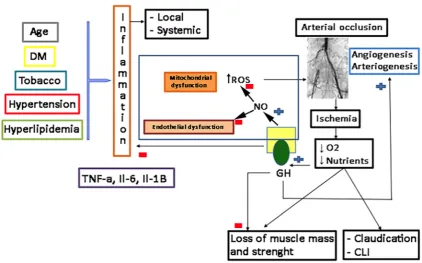
inflammatory cell recruitment and platelet activation. Figure 3 schematizes these concepts.
280
281
Figure 3. Cardiovascular risk factors converge to produce inflammation with increasing of
TNF-282
alpha, IL-6 and IL-1B, which promotes endothelial and mitochondrial dysfunction, with the overload
283
of ROS, all of them being responsible for atheroma plaque formation and arterial occlusion, that leads
284
to hypoxia and decreased nutrition of tissue. Both factors contribute to the loss of muscle mass and
285
strength and symptoms such as intermittent claudication or, critical limb ischemia. GH inhibits all
286
these deleterious effects from cardiovascular risk factors, promoting the NO pathway that
287
compensates redox imbalance, corrects endothelial dysfunction (increasing endothelial-dependent
288
vasodilation), decreases inflammation, and stimulates angiogenesis and arteriogenesis. NO: nitric
289
oxide; ROS: reactive oxygen species; TNF: tumor necrosis factor alpha; IL: Interleukin; GH: growth
290
To find biomarkers that can predict either the risk for suffering CVD or the risk for progression
292
is of high interest, but this is not the aim of this review. Thereby, we will address those biomarkers
293
related to GH that support its action and reduce the risk of CVD.
294
Most knowledge about this issue comes from the studies performed in acromegaly and GHD
295
patients. As mentioned, significant lower levels of VCAM-1 have been found in GHD patients than
296
in healthy subjects; moreover, they increase during GH treatment, as compared with patients treated
297
with placebo [24]. The development of GHD after the treatment of acromegaly affects adversely the
298
body composition and inflammatory biomarkers of cardiovascular risk [70].
299
Visceral adiposity and lipids are one of the best studied markers in CVD. The increase of NO
300
after GH administration lowers lipoxygenase activity and ox-LDL [4]. AGHD patients suffer an
301
elevated risk of CVD because of hyperlipidemia, among other factors. GH therapy in these patients
302
improves the lipid profile and decreases the vascular risk. The visceral fat is elevated in GHD children
303
and adults, perhaps because GH produces lipolysis, and when GH is administered it reverts this
304
increased adiposity [4,71]. Since GH secretion is deficient while aging, the progressive increase in fat
305
stores seen in the elderly population could be due, at least in part, to the insufficient secretion of the
306
hormone.
307
1.4. Coronary Arterial Disease (CAD) and Heart Failure
308
CAD is a broad term including several related syndromes caused by myocardial ischemia, an
309
imbalance between cardiac blood supply perfusion and myocardial oxygen and nutritional
310
requirements.
311
Cardiovascular disease (CVD) is the most important cause of death worldwide [72], and a major
312
economic global burden [73]. Despite reductions in CVD mortality in high-income countries, global
313
CVD mortality increased by 41% between 1990 and 2013, largely driven by rises in low-income and
314
lower-middle-income countries [74]. Among CVD, coronary arterial disease (CAD) is the leading
315
cause of death [72–75].
316
The majority of ischemic processes is produced by an alteration in the oxygen supply to the heart
317
due to coronary disease. The obstruction of the coronary arteries is usually of atherosclerotic origin,
318
although there are other infrequent causes such as an anomalous origin of these arteries, its
319
spontaneous dissection or embolisms [76].
320
As indicated before, atherosclerosis implies a degenerative inflammatory process where
321
different risk factors (diabetes, hypertension, dyslipidemia, smoking, obesity, sedentary lifestyle...)
322
damage the endothelium, favoring the entry of LDL particles that oxidize and initiate a complex
323
inflammatory and fibrotic process within the arterial wall that culminates with the development of a
324
plaque that can obstruct the coronary lumen, therefore preventing proper blood flow [76]. Although
325
the atherosclerotic process is usually chronic, abrupt plate instabilities can erode or ulcerate the
326
endothelium giving rise to a thrombotic phenomenon that can obstruct the coronary artery suddenly
327
causing an acute coronary syndrome (unstable angina or acute myocardial infarction). It is estimated
328
that throughout the world these processes are responsible for approximately 7 million deaths per
329
year, being the main cause of mortality in the population of industrialized countries [72–74].
330
The other major disease regarding cardiovascular system is heart failure (HF); it affects about
331
2% of the adult population worldwide. Its prevalence is clearly age-dependent, ranging from less
332
than 2% of people younger than 60 years to more than 10% of those older than 75 years, and it is
333
estimated that it will increase by 25% in the next 20 years [77–79]. The etiology of HF is diverse and
334
most patients have a history of hypertension, coronary artery disease, cardiomyopathies, or valve
335
disease, or a combination of these [77,78]. HF has a poor prognosis, with high rates of hospital
336
admission and mortality; costs related to the treatment of HF encompass 2–3% of the total
337
expenditure of healthcare systems in high-income countries, and it is believed that they will increase
338
by more than 200% in the next 20 years [79].
339
1.5. Peripheral Arterial Disease
342
Peripheral arterial disease (PAD) is the term commonly used currently to refer to the
343
atherosclerotic pathology affecting peripheral arteries of the lower extremity and compromising
344
partially or totally the flow in them. Although less frequent that the other two main CVD, cardiac
345
and cerebrovascular, it affects more than 200 million people worldwide [80]. Maybe, the spectrum of
346
symptoms may vary from none (asymptomatic PAD, 3 times more frequent) to critical limb ischemia
347
(CLI), the most severe form that threats the limb. However, the estimated prevalence depends on the
348
tools used for the diagnosis. In people aged 60-70, the prevalence is about 8 % in the Spanish
349
population [81]. For those aged > 70, it is generally accepted that the prevalence rises to 20%.
350
Additionally, PAD is an independent predictor of cardiovascular mortality and morbidity [82].
351
1.5.1. Endothelial and mitochondrial dysfunction in PAD: the role of oxidative stress
352
As stated above, oxidative stress is the key aspect in producing the endothelial dysfunction that
353
triggers the atherosclerotic process and the aging of the vascular system [56,83]. However, not only
354
vascular risk factors contribute to this phenomenon, but also the own exercise leads to generation of
355
superoxide-anion and other mediators of endothelial dysfunction, that it has been correlated with the
356
clinical severity of PAD [84]. This endothelial dysfunction is not only located in the major arteries,
357
but also in the microcirculation of the skeletal muscle [43]. Patients with PAD suffer a constant
358
ischemia-reperfusion syndrome as they walk and rest, generating reactive oxygen species (ROS) that
359
affect muscle fibers [84], and impairs mitochondrial function, reducing the energy production [85,86].
360
In fact, higher carbonyl and 4-hydroxy-2-nonenal levels have been found in calf muscle samples
361
indicating the oxidative stress [87].
362
Skeletal muscle mitochondria release free radicals during the ischemic process, including
363
superoxide-anions and some other ROS derived from the redox cascade [88,89]. Reperfusion also has
364
the same effect, increasing the oxidative stress [89]. These ROS contribute to the endothelial
365
dysfunction and the alteration of proteins in the skeletal muscle, and may lead to mitochondrial DNA
366
injury in the long term [90]. This DNA injury is also seen in less affected limbs of patients with
367
unilateral PAD, suggesting that PAD is not only a local problem, but rather a systemic one [91]
368
Mitochondrial pathways are vulnerable to free-radical injury [92], and PAD patients show
369
reduced activities of complexes I and III of the mitochondrial respiratory chain [93]. These
370
observations suggest that electron transport chain activity is impaired in PAD, probably because of
371
the ischemia-reperfusion injury and old age, which spreads the oxidative damage and the metabolic
372
dysfunction.
373
Lactate levels are also significantly elevated in PAD skeletal muscle, because of an incomplete
374
oxidation of glucose, a decreased pyruvate dehydrogenase activity, and exercise performance [94].
375
At this point it is of interest to remark that GH is a mitochondrial protector [95–97], therefore
376
suggesting that the hormone may play a positive role in this process, since GH restores the redox
377
imbalance, improving mitochondrial respiratory chain and the needed production of energy.
378
In fact, endothelial dysfunction has been evaluated in Japanese patients with AGHD in the
379
GREAT study. After 24 weeks of GH replacement therapy, the hormone significantly lowered plasma
380
diacron-reactive oxygen metabolites and improved endothelial function measured by reactive
381
hyperemia index [98]. This indicates that GH can exert a protective role in redox balance in AGHD,
382
in which predominates a pro-oxidant environment increasing the atherogenic risk; but this is
383
corrected by short-term GH administration without fully normalizing IGF-I levels [99]. Moreover,
384
GH has a role in stress resistance by altering the functional capacity of the glutathione S-transferase
385
(GST) system through the regulation of specific GST family members in long-living Ames dwarf mice.
386
The hormone also affects the regulation of Thioredoxins (TRX) and glutaredoxins (GRX), factors that
387
regulate post translational modification of proteins and redox balance, thereby further influencing
388
stress resistance [100]. However, the exact role of GH in redox balance has not been completely
389
understood, as in oxidative stress-induced conditions may enhance oxidation [101]. Thereby, both
390
GH overproduction and deficiency are tightly linked with enhanced oxidative stress.
391
1.5.2. Endothelin and PAD
393
Endothelial dysfunction might be traduced by an imbalance between the
endothelium-394
dependent vasodilation (mediated mainly by NO) and vasoconstriction (mediated by endothelin).
395
It has been well documented that vascular endothelin (ET) production is elevated in
396
atherosclerosis and influences the development of atherosclerotic lesions through a variety of
397
mechanisms [102]. ET participates in several key steps in the inflammatory component of
398
atherosclerosis, increasing various cytokines from monocytes [103], and enhancing the uptake of LDL
399
by these cells, promoting foam cells [104].
400
GH has been broadly related to an increase in the production of NO. However, GH is also related
401
to ET, as an increased secretion of GH and ghrelin have been demonstrated in cattle after the injection
402
of ET 1 and 3 [105,106]. Thus, GH increases physiologically in response to the increased level of ET.
403
Despite the relationship between GH and ET has not been well established yet in CVD, it seems that
404
GH may compensate the deleterious effects of ET, as the treatment with the hormone improves
ET-405
induced stroke in adult rats [107]. Perhaps this is due to the actions of GH on NO production.
406
2. Discussion
407
GH plays a key role for the development of a normal heart during fetal development, and plays
408
a positive role in maintaining the structure and function of the normal adult heart, by stimulating
409
cardiac growth and heart contractility [108–110].
410
It is known that myocardium and vessels have receptors for GH and IGF-I, and IGF-I can be
411
produced directly in these tissues [111,112]. Therefore, GH may exert endocrine roles on the
412
cardiovascular system, as well as endocrine or auto/paracrine effects of IGF-I can be exerted on it. On
413
the other hand, the GH/IGF-I axis can interact with the vascular system and can regulate the vascular
414
tone and thereby peripheral resistance [113].
415
The interactions between heart and GH are complex. In fact, it has recently been shown that the
416
heart may influence body growth in pediatric heart disease. In these situations, cardiomyocyte
417
synthesize and release Growth Differentiation Factor 15 (GDF-15), which inhibits liver signaling by
418
GH, therefore impeding the release of IGF-I and affecting body growth [8].
419
Life expectancy is reduced in patients with hypopituitarism as compared with healthy controls
420
(2-fold higher risk of death for CVD, higher risk in women than in men). The causes of death are
421
probably multifactorial, but GHD has been considered one of the most relevant factors of the
422
increased mortality in these patients [114,115].
423
On the other hand, patients with acromegaly, despite presenting higher incidence of other
424
cardiovascular risk factors (hypertension, insulin resistance), do not present a clear excess of CAD or
425
stroke in comparison to normal counterparts [4,111,116].
426
Nowadays, despite multiple studies about the interaction of the GH/IGF-I axis and the
427
cardiovascular system, the clinical importance of effects of GH and local and endocrine IGF-I in
428
adults remains to be clarified.
429
2.1. The role of GH in the vascular endothelium
430
Two conditions in which the effects of GH on endothelial dysfunction might provide interesting
431
data may be acromegaly and aging.
432
In the case of acromegaly, plasma levels of two biomarkers of endothelial dysfunction and
433
atherosclerosis, such as endothelin-1 (ET-1) and total homocysteine levels (tHcy), were measured in
434
patients with active acromegaly and cured disease [117]. While tHcy was similar in both groups of
435
patients, ET-1 was significantly higher in active acromegaly, suggesting that it contributes to
436
premature atherosclerosis and cardiovascular affectations observed in this pathology, although the
437
role played by IGF-I on these vascular affectations could not be discarded.
438
Particularly important, in our opinion, is the case of aging. Important changes in pituitary GH
439
secretion along the life have been widely described (for a more detailed comprehension, see
440
references [4] and [6]. An exponential decline in plasma GH concentrations starts from 18 to 30 years
441
In this situation, plasma levels of IGF-I are also low, although the liver production of this peptide
443
depends not only on GH but also on the nutritional status of the organism [4].
444
Aging is associated with an increased risk of atherosclerosis, but we know now that this disease
445
can begin earlier, during youthfulness.
446
It has been proposed that the increased risk of atherosclerosis as we age, is due to low production
447
of EPC, which makes unable to repair atherosclerotic vascular walls [118]. Treatment with GH during
448
10 days led, in middle-aged subjects to an increase in plasma levels of EPC which, moreover,
449
improved in its capacity to migrate and incorporate into tube-like structures, and showed increased
450
endothelial NO synthase (eNOS) expression up to levels equivalent to those of healthy young
451
subjects. That is, GH treatment decreased EPC senescence and increased telomerase activity. In the
452
same study, aged mice treated during 7 days with GH or IGF-I increased EPC levels and ameliorated
453
EPC functions. This was not observed when GH treatment was given during only two days. Results
454
from that study attributed to IGF-I, rather than to GH, the reversal of age-dependent EPC dysfunction
455
[118]. We do not know whether these results appear as an IGF-I age-related effect, but other studies,
456
as described before in healthy young people, indicated that GH effects on the vascular system are not
457
dependent on IGF-I, postulating that GH acts directly on GHR and eNOS in the vascular endothelium
458
[9]. These contradictory results led to suggest that GH administration during somatopause does not
459
produce clearly favorable effects on the endothelial dysfunction, while combined treatments with GH
460
plus IGF-I may produce more beneficial effects on the vascular wall in elderly individuals [119];
461
however, we do not think that this combination is advisable.
462
Preclinical studies in hypophysectomized rats also showed that the lack of GH production is
463
associated with the development of atherosclerosis [120], while GH treatment during two weeks
464
reversed several biomarkers indicative of the developing arterial disease. These researchers identified
465
in the aorta of hypophysectomized rats 18 genes regulated by GH, which most likely have a
466
physiological effect on vascular tone and atherogenesis. Among these genes, they found that GH
467
induced an increased expression of the KATP channel, which plays a key role in the regulation of
468
vascular tone, therefore involving GH in this regulation [120]. However, plasma levels of GH must
469
be within normal ranges; since, as it occurs in acromegaly, transgenic mice overexpressing bovine
470
GH develop an endothelial dysfunction, which depends on the age of the animal and the type of
471
blood vessel, indicating that the affectation in endothelial function is most likely produced by
472
increased production of mitochondrial ROS followed by many other affectations in vascular function
473
[121].
474
Curiously, similar results to these shown in transgenic mice overexpressing GH, have been
475
reported in hypopituitary Ames dwarf mice aortas in terms of enhanced production of ROS and
476
lesser expression of antioxidant enzymes (for instance, glutathione peroxidase and eNOS), therefore
477
leading to vascular oxidative stress [122], a first step, as stated above, to develop endothelial
478
dysfunction.
479
Similarly, peripubertal GHD in Lewis dwarf rats leads to a pro-oxidative cellular condition most
480
likely responsible of the development of an altered vascular phenotype (in both structural and
481
functional terms), which leads to vascular affectations, early accelerated, later in the life of these
482
animals [123]. GH treatment reverses these impairments that, interestingly, do not occur equally in
483
the cerebral vessels than in the aorta of these genetically dwarf rats [123].
484
Another model for analyzing the effects of GH on the vascular system comes from studies in
485
which rats are undernourished during pregnancy. Maternal undernutrition produces increased
486
blood pressure and endothelial dysfunction in adult offspring, but if pups receive early pre-weaning
487
GH treatment (from day 3 after birth until weaning in day 21) adult vascular function is normal; this
488
contrasts with what happens in the offspring that received saline during these days before weaning.
489
This indicates that early GH treatment can reverse the vascular alterations resulting from maternal
490
undernutrition during pregnancy, but also that there is a developmental cardiovascular
491
programming, susceptible to be reversed by early treatment with GH after delivery [124].
492
While results from both preclinical and clinical studies clearly indicate that GH plays a key role
493
GH are due to a direct action of the hormone and which are mediated by IGF-I, because this peptide
495
and its receptors (IGF-IR) are widely expressed in endothelial cells [125]. Moreover, GH induces the
496
expression of IGF-I in many territories, including the fetal brain [126]; however, GH seems to be
497
unable to increase the transcription of IGF-I in endothelial cells, and, in fact, systemic or local
498
infusions of GH lead to a prompt increase in forearm blood flow and NO release in healthy humans
499
without increasing plasma IGF-I concentrations or muscle IGF-I expression [9,127]. The fact that it
500
seems that GH is produced by endothelial cells, and endothelium-derived GH stimulates the
501
proliferation, migration, survival, and capillary formation of endothelial cells in an autocrine manner
502
[11], clearly indicates that the hormone exerts direct effects on the vascular endothelium, although
503
IGF-I is also vasoactive activating eNOS via Pi3K/Akt [127], a signaling pathway also used by GH.
504
Perhaps some of the apparently contradictory results here reported, in relation to the lack of
505
effects of the administration of GH on the vascular endothelium, in AGHD and GHD [23,26], and the
506
attribution to IGF-I rather than to GH the positive effects on the vascular wall, proceed from the
507
recently described relationships between GH, IGF-I and Klotho [7,40], but also on the effects of Klotho
508
on the vascular endothelium and aging.
509
The impact of GH on inflammatory processes is not well understood yet. Evidence shows
510
controversial data of both anti- and pro-inflammatory effects of the hormone.
511
GH therapy reduces the levels of CRP in GHD patients [128], and exerts anti-inflammatory
512
effects in different experimental models of sepsis by lowering TNF-α [129]. Exogenous GH also may
513
improve the effects of sepsis-induced IGF-I resistance [130]. Conversely, a massive increase of GH in
514
GH transgenic mice has a pro-inflammatory effect, rising pro-inflammatory cytokines [130].
515
As it has been described above, the relationship between GH and CAMs is well known. This
516
action seems to be indirect, mediated by VEGF among others. VEGF has been described as a
strong-517
inducing agent of CAM on endothelial cells during inflammation [131]. Given the fact that GH
518
directly increases VEGF levels after its administration, this peptide could be one of the main
519
mediators of the GH effects. It seems, thereby, that high supraphysiological administration of
520
exogenous GH could increase inflammation, while doses used for treating GHD, or even short-term
521
GH administration to non-GHD patients may represent a protective factor against this issue.
522
The role of CRP, IL-6 and TNF-α in CVD has also been well established [44,48,132].
523
In AGHD patients, the administration of GH decreased CRP and IL-6 levels, some that not
524
occurred when treating them with placebo [133]. However, another study in AGHD, showed that
525
GH therapy also reduced CRP, but failed in reducing TNF-α and IL-6 levels [128].
526
Pregnancy associated plasma protein A (PAPP-A) has been recently included among markers of
527
cardiovascular risk being associated both to the presence of carotid atherosclerosis and acute
528
coronary syndrome [134,135]. PAPP-A is also significantly elevated in AGHD [136], and GH
529
replacement therapy decreases this specific and not generic biomarker of CVD, although not in all
530
GHD patients [137].
531
At this point, the own GH could be a marker, as the deficit of both GH and IGF-I leads to a more
532
aggressive heart failure, with impaired functional capacity and poor outcomes [138].
533
All these data support the possible role played by GH in the correction of the state of
534
inflammation in patients suffering from CVD, at least in those with AGHD. The protective effect of
535
GH in inflammation is secondary to its action against oxidative stress, most likely because of its action
536
on NO and extracellular signal-regulated Kinase (ERK) pathway.
537
Currently, our group is conducting a phase III RCT in patients suffering PAD without options
538
for revascularization: Growth Hormone Angiogenic Study (GHAS), Eudract 2012-002228-34, approved
539
by the Spanish Agency of Drugs and Health Products (AEMPs) and the Autonomic Committee on
540
Research Ethics in Galicia (CAEIG, 2012/378), Spain, in which patients receive GH or placebo.
541
Although this study has yet finished, all the data have not yet been analyzed and therefore there
542
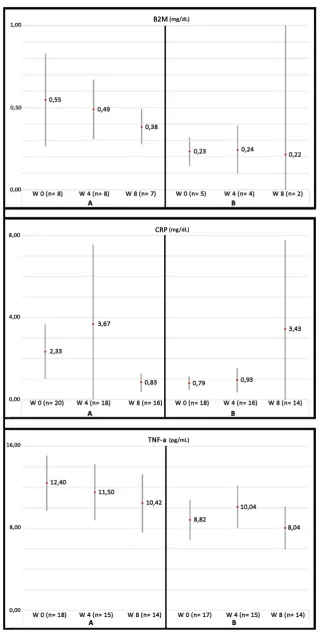
are no statistical conclusions, early results from the patients enrolled so far show that TNF-α is the
543
marker most frequently elevated in these patients (74%), followed by β2-microglobulin (B2M) (69%)
544
and C Reactive Protein (CRP) (60%). Figure 4 depicts the graphic tendencies that show these patients
545
547
Figure 4. Evolution of some biomarkers of inflammation analyzed in the GHAS study. Values are
548
shown as the mean ± SD. B2M (beta-2-microglobulin), CRP (C Reactive Protein) and TNF-α. A and B
549
represent different groups of treatment (GH or placebo, respectively). Note the tendency to decrease
550
in group A as compared with the group B. Patients from group A had significant higher basal levels
551
as compared with group B. Significant differences in the end of the study have not reached because
553
of the small sample of patients still analyzed (note the differences in n). W = weeks of treatment.
554
2.2. GH and Coronary Arterial Disease
555
The effects of GH /IGF-I in the incidence and prognosis of CAD are controversial.
556
As described before, GHD is associated with an increased prevalence of atherosclerosis, CAD
557
and stroke caused by an increased prevalence of atherosclerotic risk factors such as alterations of
558
body composition, lipid profile and coagulation pattern [4,111,116], as shown in Figure 5.
559
560
Figure 5. Effects of GH deficiency on atherosclerosis. GH: Growth Hormone, IGF-1: Insulin growth
561
factor 1. NO: nitric oxide. Blue arrows indicate the effects produced by decreased GH secretion, while
562
red arrows indicate how atherosclerosis is developed.
563
AGHD often have significant changes in their lipid profile with increased LDL, increased
564
triglycerides, decreased high-density lipoprotein (HDL) (the latter observed only in women), with no
565
differences in lipoprotein (a) [139]. GH replacement positively reverses this negative lipid profile in
566
GHD patients, decreasing LDL and total cholesterol, and increasing HDL; in addition, a decrease in
567
CRP has been observed in these GHD after GH replacement therapy, while no clear changes seem to
568
be produced in circulating triglycerides [139–141]. However, no study has determined whether GH
569
has an additive effect that optimizes statin therapy; therefore, this remains an open question.
570
Regarding hypertension and peripheral resistance conflicting results have been reported in the
571
literature [116]. Hypertension is quite frequent in GHD patients, and this condition results in
572
impaired vasodilation responses to stress and/or exercise. As described, the GH–IGF-I axis promotes
573
the synthesis of NO that reduces vascular tone, inhibiting the proliferation and migration of SMC,
574
reduces platelet adhesion, and decreases lipoxygenase activity and ox-LDL [142]. Some vasoactive
575
sympathetic nerve activity and GH replacement therapy has been shown to reduce arterial stiffness
577
and to improve vascular endothelial function [143].
578
In some AGHD (patients with high base-line diastolic blood pressure, such as elderly GHD
579
patients or those with previous Cushing´s disease), GH replacement reduces blood pressure, whereas
580
in other patients (specially in young GHD patients) no changes in blood pressure have been shown
581
[116,144].
582
Besides the cardiovascular risk factors mentioned above, GHD patients were shown to have
583
increased blood vessel intima-media thickness (IMT) that is well known to represent one of the
584
earliest morphological changes in the arterial wall in the process of atherogenesis [145].
585
A decrease in IMT has been shown in several studies after the administration of GH to GHD
586
patients [146]. Increases in IMT predict the development of symptomatic coronary disease, thus GH
587
treatment may have a significant improvement in cardiovascular outcome, but this question has not
588
yet been specifically analyzed in patients with GHD.
589
Regarding hard clinical endpoints, we previously commented on the increased risk of
590
cardiovascular mortality in GHD patients. The worse cardiac risk profile (mainly hyperlipidemia) of
591
these patients may explain part of the excess in CAD and mortality, but the studies do not allow to
592
obtain a definitive conclusion. However, as described, GHD patients present an altered body
593
composition with increased fat mass, with a preferential increase in visceral fat that decreases in
594
response to GH therapy. This change occurs within 6 months after the initiation of therapy, and it is
595
maintained if treatment is continued [146].
596
Interventional studies are old, and they did not control cardiac risk factors. There are no
597
prospective, long-term randomized studies in AGHD patients comparing GH treatment to placebo
598
on cardiovascular hard outcomes and mortality, and it is likely that there will never be such a study.
599
A more recent and prospective trial found a lower mortality in GH treated hypopituitary patients
600
compared with a retrospective analysis of patients who had not been treated with GH [147].
601
However, again the different time periods covered also included dramatic changes in the treatment
602
of risk factors such as hypertension, diabetes mellitus and hypercholesterolemia.
603
2.3. GH and heart failure
604
As stated, GH plays an important role during myocardial development that can easily be seen
605
in untreated GHD children. They present cardiac atrophy with a reduction in the left ventricle (LV)
606
mass, ejection fraction, and cavity dimensions, as well as reduced cardiac output, high peripheral
607
vascular resistance and reduced functional capacity compared with healthy controls of the same age,
608
sex, and height [148]. When GHD appears in adults, it does not produce a reduction in cardiac mass,
609
but cardiac performance and exercise capacity are impaired [149].
610
On the other hand, GH excess exerts different and opposite effects on the heart. In early-stage it
611
enhances cardiac performance, whereas it causes fibrosis and cardiac dysfunction in the
612
intermediate-late phase. This apparent discrepancy is easily clarified: a physiological GH level, or
613
short-term excess, exert positive inotropic effect; whereas long-term exposure to GH excess induces
614
cardiac dysfunction and progression to heart failure by causing morphological and functional
615
adaptive changes [150]. The most relevant histological abnormalities are interstitial fibrosis, reduced
616
capillary density, increased extracellular collagen deposition, myofibrils derangement,
lympho-617
mononuclear infiltration and myocyte death due to necrosis and apoptosis [150,151].
618
GH acts directly stimulating its own receptors or (mainly) by inducing local synthesis of IGF-I
619
and may regulate cardiac growth and metabolism by increasing protein synthesis (troponin I, myosin
620
light chain-2, and actin), and cardiomyocyte size, increasing collagen synthesis and promoting
621
cardiac hypertrophy [151–153]. There is also evidence about that IGF-I may reduce apoptosis of
622
cardiomyocyte, preventing myocyte loss [152]. The GH/IGF-I axis can also increase cardiac
623
contractility by enhancing calcium sensitivity and reducing vascular resistance [149,151].
624
Chronic heart failure (CHF) patients have a prevalence of 30% in GHD and this fact identifies a
625
subgroup of CHF patients characterized by impaired functional capacity, left ventricle remodeling
626
Several groups have studied the effects of GH and IGF-I in patients with impaired cardiac
628
function. GH replacement trials show an increase in left ventricular mass and improvement in cardiac
629
performance, diastolic filling, and systolic function after GH treatment in children or adults with
630
GHD [65,148,151]. Nevertheless, randomized placebo controlled studies show conflicting results,
631
with an increase in LV mass related to serum IGF-I levels, but no change in LV wall stress, arterial
632
blood pressure, ejection fraction, clinical status or 6-minute walking distance [155].
633
The conflicting results of the clinical trials of GH treatment may be related to the small number
634
of patients enrolled, the different dose and duration of GH treatment, the different cardiac heart
635
failure etiologies, and differences in the patients' clinical characteristics. Besides this, the
636
discrepancies may also reflect the heterogeneity of IGF-I increase in response to GH treatment. In
637
fact, a recent meta-analysis confirms that there is a clear relationship between changes in IGF-I
638
concentrations achieved and the beneficial effects of GH treatment. Only in the trials in which IGF-I
639
increased >89% vs. baseline was there a significant improvement in cardiac output,
640
echocardiographic parameters and exercise capacity, whereas in the trials in which the increase in
641
IGF-I was < 89% beneficial cardiovascular effects were not observed [156].
642
Given its possible positive effects on heart in “responders” patients, it could be speculated that
643
GH treatment might be useful in some patients with heart failure but more investigation is needed in
644
this field.
645
2.4. GH and molecular aspects of cardiovascular risk factors in PAD
646
Despite the known negative epidemiological impact of cardiovascular risk factors, its main
647
mechanism of damage is not completely clarified. However, it seems that they may modify redox
648
balance. Since the specific role of each cardiovascular risk factor in redox balance has been widely
649
described, as well as the benefit of their treatment, we will only underline the main aspect related to
650
GH and its possible role and benefit.
651
As described before, GH therapy improves arterial hypertension in GHD patients by acting on
652
the vascular smooth muscle ATP-sensitive potassium (KATP) channel [120], and on the lipid profile
653
with independence of IGF-I [157].
654
Maybe a closer attention would need diabetic patients (DM), in which a dysfunction of eNOS in
655
both endothelial cells and platelets has been found, which attenuates arterial remodeling [158,159].
656
But the latter is also affected because of the lower sensitivity to shear stress that these patients show.
657
This aspect seems to be secondary to the massive calcification and multilevel arterial disease, as well
658
as to the elevated vasomotor tone found that impairs the response to the vasodilator stimuli, and the
659
enlargement of collateral arteries [158–160]. Moreover, a quantitative and qualitative alteration in
660
EPC has been described in DM [158,161]. All these factors explain both the strong atherosclerotic
661
injury and the low capacity of compensating the latter after an arterial occlusion.
662
In addition, a high rate of patients with DM may suffer neuropathy. In these patients, the level
663
of expression of several growth factors, such as neurotrophic factors, insulin-like growth factors,
664
cytokine-like growth factors and VEGF, are altered [161].
665
GH could aid in the recovery of some of these deleterious aspects in diabetic patients, since, once
666
again, the hormone increases eNOS production, decreases vasomotor tone, and may improve the
667
nerve injury, by increasing neurotrophic factors, such as BDNF. Moreover, GH can increase
668
Substance P (SP), one of the main molecules implied in nerve damage and wound healing. SP and
669
GH are strongly related [162,163]. In fact, GH improves wound healing in diabetic rats and mice [164],
670
and SP could be one of the possible mediators. Since a high rate of diabetic patients have small vessels
671
disease, an angiogenic therapy with growth factors might be a good option for them. GH angiogenic
672
stimulation and benefit in patients suffering from PAD will be comprehensively reviewed below.
673
Although GH may cause hyperglycemia or abnormal glucose tolerance, this is not a
674
contraindication for using the hormone in diabetic patients, since clear benefit of GH therapy has
675
been described in these patients.