Quantifying cancer epithelial-mesenchymal plasticity and its association with stemness and immune response
Dongya Jia1, Xuefei Li1, Federico Bocci1,2, Shubham Tripathi3, Youyuan Deng1,4, Mohit Kumar
Jolly5, *, José N. Onuchic1,2,6,7, *, Herbert Levine8,9 *
1Center for Theoretical Biological Physics, Rice University, Houston, TX 77005, USA
2Department of Chemistry, Rice University, Houston, TX 77005, USA
3PhD Program in Systems, Synthetic, and Physical Biology, Rice University, Houston, TX 77005, USA
4PhD Program in Applied Physics, Rice University, Houston, TX 77005, USA
5Centre for BioSystems Science and Engineering, Indian Institute of Science, Bangalore 560012, India
6Department of Biosciences, Rice University, Houston, TX 77005, USA
7Department of Physics and Astronomy, Rice University, Houston, TX 77005, USA
8Department of Bioengineering, Northeastern University, Boston, MA 02115, USA
9Department of Physics, Northeastern University, Boston, MA 02115, USA
*Correspondence:
Mohit Kumar Jolly: [email protected]
José N. Onuchic: [email protected]
Herbert Levine: [email protected]
Abstract
Cancer cells can acquire a spectrum of stable hybrid epithelial/mesenchymal (E/M) states during
epithelial-mesenchymal transition (EMT). Cells in these hybrid E/M phenotypes often combine
epithelial and mesenchymal features and tend to migrate collectively commonly as small clusters.
Such collectively migrating cancer cells play a pivotal role in seeding metastases and their presence
in cancer patients indicates an adverse prognostic factor. Moreover, cancer cells in hybrid E/M
phenotypes tend to be more associated with stemness which endows them with tumor-initiation
ability and therapy resistance. Most recently, cells undergoing EMT have been shown to promote
immune suppression for better survival. A systematic understanding of the emergence of hybrid
E/M phenotypes and the connection of EMT with stemness and immune suppression would
contribute to more effective therapeutic strategies. In this review, we first discuss recent efforts
combining theoretical and experimental approaches to elucidate mechanisms underlying EMT
multi-stability (i.e. the existence of multiple stable phenotypes during EMT) and the properties of
hybrid E/M phenotypes. Following we discuss non-cell-autonomous regulation of EMT by cell
cooperation and extracellular matrix. Afterwards, we discuss various metrics that can be used to
quantify EMT spectrum. We further describe possible mechanisms underlying the formation of
clusters of circulating tumor cells. Last but not least, we summarize recent systems biology
analysis of the role of EMT in the acquisition of stemness and immune suppression.
Keywords
Epithelial-mesenchymal transition; EMT spectrum; hybrid epithelial/mesenchymal phenotypes;
Introduction
Metastasis remains the major cause of cancer-related deaths [1]. To enable successful metastasis,
cancer cells often engage a trans-differentiation program referred to as epithelial-mesenchymal
transition (EMT) in order to promote migratory and invasive properties [2]. During EMT, cells
gradually lose epithelial features such as a cobblestone-like morphology, cell-cell adhesion and
apico-basal polarity and acquire mesenchymal features such as a spindle-like morphology,
increased motility and invasiveness [2]. The concept of EMT was initially described during
embryonic development. EMT was first observed in vitro by Greenberg and Hay showing that
epithelial cells suspended in three-dimensional collagen gels lose their apical-basal polarity and
acquire characteristics of migrating mesenchymal cells [3]. Later in vivo work by Nieto et al.
argued that EMT is essential for the formation of mesoderm and the generation of the migratory
neutral crest cells during chicken embryonic development [4]. EMT also plays a critical role during
physiological wound repair and pathological fibrosis [5]. In the context of cancer metastasis, EMT
has been proposed to be typically associated with enhanced metastatic potential of cancer cells [2]
and the reverse process - mesenchymal-epithelial transition (MET) – has been considered to
facilitate effective metastatic colonization by regaining epithelial and proliferative traits that are
lost during EMT [6].
During metastasis of tumors, cells rarely undergo a complete EMT and enter a fully mesenchymal
phenotype [7]. Instead, partial EMT leading to a hybrid epithelial/mesenchymal (E/M) phenotype
has often been observed [8]. In other words, recent studies have emphasized that contrary to the
prevailing dogma of EMT being a binary process, there exists a spectrum of hybrid E/M
phenotypes characterized by varying extents of epithelial and mesenchymal features and
associated with metastatic potential and invasiveness [9,10]. Cancer cells in these hybrid E/M
phenotypes tend to combine epithelial (e.g. cell-cell adhesion) and mesenchymal (e.g. increase
motility) traits [11] and can thus migrate collectively as a cluster, which can reach and enter the
bloodstream intact. Clusters of circulating tumor cells (CTCs) or CTC clusters contribute much
more than their proportional share to forming metastases relative to individual CTCs which are
typically mesenchymal cells [12]. Clinical evidence supports the aggressiveness of CTC clusters
underlying the emergence of hybrid E/M phenotypes and the formation of CTC clusters can lead
to more effective therapeutic designs targeting metastasis.
Recently there have been ongoing debates regarding the necessity of EMT for metastasis [14].
Zheng et al. demonstrated that deletion of the EMT-inducing transcription factor (EMT-TF)
SNAIL or TWIST in genetically engineered mouse models of pancreatic ductal adenocarcinoma
(PDAC) did not cause any significant change in tumor progression and metastasis [15]. Fischer et
al. suggested that EMT inhibition by over-expression of microRNA (miR)-200 led to no obvious
change in lung metastasis development in spontaneous breast-to-lung metastasis mouse models
[16]. From our perspective, these two studies assumed that EMT can be completely repressed by
single factor manipulation, for example, deletion of SNAIL or TWIST in PDAC or
over-expression of miR-200 in breast cancer. Following these studies, Krebs et al. used the same PDAC
model [15] and showed that deletion of the EMT-TF ZEB1 significantly suppresses the
colonization capacity of tumor cells and the formation of metastases [17], indicating a
non-redundant role of EMT-TFs in regulating PDAC metastasis [18]. Moreover, another study by
Cursons et al. showed that overexpression of miR-200c in the HMLE-derived mesenchymal cells
established by exposing HMLE cells to TGF-β for 24 days can only drive a partial MET where the
canonical epithelial marker E-cadherin increases but the mesenchymal maker vimentin remains
[19]. One major contributing factor to these debates regarding the functional role of EMT in
metastasis is the lack of consistency in defining EMT itself, owing to its highly nonlinear and
multidimensional nature [20]. Thus, a rigorous quantification of the EMT status of tumor cells and
a systematic analysis of the interacting EMT regulators such as interactions between miRNAs,
EMT-TFs and epigenetic factors is urgently needed.
Notably, various extracellular biochemical and biomechanical factors can govern the induction
and maintenance of a partial or complete EMT. For instance, neighboring cells can induce EMT
through TGF-β secretion [21] or Notch signaling [22], and also the alteration of stiffness of
extracellular matrix (ECM) can trigger EMT [23]. In addition to the complexity of EMT itself,
cancer cells undergoing EMT tend to acquire “stemness” characteristics, which are believed to be
responsible for tumor-initiation ability and therapy resistance [24]. Moreover, cancer cells can
endothelial cells and immune cells [25,26]. Specifically, the co-evolution of cancer and immune
cells [27,28] has captured attentions recently due to the promising effects of cancer
immunotherapy [29,30]. Because of the typically enhanced metastatic potential and therapy
resistance of cancer cells undergoing EMT, it is natural to analyze the correlation and causal
relationship between EMT and immune response [31,32]. Furthermore, both EMT [1,2] and
immune signatures [33–39] have been shown to be prognostic indicators for various types of
cancer. A better understanding of their relationship can potentially contribute to more effective
therapeutic designs.
In this review, we focus on how a combination of theoretical and experimental efforts has led to a
better understanding of the EMT multi-stability, the characteristics of the hybrid E/M phenotypes
and the association of EMT with the acquisition of stemness and immune suppression (Figure 1).
We start with a discussion of mathematical modeling studies of EMT regulatory networks, that
elucidate mechanisms underlying EMT multi-stability and particularly the emergence of hybrid
E/M phenotypes. Then we summarize recent in vitro and in vivo experimental studies that
characterize hybrid E/M phenotypes. We then discuss non-cell-autonomous regulation of EMT by
cell cooperation and ECM. We further discuss metrics that have been developed to quantify EMT
status without pre-supposing that cells lie at the extreme ends of completely epithelial or
completely mesenchymal. Finally, we extend our discussion to the coupling of EMT with stemness
Figure 1. An overview of epithelial-mesenchymal plasticity - causes and consequences. Multiple signaling pathways driving EMT/MET tend to converge to a core regulatory circuit which involves both transcriptional and post-transcriptional regulation. The core regulatory circuit exhibits multi-stable dynamics and govern the transitions between different EMT phenotypes. The epithelial-mesenchymal plasticity enables both collective and individual cell invasion and has been implicated in multiple processes including avoiding immune destruction and heightened tumor initiation.
Emergence of hybrid epithelial/mesenchymal phenotypes
Hybrid E/M phenotypes are predicted by mathematical modeling of EMT regulation
EMT is governed by a complex gene regulatory network (GRN) including miRNAs, transcription
factors (TFs), alternative spicing factors, epigenetic modifiers, growth factors, long non-coding
RNAs and others [7,40,41]. Several groups have proposed that two microRNA families miR-200
and miR-34 interacting with two EMT-TF families ZEB and SNAIL tend to form a core EMT
regulatory network [40]. Many signaling pathways such as TGF-, WNT and Notch impinge upon
this network to regulate EMT. The miR-200 and miR-34 function as guardians of the epithelial
network that includes a detailed treatment of microRNA-mediated regulation suggests that it can
give rise to three stable states: an epithelial phenotype characterized by miR-200high/ZEBlow
/miR-34high/SNAILlow; a mesenchymal phenotype characterized by miR-200low/ZEBhigh
/miR-34low/SNAILhigh; and a hybrid E/M phenotype characterized by co-expression of miR-200 and ZEB
[42]. According to this model, the miR-200/ZEB circuit can function as a three-way
decision-making switch governing the transitions between epithelial, mesenchymal and hybrid E/M
phenotypes and the miR-34/SNAIL circuit primarily functions as a noise-buffering integrator [42].
Alternatively, a different characterization of the hybrid E/M state has been proposed: starting from
an epithelial state - miR-200high/ZEBlow/miR-34high/SNAILlow, a hybrid state can be achieved when
the miR-34/SNAIL circuit switches from miR-34high/SNAILlow to miR-34low/SNAILhigh, but the
miR-200/ZEB circuit is maintained at miR-200high/ZEBlow [43]. Despite these differences [44],
both of these mathematical models clearly indicate that EMT need not be a binary process and
instead a stable hybrid E/M state expressing both epithelial and mesenchymal traits can be the end
point of a transition.
The existence of hybrid E/M states has been further supported by other computational studies
analyzing extended versions of the core EMT regulatory network [45–47]. Steinway et al. showed
combinatorial intervention of TGF-β signal and SMAD suppression can lead to multiple hybrid
E/M states using Boolean modeling [45]. Huang et al. and Font-Clos et al. showed that the hybrid
E/M phenotypes are robust stable states emerging due to the topologies of EMT regulatory
networks [46,48–50]. Mathematical modeling approaches have been further used to characterize
phenotypic stability factors (PSFs) that can promote and stabilize hybrid E/M states. These PSFs
include the transcription factors OVOL, GRHL2, NRF2, NP63, NUMB and miR-145/OCT4
[50–54]. These PSFs can function in two related manners. First, coupling these PSFs with the
decision-making circuit of EMT - miR-200/ZEB expands the parameter space and thereby the
expected physiological conditions under which a hybrid E/M state can be attained [51–53]. In
particular, PSF coupling can create a region of parameter space in which the only stable state is a
hybrid one. Second, these PSFs increase the mean residence time of the hybrid E/M state, and thus
its expected percentage in a cell population [50]. Experimental validation for these PSFs comes
through which a stable hybrid E/M phenotype can be acquired rely on combinatorial treatments
with EMT-inducing and MET-inducing signals [54,55] or an increase of gene expression noise
[56].
In summary, these mathematical modeling studies provide insights into the multi-stable nature of
EMT, particularly the existence and characterization of hybrid E/M states. As we will now see,
these modeling-predicted hybrid E/M states have been recently characterized experimentally both
in vitro and in vivo.
In vitro characterization of hybrid E/M phenotypes
To map the EMT spectrum in ovarian carcinoma, Huang et al. analyzed the protein levels of
epithelial markers – E-cadherin (E-Cad) and pan-cytokeratin (Pan-CK) and the mesenchymal
marker – vimentin (Vim) of 42 ovarian carcinoma cell lines. Among these 42 cell lines, 9 epithelial
cell lines are characterized by E-Cad+/Pan-CK+/Vim-, 7 mesenchymal cell lines are characterized
by E-Cad-/Pan-CK-/Vim+, and 26 hybrid E/M cell lines are characterized by either E-Cad+
/Pan-CK+/Vim+ (n=18, referred to as intermediate E) or E-Cad-/Pan-CK+/Vim- (n=8, referred to as
intermediate M) [57]. The intermediate E ovarian carcinoma cell lines exhibit significantly higher
levels of SNAI1 mRNA and lower levels of ZEB1/2 mRNAs relative to the intermediate M ovarian
carcinoma cell lines. The different expression patterns of SNAI1 and ZEB1/2 in intermediate E
and intermediate M is reminiscent of the different characterizations of the hybrid E/M states by Lu
et al. [42] and Zhang et al. [43]. Interestingly, the intermediate M ovarian carcinoma cell line
SKOV3 exhibited significantly higher spheroidogenic efficiency, migratory and invasive potential
relative to the ovarian carcinoma cell lines with other phenotypes. Further studies have revealed
underlying feedback loops that can regulate such phenotypic plasticity in ovarian cancer [58].
Similarly, to characterize the EMT spectrum in lung adenocarcinoma, Schliekelman et al. analyzed
the cell morphologies and the ratios of surface localized E-cadherin to vimentin of 38 non-small
cell lung cancer (NSCLC) cell lines out of which 9 were binned as epithelial, 9 as mesenchymal
and 20 as hybrid E/M [59]. Notably, in these experiments the hybrid E/M cell lines are identified
at a population level and therefore can contain purely individually hybrid E/M cells, or
E/M NSCLC cell lines, almost all individual H1975 cells were shown to stably co-express
E-cadherin and vimentin at least for two months over multiple passages, thus representing stable
hybrid E/M cells [51]. In contrast, individual NSCLC H1944 or H2291 cells express either only
E-cadherin or only vimentin, thus these cell lines are largely a mixture of epithelial and
mesenchymal cells [60,61]. When knocking down the predicted PSFs - GRHL2, OVOL2, NUMB
or NRF2 - via siRNAs in H1975 cells, these hybrid E/M cells transition to a complete
mesenchymal phenotype [51,53,62]. Another cell line that exhibits hybrid E/M phenotype
characterized by co-expression of E-cadherin and ZEB1 at a single-cell level is human bladder
cancer (HBC) RT4. Overexpression of the PSF NRF2 in RT4 cells increases the protein levels of
both E-cadherin and ZEB1, supporting the predicted role of NRF2 in stabilizing a hybrid E/M
phenotype.
In addition to cell lines containing either individual hybrid E/M cells or a mixture of E and M cells,
there are cell lines that exhibit co-existence of hybrid E/M cells together with epithelial and/or
mesenchymal cells. For example, the NSCLC HCC827 cells contain mostly epithelial cells and a
subpopulation of individual hybrid E/M cells characterized by co-expression of epithelial markers
including E-cadherin and miR-200a/b/c and mesenchymal markers including vimentin, ZEB1 and
SNAI1 [63]. Treatment of the HCC827 cells with the epidermal growth factor receptor (EGFR)
inhibitor erlotinib induces a stably erlotinib-resistant cell population among which the percentage
of hybrid E/M cells is increased relative to their parental erlotinib-sensitive HCC827 cells [63],
indicating a correlation of hybrid E/M phenotypes with therapy resistance. As expected, these
HCC827-derived erlotinib-resistant cells exhibit collective migration and form more spheroids
relative to their parental erlotinib-sensitive HCC827 cells [63]. Other NSCLC cell lines such as
H920 and H2228 have been shown to be mixtures of hybrid E/M and epithelial cells with the
hybrid E/M ones being dominant [60]. Aside from NSCLC cells, Grosse-Wilde et al. used flow
cytometry analysis to isolate a subpopulation of breast cancer HMLER cells that are characterized
by CD24+/CD44+. Most of these CD24+/CD44+ HMLER cells co-express epithelial genes such as
CDH1 and EPCAM and mesenchymal genes such as VIM and ZEB2 and thus exist in a hybrid
E/M phenotype [64]. These hybrid E/M HMLER cells demonstrate maximum
also found emerging clinical support [65]. Later on, we will discuss how these additional properties
of hybrid cells may arise due a coupling of the EMT pathway and the network determining
“stemness”.
As already noted, hybrid E/M phenotypes can be acquired and maintained by combinatorial
treatments of EMT- and MET-inducing signals. For example, Gould et al. showed that the
epithelial colon carcinoma DLD1 cells can undergoing a partial EMT and acquire a hybrid E/M
phenotype co-expressing E-cadherin and vimentin. Such hybrid E/M DLD1 cells are driven by
simultaneous expression of the TFs - pSP1 and NFATc in response to the combined treatment of
VEGF-A and TGF-β1/2 [54]. Similarly, Biddle et al. showed that treatment of the oral squamous
cell carcinoma (OSCC) CA1 and LM cells with TGF-β and retinoic acid simultaneously can
stabilize a hybrid E/M subpopulation characterized by CD44high/EpCAMlow/−/CD24+ [55].
Additional experimental studies characterizing hybrid E/M phenotypes and their implications have
been reviewed elsewhere [66,67]. In summary, hybrid E/M phenotypes have been observed in
vitro at a single-cell level across multiple cancer types.
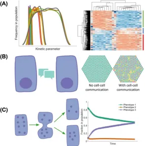
The co-existence of epithelial, mesenchymal and hybrid E/M subpopulations in a single cell line
indicates a population heterogeneity of EMT. Such heterogeneity can be generated and maintained
via multiple mechanisms all of which can contribute to the acquisition and maintenance of hybrid
E/M phenotypes (Figure 2). First, the EMT regulatory networks can be multi-stable [42,43,46,48].
Noise in the expression levels of the involved RNAs and proteins can thus cause transitions from
one stable steady state to another [68]. Such noise may arise from the inherent stochasticity of the
transcription process in cells [69] or from the random partitioning of parent cell RNAs and proteins
among the daughter cells at the time of cell division [70–72]. Since both noise sources are
cell-autonomous,, individual epithelial cells may spontaneously undergo a phenotypic transition and
acquire (or give rise to, in the second scenario) a hybrid E/M phenotype [73]. Thus, one must be
careful when choosing a cell line for experiments - a cell line known to be epithelial may include
substantial fractions of hybrid E/M and/or mesenchymal cells [55,73]. Second, the population
heterogeneity of EMT can arise via cell-cell communication. An example of such a communication
channel is Notch-Delta-Jagged signaling. Some cells with high levels of Delta/Jagged expression
This leads to a population that is inherently heterogeneous - a mix of sender and receiver cells.
Notch-Delta-Jagged signaling-mediated heterogeneity is closely tied to the emergence of hybrid
E/M phenotypes [74]. This will be discussed in more detail below. Third, population heterogeneity
of EMT can arise due to different kinetic parameters controlling gene regulation in different cells.
Due to the multitude of peripheral factors involved in governing the behavior of a core regulatory
circuit, the kinetics of the core circuit can vary from cell to cell leading to different responses to
the same external cues in different cells [46]. This might be connected to the aforementioned
random partitioning but might arise as well due to different long-lived fluctuations, perhaps related
to chromatic structure heterogeneity.
cell communication (left) mediated by, for example Notch-Delta-Jagged signaling, can lead to cells spontaneously acquiring “signal sender”, “signal receiver” and “signal sender/receiver” phenotypes in a population (right, different colors correspond to different cell phenotypes). (C) During cell division, noise can lead to asymmetric partitioning of molecules among daughter cells, thus leading to various phenotypes of daughter cells. As a result, the fractions of cells exhibiting different phenotypes can change over time.
In vivo characterization of hybrid E/M phenotypes
To identify whether cancer cells can acquire hybrid E/M phenotypes in vivo, Pastushenko et al.
used fluorescence-activated cell sorting (FACS) to screen cell surface markers of skin squamous
cell carcinoma (SCC) cells that can undergo spontaneous EMT and generate EpCAM+ epithelial
cells and EpCAM- mesenchymal-like cells in genetically engineered mouse models [10]. While
the EpCAM+ cells exhibit homogeneous expression of most of the markers, the EpCAM- cells
exhibit heterogeneous expression of 17 cell surface markers among which the most
heterogeneously expressed are CD61, CD51 and CD106. Consequently, the authors used
combinatorial multicolor FACS analysis of these three markers to further classify the EpCAM
-cells into 6 subpopulations among which the CD51−CD61−CD106−, CD51+ and CD106+
subpopulations exhibit co-expression of the epithelial marker keratin 14 (K14) and the
mesenchymal marker vimentin, thus can be tentatively considered to be hybrid E/M phenotypes.
The CD51+CD61+CD106+ and CD51+CD61+ subpopulations, on the other hand, are vimentin
positive and K14 negative and thus being mesenchymal-like. Intriguingly, the hybrid E/M
CD51−CD61−CD106− and CD106+ subpopulations generate significantly more metastases relative
to other subpopulations, though all subpopulations here share similar tumour-propagating
capacity. Similar results have also been observed in MMTV-PyMT mammary luminal tumours
[10]. Another study performed by Aiello et al. used a lineage-tracing mouse model of PDAC
showed that the PDAC cells undergoing EMT can acquire a hybrid E/M phenotype via
re-localization of epithelial proteins such as E-cadherin and claudin-7 from membrane to intracellular
foci [75]. The emergence of these hybrid E/M PDAC cells indicates that besides transcriptional
control, post-transcriptional regulation of localization can be also important to mediate the
existence of hybrid E/M phenotypes. Finally, another study by Puram et al. showed that the head
and neck squamous cell carcinoma (HNSCC) cells from patients exhibit a hybrid E/M phenotype
characterized by co-expression of epithelial markers such as EPCAM and KRT17 and
transcriptomic analysis [76]. Intriguingly, these hybrid E/M HNSCC cells tend to localize at the
leading edge of tumors close to surrounding stroma cells.
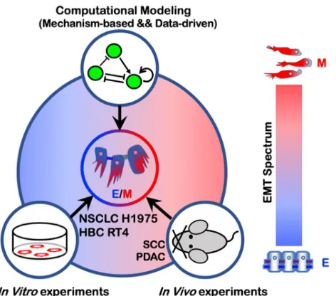
In summary, mathematical modeling together with both in vitro and in vivo experimental studies
consistently demonstrate the existence of multiple hybrid E/M phenotypes characterized by
varying extents of epithelial and mesenchymal features (Figure 3). It is worth noting that in
addition to the characterization of the hybrid E/M phenotypes, mathematical modeling of EMT
regulatory networks have generated many other interesting predictions that have been recently
validated by experimental studies. First, modeling the core EMT regulatory network –
miR-200/ZEB/miR-34/SNAIL suggests a sequential response of the EMT-TFs SNAIL and ZEB to the
EMT inducing signal TGF-β [43,44]. When treated with different levels of TGF-β, SNAIL is
predicted to precede ZEB and to be upregulated at relatively low TGF-β levels. The predicted
different responses of SNAIL and ZEB has been verified in MCF10A cells [43]. Second, another
prediction from modeling studies [42,43] is that EMT and MET are not necessarily symmetric
processes and hysteresis is expected during EMT. The predicted hysteretic behavior of EMT has
been recently demonstrated during TGF-β induced EMT of NMuMG and EpRAS cells, where the
levels of E-cadherin exhibit a bimodal transition. Such hysteretic behavior of E-cadherin is
regulated by the miR-200/ZEB1 circuit and blocking the inhibition of miR-200 by ZEB1 (referred
to in this study as mutant cells) results in only a unimodal transition of E-cadherin, though these
mutant cells can still undergo EMT with changes of EMT markers at a similar degree relative to
the wild type [77]. Moreover, TGF-β induced EMT of mutant cells exhibits significantly decreased
sphere-formation ability in vitro, decreased frequency of tumor-initiating cells and lung metastases
in vivo relative to their wild types. These results also confirm a prominent role of the miR-200/ZEB
circuit in regulating the aspects of EMT dynamics that result in a variety of functional
Figure 3. Emergence of hybrid E/M phenotypes demonstrated by a combination of theoretical and experimental efforts. The cell lines NSCLC H1975 and HBC RT4 exhibit a hybrid E/M phenotype at a single-cell level. Hybrid E/M phenotypes have also been characterized
in vivo using mouse models of SCC and PDAC.
EMT regulation by cell cooperation Notch signaling
Although EMT is fundamentally an individual cell phenomenon, it can also be regulated by cell
cooperation such as Notch signaling. Notch signaling is a cell-cell communication mechanism and
highly conserved across species. Originally characterized in Drosophila development, Notch
signaling is a conserved and well-known regulator of multiple hallmarks of cancer, including
angiogenesis and EMT [78–80].
The Notch signaling cascade is initiated by the binding of the Notch transmembrane receptor with
a ligand belonging to a neighboring cell. This binding leads to the cleavage of the Notch
intracellular domain (NICD), which is then released in the cytoplasm and transported to the cell
nucleus, where it acts as a transcriptional cofactor [81]. Notch signaling is deeply coupled to the
EMT regulatory networks discussed in previous sections. For example, on one hand,
EMT-inhibiting miRNAs miR-34 and miR-200 reduce the levels of Notch receptor and ligands [82–84]
by translational regulation [85]. On the other hand, NICD promotes the transcription of SNAIL
induce EMT in their neighboring cells through the binding of their ligands to a neighbor’s Notch
receptors.
As often seen in the developmental context, Notch signaling can give rise to different spatial
patterns of cell phenotypes due to feedback regulation between NICD and various alternate
ligands. Specifically, NICD transcriptionally represses ligands of the Delta family but activates
ligands of the Jagged family. Therefore, signaling through the Notch-Delta pathway typically
promotes opposite cell fates in neighboring cells, or ‘lateral inhibition’. This is accomplished by
amplifying initial differences in the levels of receptors and ligands, ultimately leading to one cell
with high levels of Notch receptor and low levels of Delta ligand (receiver cell) and a neighbor
cell with low Notch and high Delta (sender cell) [88,89]. Conversely, Notch-Jagged signaling
typically promotes a similar cell fate in neighboring cells, or ‘lateral induction’, because NICD
upregulates Jagged ligands [90].
(A )
(B )
N u m b e r o f c e lls
N u m b e r o f c e ll s
N o tc h -D e lta sig n a lin g
E p ith elia l
H yb rid E /M
M e se n c hym a l
N u m b e r o f c e lls
N u m b e r o f c e ll s
N o tc h -Ja g g e d sig n a lin g
E p ith elia l
H yb rid E /M
Figure 4. Notch-Delta and Notch-Jagged signaling give rise to opposite cell patterning of EMT. (A) Coupling of Notch-Delta signaling with the core EMT regulatory circuit. NICD suppresses the endogenous expression of Delta ligands upon activation of Notch receptors by exogenous Delta ligands (left, red color represents the ‘receiver phenotype’ (high notch), green color represents ‘sender phenotype’ (high delta)). Cell patterning of EMT in presence of a strong Notch-Delta signaling (right). (B) Coupling of Notch-Jagged signaling with the core EMT regulatory circuit. NICD promotes the endogenous expression of Jagged ligands upon activation of Notch receptors by exogenous Jagged ligands (left, blue color represents a ‘sender/receiver phenotype’ (high Jagged)). Cell patterning of EMT in presence of a strong Notch-Jagged signaling (right). Hexagons with different colors depict epithelial (green), hybrid E/M (yellow) and mesenchymal (red) cells. Figures in the right panel are adapted from [53].
The cell patterning mediated by Notch ligands can modulate epithelial-mesenchymal plasticity in
a tumor tissue due to the aforementioned coupling between Notch and the EMT regulatory circuits.
Intracellular Notch signaling activated by either Delta or Jagged can activate EMT. However,
mathematical modeling of the coupled regulatory networks regulating EMT and Notch signaling
suggests that Notch-Delta and Notch-Jagged signaling have dramatically different outcomes at a
multi-cellular level. While Notch-Delta signaling promotes a spatial arrangement where cells in a
partial or complete EMT are surrounded by epithelial cells, Notch-Jagged signaling can give rise
to clusters of hybrid epithelial/mesenchymal (E/M) cells [74] (Figure 4). Indeed, Jagged1 is one
of the top differentially expressed genes in collectively migrating cells of breast cancer [12,91].
These observations suggest that Jagged1 can act as an intercellular PSF that stabilizes a hybrid
E/M phenotype in a non-cell autonomous manner. Therefore, in addition to ‘conventional’ PSF
proteins that promote a partial EMT through direct crosstalk with the EMT regulatory circuitry,
such as OVOL, GRHL2, and NRF2 [51,52,62], other PSFs can facilitate a partial EMT and the
formation of CTC clusters by activating Notch-Jagged signaling and/or inhibiting Notch-Delta
signaling. For example, NUMB/NUMBL that forms a negative feedback loop with Notch [92–94]
can prevent a complete EMT, and consistent with that identification, knockdown of NUMB/
NUMBL result in a mesenchymal phenotype and enables individual migration of the hybrid E/M
NSCLC H1975 cells that typically migrate collectively [53].
Interaction among epithelial, hybrid E/M, and mesenchymal cells
Similar to bidirectional interactions among epithelium and mesenchyme during organ
development [95], there may be crosstalk and cooperation among cells exhibiting varying extents
crosstalk, due to their plasticity that can generate and maintain epithelial-mesenchymal
heterogeneity at a population level; such plasticity is limited for cells on either end of the EMT
spectrum. Moreover, hybrid E/M cells can maintain cell-cell adhesion via E-cadherin, and thus
potentially enabling formation of heterotypic clusters of CTCs with cells of varying EMT statuses
[99]. Last but not least, a recent study highlighted that the hybrid E/M HMLER cells, but not a
mixture of nonplastic ‘extremely epithelial’ (xE) cells and nonplastic ‘extremely mesenchymal’
(xM) cells, account for high tumorigenicity in vivo [100]. Notably, the nonplastic xE cells are
created by ZEB1 knockdown and the nonplastic xM cells are created by constitutive ZEB1
expression, which supports the role of ZEB1 in mediating EMT [42,44].
Whether a mixture of E and M cells is sufficient to initiate the metastatic cascade and successfully
form metastases remains to be resolved. Few molecular mechanisms enabling crosstalk between
E and M cells have been elucidated recently. Tsuji et al. demonstrated that when EMT and
non-EMT cells are inoculated subcutaneously in mice, they both establish primary tumors, but neither
of them form lung metastases [96]; the ability to invade local tissues and enter circulation is
demonstrated only by EMT cells. Further, when both cell types are injected intravenously,
non-EMT cells form overt metastases, but the metastatic ability of non-EMT cells is compromised. Finally,
when a mixture of EMT and non-EMT cells is subcutaneously injected, both cell types can
intravasate but only non-EMT cells form lung metastases. Put together, these observations indicate
a possible cooperation between EMT and non-EMT cells – while EMT cells can cleave the matrix
to make way for both cell populations to intravasate, the non-EMT cells can colonize distant organs.
This study did not identify any juxtacrine and/or paracrine signaling underlying this cooperation,
but recent in vitro co-culture experiments of EMT and non-EMT cells have identified a few players
that can mediate this crosstalk. HMLER cells overexpressing TWIST or SNAIL have been shown
to impart migratory and invasive traits in vitro to control HMLER cells via paracrine Hedgehog
signaling, but without explicitly inducing any morphological or molecular changes associated
typically with EMT [97]. The authors also demonstrated that the EMT cells are able to increase
the metastatic propensity of non-EMT cells in vivo, thus lending further credence to the notion that
EMT cells can stimulate the migration of non-EMT cells. This idea is further strengthened by
invasive potential which persisted for around 7 days after the co-culture but eventually decline
after being segregated from PC-3/S cells [101]. This increased invasive response is also observed
upon co-culture of PC-3/Mc cells with NIH3T3 fibroblasts, suggesting that the invasiveness of
non-EMT cells can be increased by both tumor and non-tumor mesenchymal cells [101]. In vivo
experiments for individual or co-injection of PC-3/S and/or PC-3/Mc cells corroborate previous
observations that the post-EMT cells had little, if any, contribution to distant organ colonization
[101]. Put together, these findings are reminiscent of in vivo studies showing that a persistent EMT
activation can reduce metastases formation [6,102], and clinical evidence that carcinoma
metastases are largely epithelial [103].
Reversible transitions among epithelial and mesenchymal phenotypes of disseminating cells has
been dogmatically considered as the driving engine of metastasis for a long time [1,104,105] (also
referred to as the ‘sequential metastasis’ model) [98], but with the proposed key role of EMT and
MET being relooked at more carefully [20], the possibility of a cooperative journey taken together
by epithelial and mesenchymal cells where they necessarily do not change their phenotypes cannot
be ruled out (also referred to as the ‘cooperative metastasis model’) [98]. Multiple possibilities
may underlie this cooperation: a) epithelial cells facilitate MET of mesenchymal cells, b)
mesenchymal cells facilitate the survival, persistence and re-adhesion of epithelial cells during
colonization, c) epithelial and mesenchymal cells exchange survival signals, d) a combination of
the above. Nevertheless, collective transport of epithelial and mesenchymal cells is likely to be
more effective for colonization as compared to that of one population alone. However, the role of
hybrid E/M cells in this cooperative metastasis model remains to be explored.
What mechanisms may allow such collective migration of epithelial and mesenchymal cells? In
developmental contexts, epithelial or mesenchymal cells have been seen to migrate collectively
through respective cell-cell contacts mediated by E-cadherin or N-cadherin [106,107]. Recently, a
N-cadherin/E-cadherin mechanically active heterophilic adhesion among the cancer-associated
fibroblasts and cancer cells was reported to guide collective migration of tumor cells [108]. Given
that the heterophilic E-cadherin/N-cadherin interaction has been proposed to be of similar affinity
as that of homophilic E-cadherin interactions [109], collective cell migration can be expected to
a short-ranged interaction via EGF/CSF-1 paracrine axis to mediate macrophage-driven tumor cell
migration [110,111]. CSF-1/CSF-1R axis has been recently proposed to associate with a hybrid
E/M phenotype in inflammatory breast cancer [108] - a highly aggressive breast cancer subtype
that metastasizes via clusters or emboli of circulating tumor cells [91]. These mechanisms may
mediate, at least in part, a collective cooperative migration of cells in varying hues of EMT.
Increased plasticity of hybrid E/M cells may be necessary and sufficient to maintain and propagate
the non-genetic heterogeneity in terms of EMT status in a given isogenic cancer cell population.
Mechanical control of EMT by ECM
In addition to chemical communication, the stiffness of ECM also plays a key role in regulating
EMT. For example, cancer cells when cultured in stiffer substrates exhibit increased migratory and
invasive ability and become more mesenchymal-like [112]. Alternation in ECM stiffness can
trigger multiple signaling pathways to regulate EMT, such as TWIST1-G3BP2 [23], HA-CD44
[113] , MRTF-A [114], PI3K/Akt [115] and YAP/TAZ [116]. Yet, the reversibility of ECM
stiffness-induced EMT can be cell line-dependent. For example, the mammary epithelial cells that
have undergone EMT in a stiff substrate partially revert to epithelial phenotype [117], while the
colon carcinoma HCT-8 cells can retain their mesenchymal-like phenotype, all after being
re-cultured in the compliant substrate [118]. It is worth noting that cells undergoing EMT can in turn
regulate ECM. For example, the LOX-family enzymes are upregulated in fibrosis and upregulation
of LOX-family enzymes can directly increase connectivity of collagen fibers, stabilize and stiffen
the collagen networks [119,120]. Given the importance of mechanical regulation of EMT,
mathematical models that integrate mechanical with chemical signaling networks need to be
developed to better understand EMT-ECM dynamics [121].
Quantification of the EMT spectrum
Our discussion so far has hopefully made it clear that cells undergoing EMT can acquire a spectrum
of hybrid E/M states both in vitro and in vivo. However, the lack of a rigorous quantification of
the EMT spectrum, namely, the exact proportions of epithelial, mesenchymal and hybrid E/M
subpopulations of cell lines and clinical samples, can lead to potentially contradictory conclusions
Cells undergoing EMT typically alter both their omics profiles and morphologies. Therefore, in
principle, the EMT spectrum can be quantified via evaluating the change of cell morphology and/or
their omics profiles. To classify epithelial and mesenchymal phenotype at a single-cell level,
Leggett et al. developed a probabilistic classification scheme using Gaussian mixture model
(GMM) focusing on 4 morphological features of single cells – maximum radius of the nucleus,
vimentin area, cytoplasm form factor and maximum feret diameter of cytoplasm [124]. The GMM
is trained using the morphological features of DMSO-treated (epithelial) and 4-hydroxytamoxifen
(OHT)-treated (mesenchymal) human mammary MCF-10A cells which are transfected with an
inducible Snail construct, referred to as MCF-10A Snail cells. The probabilistic GMM model has
revealed various EMT kinetics of MCF-10A Snail cells when induced by TGF-β1, plating density
and the microtubule inhibitor Taxol respectively. This GMM model also provides insight into the
EMT status of individual cells which may be overlooked by population-average analysis.
However, this method only focuses on a binary classification of EMT – epithelial or mesenchymal
and one missing piece of this model is the classification of hybrid E/M phenotypes.
To quantitatively measure the EMT status of cell lines with specific attention to a hybrid E/M
phenotype, George et al. developed an EMT scoring metric to calculate the probability of a given
sample to be hybrid E/M phenotype and assign a score between 0 and 2 with 0 being fully
epithelial, 2 being fully mesenchymal and 1 representing hybrid E/M [125]. Using the gene
expression data of NCI-60 human tumor cell lines as the training set, the ratio of VIM to CDH1
together with CLDN7 expression are identified as the best-fit pair of predictors to classify EMT
phenotypes. This EMT scoring metric has been used to characterize multiple hybrid E/M cancer
cell lines including A549 and DU145. Furthermore, this EMT scoring metric has been extended
to distinguish hybrid E/M cells from mixtures of epithelial and mesenchymal cells [60]. Another
EMT scoring metric developed by Tan et al. assigns a score between -1 and 1 to a given sample
with -1 being fully epithelial and 1 being fully mesenchymal [126]. Both George et al. and Tan et
al. demonstrated that patient samples that are more mesenchymal-like do not necessarily correlate
with worse overall and disease-free survival results and do not always show resistance to
chemotherapy, indicating a subtype-dependent role of EMT in cancer progression and therapy
resistance. In summary, these methods to quantify EMT status help address the multifaced roles
EMT and CTC clusters
Cancer cells that acquire a hybrid E/M phenotype maintain both epithelial (e.g. cell-cell adhesion)
and mesenchymal (e.g. migration) features thus can migrate collectively as a cluster. Such clusters
of migrating tumor cells have been shown to be one of the primary instigators of metastases
[12,127]. The experimental studies supporting this notion are discussed below.
Using multicolor lineage tracing, Cheung et al. showed by two sets of experiments that the CTC
clusters are mainly formed by multi-cellular clusters from the primary tumors. In the first set of
experiments, two differently colored populations - mTomato+ and CFP+ MMTV-PyMT tumor
organoids were respectively transplanted into the right and left flanks of a nonfluorescent mouse.
After 6 to 8 weeks, only single-colored metastases were observed in lung. In the second set of
experiments, mTomato+ single cells isolated by FACS were transplanted into a nonfluorescent
mouse via tail-vein injection and 2 days later FACS isolated CFP+ single cells were injected into
the same mouse. After 2 days, exclusively single-colored metastases were observed in lung. These
two sets of experiments suggest that the polyclonal metastases in lung is more effectively
generated by multicellular seeds and not by serial aggregation of single tumor cells [12]. The CTC
clusters exhibit enriched expression of an epithelial cytoskeletal protein K14 that is required for
the collective invasive behavior and distant metastasis. Another study supporting the idea that CTC
clusters can arise as oligoclonal groups of cells detached from the primary tumors is Aceto et al.
who identified plakoglobin as a key mediator for CTC cluster formation [128].
An alternative mechanism – that CTC clusters can be formed via aggregation of single CTCs in
circulation - has been recently demonstrated by Liu et al. using intravital multiphoton microscopic
imaging in both patient-derived xenograft (PDX) models of triple negative breast cancer (TNBC)
and PyMT transgenic mouse models [127]. Liu et al. first showed that in
chemoattractant-containing matrigel about 20% of invasion events of the tumor cells collected from the PDX
models occur as multicellular aggregates, and in suspension culture individual CTCs derived from
a breast cancer patient aggregate into clusters within 1 to 2 hours. These results indicate that single
MDA-aggregates within 2 hours. The percentage of the dual-colored MDA-aggregates is affected by the timing
of the eGFP+ and tdTomato+ entering blood vessels. Consequently, sequential infusion of the
tdTomato+ cells 5 minutes, 10 minutes and 2 hours after the infusion of the eGFP+ cells result in
gradually decreased percentages (27%, 16% and 10%) of the dual-colored aggregates. The authors
further showed that the CTC clusters exhibit notably enriched expression of CD44, and that the
intercellular CD44 homophilic interaction is responsible for the aggregation of single CTCs.
One major difference in the studies by Cheung et al. and Liu et al. is the timing of the
second-colored single cells entering the blood vessels. Cheung et al. injected the second-colored cells into
the tail vein 2 days after the injection of the first-colored cells while Liu et al. waited for at most
2 hours to inject the second-colored cells. As discussed by Liu et al., the timing of the
second-colored cells entering the blood vessel could have significant effect on the percentages of
dual-colored lung metastases. From our perspective, the key issue in these two studies seems to be the
lifetime and density of the injected single cancer cells. Since it is not clear how often the injected
cells are expected to reach the bloodstream, it is hard to evaluate which protocol is a better match
to reality. The experiments by Cheung et al. where two differently colored populations of tumor
organoids are injected respectively into the right and left flanks of nonfluorescent mice seems to
be a better approach since the interaction of cells in the bloodstream is determined naturally rather
than by the experimenters. Nonetheless, both studies showed that the CTC clusters significantly
promote colony formation and lung polyclonal metastases in vivo. Liu et al. showed through
CellSearch platform-based blood analysis that the breast cancer patients with CTC clusters exhibit
significantly worse overall survival results relative to those with only single CTCs. And, CTC
clusters in different contexts can be different. For example, Khalil et al. showed that though
invasive ductal carcinoma (IDC) and invasive lobular carcinoma (ILC) exhibit collective invasion
patterns, the collective invasion in IDC lesions maintains intercellular E-cadherin while collective
invasion in ILC lesions loses intercellular E-cadherin but retains CD44 for intercellular junctions
[129]. All together, these studies suggest that CTC clusters can in principle be generated both by
cohesive shedding from the primary tumors and/or serial aggregation of single cells in the
To model the migration of CTC clusters, Bocci et al. proposed a simple biophysical model where
cancer cells can undergo a partial EMT that allows both single-cell and clustered-cell migration,
or a complete EMT that only allows single-cell migration [130]. According to this reduced physical
model, a tumor can undergo a transition from primarily single cell-based invasion to collective
invasion. Strikingly, this theoretical framework reproduces multiple CTC cluster size distributions
measured in patients and mouse models across cancer types, hence suggesting the existence of a
unifying set of principles governing cancer cell migration [130].
Adding to the complexity of the problem, CTC clusters can associate with non-cancer cells
including platelets and/or immune cells such as tumor-associated macrophages and neutrophils
[131–133]. In particular, it is well recognized that interactions between macrophages and tumor
cells are very important for tumor cell intravasation and extravasation, though the detailed
mechanisms have not been elucidated [134,135]. In addition, the CTC-neutrophil clusters lead to
more efficient metastasis relative to CTCs alone [131]. Future studies need to be performed to
elucidate the cross-talk between CTC clusters and different types of immune cells.
EMT and stemness in tumor microenvironment
We have already come across the fact that besides the migratory and invasive traits conferred by
EMT, some cancer cells undergoing EMT can also acquire an enhanced ability to drive tumor
initiation and an enhanced therapy resistance. These properties are associated with the notion of
‘stemness’, and such cells are sometimes referred to as cancer stem cells (CSCs). The connection
between EMT and stemness was first proposed by Brabletz et al. [24] more than a decade ago
based on the premise that EMT and stemness cannot explain the different steps of the metastatic
cascade when considered as separate and independent processes [24]. Indeed, increasing
experimental evidence suggests a strong association of EMT with stemness. For instance, human
mammary epithelial cells (HMLEs) undergoing EMT can express stem cell markers and exhibit
increased mammosphere formation [122]. Moreover, reversing EMT via knockdown of SNAIL
represses stemness and tumor growth in ovarian cancer [136]. This and other evidence suggests
that the activation of the EMT program leads to the acquisition of stemness [137]. This correlation,
Revisiting the EMT-CSC connection through the lenses of epithelial-mesenchymal plasticity and
hybrid E/M phenotypes, however, offers a more consistent picture. Mathematical modeling
approaches have been applied to decode the connection of EMT with stemness via simulating the
coupled decision-making regulatory networks of EMT and stemness. Specifically, EMT activation
can downregulate let-7, a miRNA typically associated with repressing stemness. Let-7 can bind to
ZEB and promote its degradation. These two processes can settle at an intermediate level of let-7
and ZEB leading to hybrid E/M CSCs [139]. This association, however, can vary depending on
the coupling between the modules governing EMT and stemness, allowing for CSCs in an
epithelial, mesenchymal or hybrid E/M phenotype [139]. In addition, local perturbations in the
tumor microenvironment such as TGF-β or Notch signaling also can modulate the association of
CSCs with different EMT phenotypes [140,141]. All told, emerging evidence from theoretical and
experimental studies tends to associate the hybrid epithelial/mesenchymal phenotypes with
stemness [139,140,142–145].
Along these lines, the association of hybrid E/M phenotypes with stemness can be promoted by
the PSFs – OVOL and Jagged1. Indeed, Jagged1 is typically overexpressed in CSC populations as
compared with non-CSCs in multiple types of cancer including glioblastoma, pancreatic cancer,
colon cancer, and breast cancer [22,146–148]. Through modeling the coupling of the regulatory
networks governing EMT, stemness and Notch signaling, Bocci et al. proposed that Jagged1
facilitates a ‘window of opportunity’ that confers maximal invasion potential in terms of EMT and
stemness [140,145]. A follow-up work showed that knockdown of Jagged1 impairs breast
organoid formation in vitro, therefore highlighting a causal relationship between Jagged1 and
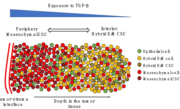
Figure 5. The spatial patterning of CSCs with different EMT phenotypes. Tumor-stroma interactions can give rise to a gradient of TGF- β (top, blue scale). In the periphery of the tumor, most cells are mesenchymal (red spheres), while the interior is mostly composed by hybrid E/M and epithelial cells (yellow and green spheres, respectively). CSCs are mostly mesenchymal in the periphery (black-dotted red spheres) and mostly hybrid E/M in the interior (black-dotted yellow spheres).
Subpopulations of CSCs with different EMT phenotypes can be representative of the spatial
organization of a tumor tissue. In breast cancer, CD24-/CD44+ mesenchymal CSCs are typically
located toward the invasive edge of the tumor by the tumor-stroma interface, while ALDH1+ CSCs
are found in the more interior region [149]. Interestingly, ALDH1+ CSCs were originally
considered as epithelial-like, but their RNA sequencing (RNA-Seq) data has later shown that these
cells lean more toward a hybrid E/M phenotype and share several genes with TNBC signature
[150]. Recently, Bocci et al. have argued that this spatial segregation can be qualitatively explained
by the interplay of cancer cells with the tumor microenvironment [141]. Cytokines such as the
EMT-inducer TGF-β are typically secreted at the tumor-stroma interface and give rise to a gradient
of EMT-inducing signal throughout the tumor tissue. Therefore, CSCs at the invasive edge are
highly exposed to TGF-β and tend to undergo a complete EMT leading to a fully mesenchymal
phenotype, while CSCs in the interior maintain a hybrid E/M phenotype [141] (Figure 5).
Consistently, in non-small cell lung CSCs, a subpopulation of mesenchymal CSCs exhibits high
expression of TGF-β targets such as SNAI1 and ZEB1, as compared to a hybrid E/M CSC
population [140].
E p ith e lia l c e ll H yb rid E /M c e ll M e se n c h ym a l c e ll
P e rip h e ry M e se n c h ym a l C S C
In te rio r H yb rid E /M C S C
D e p th in th e tu m o r tissu e Tu m o r-stro m a
in te rfa c e
H yb rid E /M C S C M e se n c h ym a l C S C
In summary, EMT and CSC formation represent two essential axes of tumor progression, whose
connection is modulated by factors including tumor microenvironment and cell-cell signaling
[141,149,151,152]. The stem cell paradigm inherited from developmental biology implies a
hierarchical lineage of cells that gradually but irreversibly differentiate [153,154]. In the context
of cancer, however, stemness has proven to be a dynamical property that cells can gain and lose
[155–157]. The complex interplay between EMT, stemness and tumor microenvironment gives
rise to tumor heterogeneity that still represents the major challenge hindering metastasis and
therapy resistance [151].
EMT and immune suppression
In addition to cancer cells, a solid tumor harbors other types of cells which form the tumor
microenvironment and strongly affects cancer outcome [26]. Specifically, many groups have
investigated the roles of immune cells in cancer progression. Certain types of immune cells, such
as macrophages and T cells, can comprise up to 50% of the cells in a solid tumor [27]. These
immune cells usually polarize into phenotypes, such as M2-like macrophages and regulatory T
helper cells (Tregs), that promote tumor progression via suppressing the activity or viability of
anti-cancerous immune cells, e.g., M1-like macrophages and cancer-killing T effector cells
[26,27,158]. Our goal here is to focus on the role of EMT in this tumor-immune interplay.
A series of mathematical models have been proposed to understand the roles of tumor-immune
interactions in polarizing the cytokine-immune cell network into states dominated by either
immune-promoting or immune-suppressing populations [159–164]. Such modeling frameworks
can help to design effective perturbations to revert the immune microenvironment from an
immune-suppressing to an immune-promoting one. Many of these models consider the fact that
macrophages and cancer cells can directly interact with each other and regulate the behaviors of
one another, as shown by many in vitro experiments [165–170]. The interactions between
macrophages and cancer cells are formidably complex, and the emergent dynamics can be
non-intuitive. A series of mathematical models capturing these interactions suggests that cancer cells
in the epithelial-like state (less aggressive) and M1-like macrophages might form a stable
ecosystem, whereas cancer cells in the mesenchymal-like state (more aggressive) and M2-like
The question of establishing an immune-dominated versus immune-suppressed microenvironment
should have an effect on the activity of cd8+ effector T-cells. There have been studies suggesting
that T cells tend to be excluded from tumors enriched by mesenchymal-like cancer cells [172,173];
we will discuss this further below. Combining this data with the modeling results, one could argue
that interactions between macrophages and cancer cells drive the macrophages to be M2-like and
the cancer cells to be mesenchymal-like; and due to the effects of M2-like macrophages, T cells
may be excluded from tumor areas enriched with mesenchymal-like cancer cells. Since both the
infiltration of cancer-killing immune cells [33–39] and the epithelial-mesenchymal plasticity of
cancer cells are important for the prognosis of cancer patients, it is clearly valuable to evaluate the
association and ultimately the casual relationship between the two.
In the following, we will first describe in greater detail the corresponding in vitro and in vivo
experiments as well as analyses of gene expression data from The Cancer Genome Atlas (TCGA)
(Figure 6). Finally, we discuss the potential causal relationship between the EMT status of cancer
cells and the infiltration of cancer-killing immune cells.
mou se
mod el
in vitro co
-cu ltu
re
TCGA dat
a
anal ysis
Ou r m
od eling effo
Figure 6. The relationship between EMT and immune response as shown by various approaches.
In vitro characterization of the immune-suppressing role of mesenchymal-like cancer cells
For the interaction between mesenchymal-like cancer cells and immune cells, TGF-β signaling has
been studied extensively. TGF-β is a well-known EMT inducer [21] and can be secreted by
tumor-associated fibroblasts [174,175] and tumor-tumor-associated macrophages [176]. TGF-β signaling can
impair maturation, differentiation, and/or activation of both innate and adaptive immune cells
[31,177]. Specifically, TGF-β can inhibit the functions of cytotoxic T-cell functions [178,179]. In
the co-culture experiments, Joffroy et al. showed that TGF-β secreted by cancer cells induces the
differentiation of CD4+ T cells into Treg (Foxp3+) cells, which are immune-modulating cells [178].
Similar cancer-cell dependent expansion of CD4+Foxp3+ T cells is also shown by Kudo-Saito et
al. when co-culturing SNAIL-enhanced mouse melanoma B16F10 cells with CD4+ T cells [172].
Furthermore, TGF-β can downregulate the MHC class I proteins as shown in prostate cancer [180]
as well as NSCLC cell lines [181].
Aside from TGF-β, the PD-L1/PD-1 axis has also attracted attentions due to its implications in the
immune-checkpoint blockade therapy. Presumably, PD-L1 expressed by cancer cells can bind to
PD-1 expressed on the surface of cancer-killing immune cells, which will modulate their cytotoxic
functions. It is shown that when driving EMT via downregulating miR-200 and overexpressing
ZEB1, PD-L1 expression by cancer cells is upregulated [182]. Therefore, mesenchymal-like
cancer cells could be more resistant to cancer-killing immune cells by upregulating PD-L1.
Interestingly, expression levels of both TGF-β1 and PD-L1 by cancer cells can be induced under
hypoxic conditions [183–185]. The hypoxia-induced EMT can promote immunosuppression via
induced expression of indoleamine 2, 3-dioxygenase (IDO, another T-cell suppressing factor) in
monocyte-derived macrophages [186].
EMT can also be induced when cancer cells are challenged by immune cells or inflammatory
cytokines [187]. In addition, cancer cells can be equipped with immunomodulatory effects which
interfere with proliferation, differentiation and apoptosis of NK, T-cell, and B-cell populations
effects on T cells after inflammation-induced EMT [187]. These experimental results are
potentially helpful for establishing a potential causal relationship between EMT of cancer cells
and the infiltration of cytotoxic immune cells.
In vivo characterization of the immune-suppressing role of mesenchymal-like cancer cells
Generally, in vivo experiments using mouse models show that tumors formed by
mesenchymal-like cancer cells are less infiltrated with cancer-killing immune cells and/or more infiltrated by
immune-suppressing immune cells. For example, Kudo-Saito et al. showed that in mice, compared
with tumors formed by mock-transfected B16F10 cells, tumors derived from Snail-transduced
B16F10 cells exhibit less tumor-infiltrating CD8+ T cells, while more Tregs, and form more lung
metastases [172]. In the same mouse model, the chemokine CCL2 can recruit immune-modulating
populations such as MDSC and macrophages [188], which are responsible for creating the
immune-suppressing microenvironment. In addition, in a mouse model of breast cancer [173],
tumors formed by mesenchymal carcinoma cell lines contained more Tregs, M2 macrophages and
exhausted CD8+ T cells, whereas tumors formed by more epithelial carcinoma cell lines contained
CD8+ T cells and M1 macrophages. Furthermore, using transgenic mouse models, Spranger et al.
showed that an active β-catenin signaling in cancer cells contributes to a lack of T cell infiltration
in tumor sites and resistance to anti-PD-L1 and/or anti-CTLA4 mAb therapy [189].
In summary, from the perspective of direct experiment in vitro or in preclinical models it is
becoming clear that mesenchymal-like cancer cells can directly suppress the function of
cancer-killing immune cells as well as promote the immune-suppressing microenvironment by recruiting
or polarizing immune-suppressing immune cells. Following this logic, one would expect a lower
presence of functional cancer-killing immune cells in the tumor area enriched with
mesenchymal-like cancer cells.
Gene expression data analysis
What about the results for patients? Unfortunately, the overall picture here as it emerges from
TCGA data analyses tends to suggest that while mesenchymal tumors are generally enriched with
samples with high EMT scores correlate with high expression of several immune checkpoints
including PD-1, PD-L1, CTLA4, OX40L, and PD-L2 [190]. Lou et al. observed similar trend in
lung cancer patients with early or advanced NSCLC adenocarcinomas [191]. Aside from the
immune checkpoint markers, Lou et al. also found that the lung tumors that displayed an EMT
phenotype also have a higher infiltration of Tregs. Interestingly, in their work, some immune
costimulatory molecules such as CD80 and CD86 as well as IFN- signals are more highly
expressed in “mesenchymal” lung adenocarcinoma. Reports also suggest that, in the claudin-low
subtype of breast and bladder cancer, which are mesenchymal-like, tumors are generally
well-inflamed by immune-promoting immune cells but these cells are under active immunosuppression
[192].
It should be noted that data in TCGA is rarely from tumor cells exclusively, thus the mesenchymal
features seen there may be a consequence of higher infiltration of stromal cells. Conversely,
analysis of epithelial markers [193] may not be biased by stromal cells. When investigating the
epithelial marker ESRP1 for melanoma samples in TCGA dataset, Yao et al. found that a high
infiltrating lymphocyte activity is enriched in ESRP1-low melanoma samples which have
enhanced mesenchymal features [193]. The lymphocyte activity was evaluated by the gene
expression of two cytotoxic agents - perforin (PRF1) and granzyme A (GZMA). Considering the
use of an epithelial marker instead of mesenchymal markers, this work may be a strong piece of
evidence, since the contamination by mesenchymal markers from non-cancer cells is supposed to
be low. However, the immune infiltration still needs to be defined rigorously. It is possible that
cytotoxic immune cells are constrained to lie in the tumor stroma instead of infiltrating into the
tumor islets, though many of them infiltrate into the core of the tumor. For these tumors, the bulk
tumor gene expression data will give a high infiltration score of cytotoxic immune cells. If these
tumors happen to be ESRP1-low tumors, we are likely to conclude that a higher infiltration of
cytotoxic immune cells is associated with more mesenchymal-like cancer cells.
Although the above-mentioned evidence tends to suggest that a higher infiltration of cytotoxic
immune cells associates with the mesenchymal features of tumor samples, the jury is still out, and
an opposite trend has been reported elsewhere. For example, Chae et al. reported that EMT is