Modulation of the AMPA Receptor by Membrane
cholesterol and neuroactive steroids
A thesis submitted for the Degree o f Doctor o f Philosophy, University o f London
By
Farnosh Fariba BPharm MRPharmS
Department o f Pharm acology The School o f Pharmacy
July 20 03 U niversity o f London
ProQuest Number: 10104718
All rights reserved
INFORMATION TO ALL USERS
The quality of this reproduction is dependent upon the quality of the copy submitted.
In the unlikely event that the author did not send a complete manuscript and there are missing pages, these will be noted. Also, if material had to be removed,
a note will indicate the deletion.
uest.
ProQuest 10104718
Published by ProQuest LLC(2016). Copyright of the Dissertation is held by the Author.
All rights reserved.
This work is protected against unauthorized copying under Title 17, United States Code. Microform Edition © ProQuest LLC.
ProQuest LLC
789 East Eisenhower Parkway P.O. Box 1346
Abstract
Cholesterol in the plasma membrane of cells has been found to modulate the actions of
various proteins in the membrane. This thesis describes the influence of membrane
cholesterol on the excitatory AMPA receptor in acutely dissociated rat hippocampal
neurones. Non-genomic influences of acute exposure to neuroactive steroids on the
function of the AMPA receptor have also been investigated.
Neuronal membrane cholesterol levels were altered by means of the cholestrol carrier
methyl-P-cyclodextrin. Using the patch clamp technique, whole cell current responses
to local applications of AMPA were recorded. Depletion of cholesterol to 62% control
increased the potency of AMPA, whilst enrichment to 205% control decreased the
potency of AMPA and the maximum response obtainable. The AMPA responses
showed rapid desensitization, which was reduced in cholesterol-depleted neurones and
increased in cholesterol-enriched neurones. NBQX antagonised responses to AMPA
competitively but failed to affect the relationship between desensitization and AMPA
concentration. GYKI 52466 reduced the maximum response to AMPA and shifl;ed the
desensitization profile to higher AMPA concentrations
The neuroactive steroids pregnenolone sulphate, deoxycorticosterone, corticosterone and
progesterone reduced the maximum response to AMPA and reduced its potency whereas
tetrahydrodeoxycorticosterone, allopregnanolone and epiallopregnanolone simply
reduced the potency of AMPA. The 5P isomers of the former two steroids were
inactive. Cholesterol depletion enhanced and enrichment diminished the reductions in
Possible mechanisms underlying these novel observations with cholesterol and the
neuroactive steroids include membrane fluidity changes and specific modulatory sites
CONTENTS
Page
Abstract... i
Contents... iii
List of Figures...ix
List of Tables...xvi
List of Publications... xxi
Chapter 1. General Introduction 1.1 Aims and Objectives... 2
1.2 Plasma membrane of neuronal cells... 3
1.3 Cholesterol and biological membranes... 4
1.3.1 Location of cholesterol in the membrane...4
1.3.2 Cholesterol absorption and synthesis... 7
1.3.3 Cholesterol effects on the physical properties of the membrane... 8
1.3.4 Cholesterol influence on the functions of membrane proteins...9
1.4 Glutamate and excitatory receptors... 11
1.5 The AMPA receptor... 13
1.5.1 AMPA modulation of cationic channels... 15
1.5.2 Divalent permeability...15
1.5.3 The number of agonist binding sites... 16
1.5.4 Association of glutamate receptors with intracellular proteins... 17
1.5.5 Internal polyamine block of AMPA receptors... 18
1.5.7 Chemical modulators o f AMPA receptors... 23
1.6 AMPA receptors and the Hippocampus...25
1.7 Developmental regulation of AMPA receptor... 29
1.8 AMPA receptors in synaptic plasticity and development... 30
1.9 Mechanism of excitotoxicity and neuronal damage... 31
1.10 Steroids and neuronal modulation...33
1.10.1 Biosynthesis of neuroactive steroids... 34
1.10.2 Modulation of neurotransmitter receptors by neuroactive steroids... 37
1.10.3 Modulation of AMPA receptors by neuroactive steroids... 39
Chapter 2. Materials and Methods 2.1 Introduction... 42
2.2 Dissociation of Hippocampal Neurones... 42
2.3 Preparation of methyl-p-cyclodextrin solution and the cholesterol inclusion complex... 44
2.4 Preparation of epicholesterol-methyl-p-cyclodextrin inclusion complex...44
2.5 Manipulation of membrane cholesterol using methyl-p-cyclodextrin and its cholesterol and epicholesterol inclusion complex...45
2.6 Cholesterol and protein assay of hippocampal neurones... 46
2.7 Electrophysiological measurements... 50
2.8 Construction of a U-tube...51
2.9 Standardisation of AMPA current responses... 54
2.10 Drugs and chemicals...55
2.11 Data analysis... 56
Chapter 3. Manipulation of Membrane Cholesterol
3.1 Introduction... 60
3.2 The viability of hippocampal neurones... 61
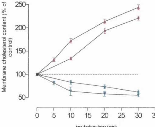
3.3 Alteration of membrane cholesterol content by methyl-P-cyclodextrin and its cholesterol inclusion complex... 61
3.4 Effect of time elapsed on membrane cholesterol content of neurones... 65
3.5 Discussion... 66
Chapter 4. Effect of Membrane Cholesterol on the Sensitivity of AMPA receptors to AMPA 4.1 Introduction... 69
4.2 Effect of membrane cholesterol on AMPA currents... 70
4.2.1 Effect of membrane cholesterol on peak AMPA currents... 72
4.2.2 Effect of membrane cholesterol on plateau AMPA currents...75
4.2.3 Effect of cholesterol on the decay from peak to plateau AMPA currents... 78
4.2.4 Effect of cholesterol on the rate of current decay from peak... 79
4.3 Effect o f membrane cholesterol on the current-voltage relationship...81
4.4 Restoration of membrane cholesterol following depletion... 83
4.5 Effect of acute application of cholesterol on AMPA currents... 86
Chapter 5. Effect of Epicholesterol on the Sensitivity of AMPA receptors to AMPA
5.1 Introduction...95 5.2 Effect of incubation with epicholesterol on AMPA responses... 97
5.2.1 Effect of epicholesterol enrichment on peak AMPA
currents... 98 5.2.2 Effect of epicholesterol enrichment on plateau AMPA
currents...101 5.2.3 Effect of epicholesterol on the decay from peak to plateau
AMPA currents... 103 5.3 Discussion... 104
Chapter 6. Effect of Neuroactive steroids on the Sensitivity of AMPA Receptors to AMPA
6.1 Introduction... 108 6.2 Selection of AMPA receptor modulators... 109 6.3 Effect of cortisol on AMPA receptor responses...I l l 6.4 Modulation of AMPA receptor responses by pregnenolone
sulphate... 112 6.5 Modulation of AMPA receptor responses by deoxycorticosterone
and corticosterone...118 6.6 Modulation of AMPA receptor responses by progesterone... 123 6.7 Modulation of AMPA receptor responses by THDOC... 125 6.8 Modulation of AMPA receptor responses by allopregnanolone and
epiallopregnanolone...130 6.9 Comparison of the influence of neurosteroids on the rate of decay of
AMPA currents from peak... 135 6.10 Further investigation of the neuromodulatory site/s on the AMPA
6.11 Discussion... 140
Chapter 7. Effect of Membrane Cholesterol on the Modulation of AMPA Currents by Neuroactive Steroids
7.1 Introduction... 148 7.2 Effect of the manipulation of plasma membrane cholesterol on the
modulatory influence of 10 pM pregnenolone sulphate... 150 7.3 Effect of the manipulation of plasma membrane cholesterol on the
modulatory influence of 1 pM deoxycorticosterone... 154 7.4 Effect of the manipulation of plasma membrane cholesterol on the
modulatory influence of 10 pM THDOC...157 7.5 Effect of epicholesterol enrichment... 160 7.6 Discussion... 163
Chapter 8. Effect of Membrane Cholesterol on the Modulation of AMPA Currents by Receptor Antagonists
8.1 Introduction... 167 8.2 NBQX antagonism...169 8.2.1 Effect of NBQX on AMPA currents... 170 8.2.2 Effect of NBQX on the decay from peak to plateau AMPA
currents...173 8.2.3 Effect of the manipulation of plasma membrane cholesterol
on NBQX antagonism... 174 8.3 GYKI 52466 antagonism... 179
8.3.1 Effect of GYKI 52466 on AMPA EC50... 179 8.3.2 Effect of GYKI 52466 on the decay from peak to plateau
AMPA currents... 181 8.3.3 Effect of the manipulation of plasma membrane cholesterol
8.4 Discussion...189
Chapter 9. General Discussion 9.1 The influence of membrane cholesterol on AMPA... 193
9.2 Comparison with the effect of membrane cholesterol on other receptors...195
9.3 The influence of neurosteroids on AMPA...196
9.4 Cholesterol interaction with modulators of the AMPA receptor... 200
9.5 Sites of cholesterol-neuroactive steroid interaction...203
9.6 In-Vivo implications of AMPA receptor modulation...207
a) Cholesterol implications... 207
b) Neurosteroid implications...210
9.7 Conclusion... 214
List of Figures
1.1 Structure of cholesterol (5-cholesten-3 3-ol)... 4
1.2 Model of synaptic plasma membrane lipid domains...6
1.3 Schematic representation of the AMPA receptor subunits... 14
1.4 The proposed domain structure of iGluRs... 21
1.5 The chemical structure o f DL-a-amino-3-hydroxy-5-methyl-4-isoxazole propionic acid (AMPA)...23
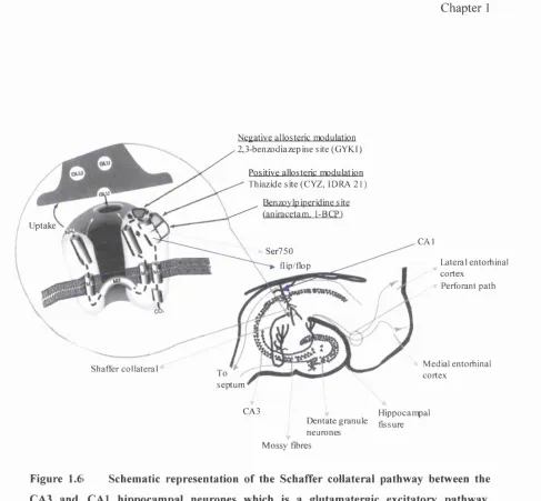
1.6 Schematic representation of the Schaffer collateral pathway between the CA3 and CAl hippocampal neurones which is a glutamatergic excitatory pathway... 28
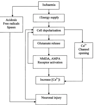
1.7 Potential paths leading to neuronal injury resulting from an episode of ischaemic insult...32
1.8 The lettering of the steroid rings and numbering of the carbon atoms in the steroidal structure, showing pregnanolone... 35
1.9 Outline of the synthetic pathways for neuroactive steroid synthesis in the nervous system... 36
2.1 True phase photomicrograph of an acutely dissociated hippocampal neurone, prior to cholesterol manipulation procedure... 43
2.2 Standard curve of cholesterol measured by cholesterol diagnostic kit (Sigma, US)...49
2.3 Standard curve of protein measured by BioRad Detergent Compatible Protein Assay kit from BIO-RAD (Germany)... 49
2.4 Schematic diagram showing the stages o f U-tube fabrication process... 53
2.5 Schematic diagram showing AMPA receptor response to a 1.0 second application of AMPA in comparison to G ABA receptor response to G ABA...54
4.1 The effect of membrane cholesterol alterations by methyl-p-
cyclodextrin on AMPA currents in acutely dissociated hippocampal
neurones...71 4.2 AMPA dose-response relationships showing the whole cell peak
current responses to AMPA recorded from acutely dissociated
hippocampal neurones voltage clamped at -40mV... 73 4.3 AMPA dose-response relationships showing whole cell peak current
responses to AMPA recorded from cholesterol-enriched acutely
dissociated hippocampal neurones clamped at -40mV... 74 4.4 Whole cell plateau current responses to AMPA recorded from
cholesterol-depleted acutely dissociated hippocampal neurones
clamped at -40mV... 74 4.5 AMPA dose-response relationships showing whole cell plateau
current responses to AMPA recorded from cholesterol-enriched acutely
dissociated hippocampal neurones clamped at -40mV... 76 4.6 Whole cell plateau current responses to AMPA recorded from dissociated
hippocampal neurones clamped at -40mV...77 4.7 The percentage desensitization of AMPA receptor responses in
control, cholesterol-enriched and depleted neurones... 78 4.8 AMPA (300pM) evoked whole cell currents in acutely dissociated
hippocampal neurones showing the degree of decay in response in (A) control neurone vs enriched and (B) control vs depleted neurone. (C) The decay of a control current shown on an expanded time scale,
fitted by a single exponential with a time constant (td)... 80 4.9 The peak and steady-state current-voltage (I/V) relation of AMPA
receptor mediated responses in whole cell recordings of hippocampal
neurones... 82 4.10 Whole cell peak current responses to AMPA recorded from control,
cholesterol-depleted and cholesterol-repleted hippocampal neurones
4.11 The effect of acute application of cholesterol on the AMPA
concentration-response curves... 86 4.12 Schematic model demonstrating a possible method for the relief of
the conformation-induced bending strain in the lipid bilayer experienced upon binding of the agonist in cholesterol-depleted neurones... 91
5.1 The cholesterol content of neurone samples following incubation at 31°C for 30 minutes with 5mM methyl-p-cyclodextrin (MPC) (depletion) or epicholesterol-methyl-P-cyclodextrin complex (Ch-MpC) at a final concentration of 0.15mM epicholesterol +
l.SmMMpC (enrichment)... 98 5.2 Whole cell peak current responses to AMPA recorded from
dissociated hippocampal neurones clamped at -40mv...100 5.3 Whole cell peak current responses to AMPA recorded from
dissociated hippocampal neurones clamped at -40mv... 100 5.4 Whole cell plateau current responses to AMPA recorded from
dissociated hippocampal neurones clamped at -40mV...101 5.5 The percentage desensitization of AMPA receptor responses in
depleted and epicholesterol-repleted neurones...103
6.1 The structures of the main neuroactive steroids outlined in the chapter... 110 6.2 The effect of application of lOpM cortisol on the AMPA concentration
-response curve in acutely dissociated hippocampal neurones... 111 6.3 AMPA (30 pM) evoked whole cell currents in a control acutely
dissociated neurone (A), in the presence of varying concentrations
o f PS (B-D), and following a washout period of 3 minutes (E)... 114 6.4 AMPA dose-response relationships showing whole cell peak current
responses to AMPA alone and in presence of 0.1 pM PS, 1 pM PS and 10 pM PS, recorded from dissociated hippocampal neurones clamped
as the percentage desensitisation in control neurones (dotted line),
and control neurones + lOpM PS... 116 6.6 The peak current-voltage (l/W) relation of AMPA receptor mediated
responses in whole cell recordings of control hippocampal neurones
and in the presence of 10 pM pregnenolone sulphate... 117 6.7 AMPA (30 pM) evoked whole cell currents in a control acutely
dissociated neurone (A), in the presence of varying concentrations of deoxyorticosterone (DOC) (B-D), and following a washout period of
3 (E) and Sminutes (F)...119 6.8 AMPA dose-response relationships showing whole cell peak
current responses to AMPA alone and in presence of 0.1 pM DOC, IpM DOC and lOpM DOC recorded from dissociated hippocampal
neurones clamped at -40mV... 121 6.9 The degree of fade to plateau of control AMPA receptors expressed
as the percentage desensitisation in control neurones (dotted line),
and control neurones + lOpM DOC... 121 6.10 AMPA dose-response relationships showing whole cell peak
current responses to AMPA alone and in presence of 20pM progesterone, recorded from dissociated hippocampal neurones
clamped at -40mV...124 6.11 AMPA (30 pM) evoked whole cell currents in a control acutely
dissociated neurone (A), in the presence of varying concentrations
of THDOC (B-D), and following a washout period of 3 minutes (E)...127 6.12 AMPA dose-response relationships showing whole cell peak current
responses to AMPA alone and in presence o f 0.1 pM THDOC, IpM THDOC and 10 pM THDOC, recorded from dissociated hippocampal
neurones clamped at -40mV... 129 6.13 The degree o f fade to plateau of control AMPA receptors expressed
as the percentage desensitisation in control neurones (dotted line),
responses to AMPA in presence o f 1 and lOyM Allopregnanolone,
recorded from dissociated hippocampal neurones clamped at -40mV... 134 6.15 AMPA dose-response relationships showing whole cell peak current
responses to AMPA in presence o f lOpM epiallopregnanolone,
recorded from dissociated hippocampal neurones clamped at -40mV... 135 6.16 AMPA (300pM) evoked whole cell currents in acutely dissociated
hippocampal neurones showing the degree of decay of response in
the presence of A) pregnenolone sulphate (10 pM) and deoxycorticosterone (10 pM), B) THDOC (10 pM )...136 6.17 AMPA dose-response relationships showing whole cell peak current
responses to AMPA for control, in presence of 10 uM THDOC, 6 |iM NBQX and 10 pM THDOC + 6 pM NBQX recorded from
acutely dissociated hippocampal neurones clamped at -^OmV... 139 6.18 AMPA dose-response relationships showing whole cell peak current
responses to AMPA for control, in presence of 10 pM cortisol, 10 pM pregnenolone sulphate and 10 pM cortisol +10 pM pregnenolone sulphate recorded from acutely dissociated hippocampal neurones
clamped at —40mV...139 6.19 Chemical structures of i) 5a- and ii) 53-THDOC... 141
7.1 The effect of membrane cholesterol alterations on the concentration-response relationship of peak AMPA currents and their modulation by lOpM pregnenolone sulphate. A) cholesterol depletion. B) cholesterol
enrichment... 153 7.2 The effect of membrane cholesterol alterations on the
concentration-response relationship of peak AMPA currents and their modulation by IpM deoxycorticosterone. A) cholesterol depletion. B) cholesterol
enrichment... 156 7.3 The effect of membrane cholesterol alterations on the concentration-
response relationship of peak AMPA currents and their modulation by
7.4 The effect of membrane epicholesterol enrichment on the concentration-response relationship for the modulation of peak AMPA currents. A) In the presence of lOpM pregnenolone sulphate. B) In the presence
of lOpM THDOC... 162
8.1 Peak current responses to 30pM AMPA in the presence of varying concentrations of NBQX, recorded from acutely dissociated
hippocampal neurones clamped at -40mV... 169 8.2 AMPA currents in a control (A) acutely dissociated hippocampal neurone,
and in the same neurone, in the presence of 6pM NBQX (B)... 171 8.3 AMPA dose-response relationships showing whole cell peak current
responses to AMPA in presence of 6pM NBQX, recorded from dissociated hippocampal neurones clamped at -40mV (n=13)...171 8.4 The degree of fade to plateau of control AMPA receptors expressed
as the percentage ratio of steady-state plateau current (Iss) to peak
current (Ip) of control neurones and in the presence of NBQX... 173 8.5 The effect of membrane cholesterol alterations by methyl-p-
cyclodextrin on the concentration-response relationship for the antagonism of AMPA currents by 6pM NBQX. A) cholesterol
enrichment. B) cholesterol depletion... 177 8.6 Peak current responses to AMPA in the presence of varying
concentrations of GYKI52466, recorded from acutely dissociated
hippocampal neurones clamped at -40mV... 180 8.7 The antagonism of AMPA currents in control acutely dissociated
neurones (A) in the presence of 15pM GYKI52466 (B)...180 8.8 AMPA dose-response relationships showing whole cell peak current
responses to AMPA in presence of ISpM GYKI 52466, recorded from
dissociated hippocampal neurones clamped at -40m V ...182 8.9 AMPA dose-response relationships showing whole cell plateau current
responses to AMPA in presence of 15pM GYTG 52466, recorded from
8.10 The degree o f fade to plateau of control AMPA receptors expressed as the percentage ratio of steady-state plateau current (Iss) to peak
current (Ip) control neurones in the presence of 1 SyM GYKI52466...183 8.11 The effect of membrane cholesterol alterations by methyl-(3-
cyclodextrin on the concentration-response relationship for the antagonism of AMPA currents by GYKI 52466. A) cholesterol
enrichment. B) cholesterol depletion... 186
9.1 A proposed model (not to scale) for the possible modulation sites
List of Tables
1.1 Expression of AMPA subtypes in the rat hippocampus... 27 1.2 Neurotransmitter receptors affected by neurosteroids... 38
3.1 Alterations of membrane cholesterol by methyl-P-cyclodextrin
in acutely dissociated hippocampal neurones...62 3.2 Time elapsed following the incubation of neurone samples at
31®C for 30 minutes with 5mM methyl-p-cyclodextrin (MpC) (depletion) or cholesterol-methyl-p-cyclodextrin complex (Ch-MpC)
at a final concentration of 0.15mM cholesterol (enrichment)...65
4.1 The incubation of neurone samples at 31°C for 30 minutes with 5mM methyl-p-cyclodextrin (MpC) (depletion) or cholesterol- methyl-P-cyclodextrin complex (Ch-MpC) at a final concentration
of 0.15mM cholesterol + 1.5mM MpC (enrichment)... 70 4.2 The effect of membrane cholesterol alterations on peak AMPA
dose-response relationships of acutely dissociated neurones... 73 4.3 The effect of membrane cholesterol alterations on plateau AMPA
dose-response relationships of acutely dissociated neurones... 76 4.4 The effect of membrane cholesterol on AMPA current decay,
shown as the rate constant (td) of a single exponential fit... 81 4.5 The incubation of neurone samples with 5mM methyl-P-cyclodextrin
(MpC) (depletion) or depletion followed by enrichment with cholesterol- methyl-p-cyclodextrin complex (Ch-MpC) at a final concentration of
0.15mM cholesterol + 15mM MpC... 84 4.6 The effect of membrane cholesterol depletion followed by enrichment
on peak AMPA dose-response relationships o f acutely dissociated
neurones...85
5.1 The incubation of neurone samples at 31 °C for 30 minutes with 5mM methyl-p-cyclodextrin (MpC) (depletion) or epicholesterol- methyl-P-cyclodextrin complex (Ch-MpC) at a final concentration
of 0.15mM epicholesterol + 15mM MpC (enrichment)... 97 5.2 The effect of membrane epicholesterol enrichment on peak AMPA
dose-response relationships of acutely dissociated neurones...99 5.3 The effect of membrane cholesterol depletion and depletion followed
by epicholesterol enrichment (10 and 20 minutes) on peak AMPA
dose-response relationships o f acutely dissociated neurones...99 5.4 The effect of membrane epicholesterol enrichment on plateau
AMPA dose-response relationships of acutely dissociated neurones...102 5.5 The effect of membrane cholesterol depletion and depletion followed
by epicholesterol enrichment (10 and 20 minutes) on plateau AMPA
dose-response relationships of acutely dissociated neurones...102
6.1 The effect of PS on peak AMPA dose-response relationships of acutely
dissociated neurones... 115 6.2 The effect of PS on plateau AMPA dose-response relationships of acutely
dissociated neurones... 115 6.3 The effect of DOC on peak AMPA dose-response relationships of acutely
dissociated neurones... 120 6.4 The effect of DOC on plateau AMPA dose-response relationships of
acutely dissociated neurones... 120 6.5 The effect of THDOC on peak AMPA dose-response relationships of
acutely dissociated neurones... 128 6.6 The effect of THDOC on plateau AMPA dose-response relationships of
acutely dissociated neurones... 128 6.7 The effect of Allopregnanolone on peak AMPA dose-response
6.8 The effect of epiallopregnanolone on peak AMPA dose-response
relationships of acutely dissociated neurones...132 6.9 The effect of Allopregnanolone on plateau AMPA dose-response
relationships of acutely dissociated neurones...133 6.10 The effect of epiallopregnanolone on plateau AMPA dose-response
relationships of acutely dissociated neurones...133 6.11 The effect of neurosteroids on AMPA current decay, shown as the
rate constant of a single exponential fit (td) ...135 6.12 The effect of combined presence of 10 p,M THDOC and 6 pM
NBQX on AMPA EC50 values, compared to individual exposures... 137
7.1 The effect of membrane cholesterol enrichment and depletion on modulation of peak AMPA currents by lOpM pregnenolone sulphate
in acutely dissociated neurones... 152 7.2 The effect of membrane cholesterol enrichment and depletion on
modulation of plateau AMPA currents by lOpM pregnenolone
sulphate in acutely dissociated neurones... 152 7.3 The effect of membrane cholesterol enrichment and depletion on
modulation of peak AMPA currents by 1 pM deoxycorticosterone
in acutely dissociated neurones... 155 7.4 The effect of membrane cholesterol enrichment and depletion on
modulation of plateau AMPA currents by 1 pM deoxycorticosterone
in acutely dissociated neurones... 155 7.5 The effect of membrane cholesterol enrichment and depletion on
modulation of peak AMPA currents by lOpM THDOC in acutely
dissociated neurones...158 7.6 The effect of membrane cholesterol enrichment and depletion on
modulation of plateau AMPA currents by lOpM THDOC in acutely
enriched neurones... 161 7.8 Comparison of the effect of 10 |iM THDOC on AMPA peak
responses in control, cholesterol-enriched and epicholesterol-
enriched neurones... 161
8.1 The effect of NBQX on peak AMPA dose-response relationships
of acutely dissociated neurones...172 8.2 The effect o f NBQX on plateau AMPA dose-response relationships
of acutely dissociated neurones...172 8.3 The incubation of neurone samples at 3 1®C for 30 minutes with
5mM methyl-p-cyclodextrin (MpC) (depletion) or cholesterol-methyl- p-cyclodextrin complex (Ch-MpC) at a final concentration of 0.15mM
cholesterol + 15mM MpC (enrichment)... 174 8.4 The effect o f membrane cholesterol enrichment on antagonism of peak
AMPA currents by NBQX in acutely dissociated neurones... 176 8.5 The effect of membrane cholesterol depletion on antagonism of peak
AMPA currents by NBQX in acutely dissociated neurones... 176 8.6 The effect of membrane cholesterol enrichment on antagonism of
plateau AMPA currents by NBQX in acutely dissociated neurones... 178 8.7 The effect of membrane cholesterol depletion on antagonism of
plateau AMPA currents by NBQX in acutely dissociated neurones... 178 8.8 The effect of GYKJ52466 on peak AMPA dose-response relationships
of acutely dissociated neurones...181 8.9 The incubation of neurone samples at 31°C for 30 minutes with
5mM methyl-p-cyclodextrin (MpC) (depletion) or cholesterol-methyl- p-cyclodextrin complex (Ch-MpC) at a final concentration of 0.15mM
cholesterol + 15mM MpC (enrichment)... 185 8.10 The effect of membrane cholesterol enrichment on antagonism of peak
8.12 The effect of membrane cholesterol enrichment on antagonism of
plateau AMPA currents by GYKI 52466 in acutely dissociated neurones...188 8.13 The effect of membrane cholesterol depletion on antagonism of plateau
List of Publications
Fariba F. and Simmonds M.A. (2001) Some effects of membrane cholesterol on
AMPA receptors in acutely dissociated hippocampal neurones. British Journal o f
Pharmacology - Proceedings Supplement. 133: 150P.
Simmonds M.A. and Fariba F. (2003) Modulation of neuronal responses to
AMPA by membrane cholesterol. British Journal o f Pharmacology
Chapter 1
Chapter 1
1.1
Aims and objectives
Although much interest has been focused in the public domain on the association of
cholesterol to conditions of atheroscelosis and its sequelae, cholesterol is, of course, an
essential component of all mammalian cells. In neurobiology, three quite different roles
of cholesterol can be discerned: as a structural component of plasma membranes of
neurones and glia; as a precursor for bile acids and lipoproteins as well as a cascade of
steroidal hormones and mediators affecting both genomic and non-genomic processes in
neurones; and as a direct modulator of the functions of certain plasma membrane
proteins.
The work in this thesis concentrates on the role that membrane cholesterol and steroidal
metabolites exert on the AMPA subtype of the glutamatergic system. Detailed functional
studies on such interactions involving the AMPA receptors have not, to my knowledge,
been previously reported.
The aim of this project has therefore been to investigate the AMPA receptor response
under various conditions. These studies were hoped to yield:
i) An understanding of the relationship between neuronal bilayer composition
and structural properties, together with their associated influences on AMPA
receptor behaviour which may then be developed to yield information about
regulatoiy mechanisms at work in biomembranes.
ii) A more extensive research on the non-genomic influence of neuroactive
steroids on the quantitative pharmacological properties of a native population
Chapter 1
1.2
Plasma membrane of neuronal ceils
The plasma membrane of the cell is a highly differentiated structure which acts to
separate the cell from the environment and affords a reactive interface between the
external environment and the cytoplasm. The associated specific molecular pumps and
gates allow a highly selective permeability barrier which regulates the molecular and
ionic composition of the intracelluar medium. All membranes contain a substantial
proportion of phospholipids, such as phosphatidylcholine, phosphatidylserine,
phosphatidylethanolamine, sphingomyelin, and phosphatidylinositol. Cholesterol is
especially abundant in the mammalian plasma membrane. Rat synaptic plasma
membranes consist of phospholipids, representing about 74% of the total membrane
lipids, and cholesterol about 19% of the total membrane lipids (Cotman et al., 1969).The
ratio by weight of protein to lipid in neuronal plasma membranes is close to 1.0. This
ratio is 0.9 in rat synaptic plasma membrane (Cotman et al., 1969).
In 1972, S. Jonathan Singer and Garth Nicolson proposed a fluid mosaic model for the
overall organization of biological membranes. The essence of their model is that for
most phospholipids and glycolipids in aqueous media, the favoured structure is a
bimolecular sheet rather than a micelle because their two fatty acyl chains are too bulky
to fit into the interior of a micelle. The crucial property of this molecular assembly is the
bilayer fluidity that assures sufficient lateral mobility of the membrane components to
support their biological function.
Towards the cell interior, the lipid bilayer is attached to a cytoskeleton of extended
Chapter 1
exterior side, the membrane is covered by a glycocalyx that is made up by the
carbohydrate moieties of membrane proteins and lipids in the form of glycolipids, such
as gangliosides, or glycoproteins. The interactions between the molecules of the bilayer,
such as lipid-lipid, lipid-protein and lipid membrane-cytoskeletal, lead to a lateral
organisation of the membrane often characterized by a substantial degree of static and
dynamic membrane heterogenity (Mouritsen and Biltonen, 1993).
1.3
Cholesterol and biological membranes
1.3.1 Location of cholesterol in the membrane
Cholesterol is one of a family of neutral lipids with a common chemical structure: the
steroid ring system. Sterols are found in most plant and animal cells. However, each of
these phyla requires its own specific sterol. In the animal kingdom, including humans,
cholesterol is the most common essential sterol (Yeagle, 1991).
The nature of the cholesterol molecule is dominantly hydrophobic, with the exception of
the hydroxyl groups (Figure 1.1). Therefore, as is typical for amphipathic compounds,
cholesterol must be found in hydrophobic environments with a polar interface, such as is
offered by lipid bilayers: both simple bilayers and lipid bilayers that are part of the cell
membranes.
OH
CH3
OK
HO
Chapter 1
Establishing that as much as 90% of the total cellular cholesterol is found in the plasma
membrane of the cell (Lange and Ramos, 1983), therefore means that when searching
for the role of cholesterol in mammalian cells, the focus of such investigation would lie
fundamentally on the influence of cholesterol on plasma membrane properties and
functions.
The largest pool and concentration of cholesterol in the body is found in the brain. Being
a constituent of myelin and cell membranes, cholesterol is important for the function of
this organ and inborn defects in cholesterol synthesis or metabolism such as Niemann-
Pick C disease are associated with serious neurological and mental dysfunction (Tint et
al., 1994; Wood et al., 1999).
Different types of brain membrane lipid domains, dissimilar in location, structure and
function have been described and those domains are shown in Figure 1.2. The exofacial
or outer leaflet and the cytofacial or inner leaflet are two major domains of plasma
membrane. The lipids in the plasma membrane show a clear asymmetric arrangement,
with the majority of the cholesterol found in the cytofacial rather than exofacial leaflet
(Devaux, 1991; Zachowski, 1993; Liscum and Underwood, 1995), although many
questions still remain as to the mechanisms responsible for generating and maintaining
this organization (Wood et al., 1999; Pomorski et al., 2001). Lateral lipid domains
comprise a group that includes lipid rafts, caveolae, annular lipid domains, and
Chapter
L ip id RaCtfe
Exofacial Leaftlet m PC, S M , PE
IT
Cytofacial Leaftlet PE, PS. PI
I Annular Lipid
ÎÎ
m
N on-exchangeable C holesterol
Pools Exchangeable C holesterol
Pools
Figure 1.2 Model of synaptic plasma membrane lipid domains (adapted from Wood et
al., 2002): C, cholesterol ; GPl, glycosylphosphatidyl inositol-anchored protein; PC,
phosphatidylcholine; PE, phosphatidylethanolamine; PI, phosphatidylinositol; PS,
phosphatidylserine; SM, sphingomyelin.
Cholesterol can m ove (flip /flop ) betw een outer and inner leaflets o f the m em branes with a migration tim e ranging from a few m inutes (ti/2 between 1-6 m inutes) in biological mem branes (Schroeder et al., 1991) to several hours (ti/2 between 9 -38 hours) in liposom al m em branes (R odrigueza et al., 1995).
Chapter 1
lipid particles. De-esterification processes come into play to release sterol to the various
membrane systems as needed (Menon, 2002).
1.3.2 Cholesterol absorption and synthesis
Sterols such as cholesterol are widely found to be a requirement for cell growth and
differentiation. They are synthesized according to long, complicated, and energetically
expensive pathways, independent of those for common lipids. To synthesize cholesterol
from acetyl-CoA requires about 30 enzymatically catalyzed steps, an extensive use of
reducing equivalents (NADH and NADPH), a number of enzymes and co-factors (all of
which must be synthesized by the cell), and molecular oxygen. The expense of such a
considerable amount of cellular energy would lead to the deduction that sterols are
crucial to cell survival, and that only a particular sterol structure is maximally useful to a
particular cell type (Yeagle, 1993). Yet the accumulation of the mammalian sterol,
cholesterol, in the form of cholesterol ester and free cholesterol in atherosclerotic
plaques is deadly to the human organism. Thus, there is an apparent dichotomy between
the essential and lethal natures of this natural compound.
In humans, dietary cholesterol is effectively absorbed, while dietary sitosterol,
campesterol, and stigmasterol are not efficiently absorbed (less than 10% of the
absorption efficiency o f cholesterol) (Yeagle, 1993). There is obviously a selectivity for
sterols based on their chemical structure, even though they are all largely hydrophobic
and will partition into membranes.
Cholesterol cannot substitute for ergosterol in an ergosterol-requiring cell (for example,
Chapter 1
reasonable to suggest, therefore, that while the important roles, which sterols play in cell
biology, may be analogous from one cell type to another, finely tuned recognition
systems are involved so that subtle changes in the chemical structure of the sterol, render
the compound incompetent for its essential role in a foreign cell type.
Mammalian cells obtain cholesterol by internalization of low density lipoproteins (LDL)
following receptor-mediated endocytosis and subsequent hydrolysis in lysosomes
(Brovm and Goldstein, 1986) or by d e n o v o biosynthesis in the endoplasmic reticulum
(Reinhart et al., 1987; Neufeld et al., 1996).
Very little cholesterol is taken up into the brain from circulating lipoproteins due to the
efficient blood-brain barrier (Dietschy, et. al, 1993). The local synthesis of cholesterol in
brain cells is also very low, and it has been reported that only about 0.1% of newly
synthesized cholesterol in adult monkey is present in the brain (Spady & Dietschy,
1983). If this is also valid in adult humans, only 1-2 mg of cholesterol would be
synthesized each day. From in vitro experiments on slices of rat brain, it was calculated
that the half-life of cholesterol is about 6 months (Andersson et.al, 1990). However,
even the very low uptake and synthesis of cholesterol in the brain must be balanced by
some mechanism for removal of cholesterol. Although very little high-density
lipoprotein-dependent cholesterol transport occurs, it must be noted that some of the
cholesterol is also utilized for conversion to other metabolites.
1.3.3 Cholesterol effects on the physical properties of the membranes
The presence of cholesterol in the mammalian membranes reduces the conformational
Chapter 1
average conformation in which most carbon-carbon single bonds are in the t r a m (more
ordered) configuration (Yeagle, 1991).The increase in the ordering o f the lipid
hydrocarbon chains leads to more effective packing of the lipids in the bilayer and
consequent reduction in the bilayer packing defects.
The presence of sterols in phospholipid bilayers, therefore, profoundly alters bilayer
properties resulting in the retention of a fluid-like microviscosity over a wide
temperature range, elimination o f the gel-liquid phase transition, and reduction in
membrane permeability to small molecules. These effects are particularly evident in the
plasma membrane of eukaryotic cells, since these membranes contain the highest sterol
concentration of all cellular membranes (Menon, 2002).
The structural requirements for a sterol to produce this special ordering effect include a
planar steroid ring system, a hydroxyl group at position 33, and a hydrophobic
cholesterol-like tail at position 17 (Demel et al., 1972).
1.3.4 Cholesterol influence on the functions of membrane proteins
Cholesterol is found in varying proportions in different membranes. Compositional
heterogeneity of the lipid fraction of biological membranes, in this case neurones,
therefore offers a means for regulating the physical properties of the bilayer, which in
turn, can have an influence in mediating the functional responses of membrane proteins.
It is hence o f interest to characterize, in a controlled manner, the influence of variation in
bilayer cholesterol content on the membrane receptor protein channels.
Variations in the composition of cell membranes can strongly affect the behaviour of
Chapter 1
other membrane soluble molecules) and proteins can be distinguished on the basis of
“specificity”: the former can influence the latter either through direct binding to
localized protein sites, or indirectly, by altering the structural, thermodynamic, or
dynamic properties of the bilayer, which in turn modulates protein behaviour (Cantor,
1999).
Several membrane receptor proteins have been reported to depend upon cholesterol for
their functional activities, for example, the nicotinic acetylecholine receptor (Jones and
McNamee, 1988; Femandez-Ballester, et. al, 1994), the GABAa receptor (Sooksawate
& Simmonds, 1998), neuronal oxytocin receptors (Gimpl et al., 1997), and rhodopsin
(Mitchell et al., 1990; Albert et al., 1996). Other plasma membrane proteins have also
been reported to be modulated by cholesterol, such as, the Na^-K^ ATPase, (Yeagle et
al., 1988; Vermuri and Philipson, 1989), the Na^-Ca^^ exchanger (Vermuri and
Philipson, 1989), adenylyl cyclase (Whetton et al., 1983), the GABA transporter and the
L-glutamic acid transporter from rat brain (Shouffani and Kanner, 1990). Cholesterol
likely modulates the function of these membrane proteins by more than one mechanism,
e.g. alteration of the bulk biophysical properties such as membrane fluidity and/or by a
specific cholesterol-protein interaction (Yeagle, 1993; Sooksawate and Simmonds,
1998). Cholesterol alters membrane physical properties by increasing the ordering o f the
lipid hydrocarbon chains of phospholipids and, thereby, reducing the free volume
available to membrane proteins for the conformational changes that may be necessary
for binding to specific sites on the membrane proteins or have an influence at the lipid-
protein interface to modulate protein function. Those latter mechanisms are likely to be
Chapter 1
1.4
Glutamate and excitatory receptors
Glutamate receptors are the primary excitatory neurotransmitter receptors in the
mammalian CNS. They are classified according to the signal transduction mechanism,
with metabotropic glutamate receptors operating through second messenger systems and
ionotropic glutamate receptors (iOluRs) as ligand-gated cation channels which convey
most o f the fast excitatory transmission.
The role o f these receptors in the CNS development and in forms of synaptic plasticity
underlying learning and memory has also been recognised (Maren & Baudry, 1995;
Asztely & Gustafsson, 1996; Turrigiano, 2000). The excitatory amino acid receptors
have been implicated in a variety of neurological disorders such as epilepsy, ischaemic
brain damage, and more speculatively, neurodegenerative disorders such as Parkinson’s
and Alzheimer’s diseases, Huntington’s chorea, and amyotrophic lateral sclerosis
(Dingledine et al., 1999).
The iGluRs have been classified into three broad subtypes based on pharmacological
and electrophysiological criteria: DL-a-amino-3-hydroxy-5-methyl-4-isoxazole
propionic acid (AMPA), kainate and N-methyl-D-aspartate (NMDA) receptors
(Monaghan et al., 1989; Hollman and Heinemann, 1994; Fletcher and Lodge, 1996).
In terms of functional assignments, it is recognized that AMP A receptors mediate most
of the rapid excitatory neurotransmission. NMDA receptor channels display a prominent
voltage-dependent Ca^^ permeability that is thought to be a prerequisite for use-
dependent synaptic plasticity. Considerable evidence for a mutual interaction between
Chapter 1
conductance response evoked by AMP A has been analysed, it is competitive with
kainate, not additive.
Hydrophobicity plots predict four membrane spanning segments in the subunits of the
iGluRs, but these subunits are about twice as large as those of the nicotinic, GABAa,
glycine, and 5HT] receptors, and clearly form a separate family. They have a large
extracellular amino terminal domain that probably contributes to the ligand-binding site,
and the region of M2 is not believed to span the membrane completely, but rather dip
into the membrane and return to form a potassium-channel-like pore domain (Figure
1.3). According to this model, the large loop between M3 and M4 is extracellular,
whereas it is thought to be intracellular in the other ligand-gated ion channels.
Both kainate and AMP A receptors possess low-affinity binding sites for the endogenous
transmitter glutamate, and thus are thought to deactivate quickly because of the brief
mean boundtime of the agonist. AMP A receptors, however, recover from desensitization
with time constants that are approximately 10-fold faster than kainate receptors
(Dingledine et al,, 1999).
Chapter 1
1.5
The AMPA Receptor
AMPA receptors are widespread throughout the CNS and believed to serve as synaptic
receptors for fast glutamate-mediated excitatory transmission (Mayer and Westbrook,
1987; Nicoll et al., 1990; Settler and Mulle, 1995).
Molecular cloning techniques have shown that the AMPA receptor family is composed
of four different subunits designated GluRl-4 or GluRA-D (Hollmann et al., 1989;
Keinânen et al., 1990). AMPA receptors are most likely to be tetramers that are
generated by the assembly of one or more of these subunits, yielding homomeric or
heteromeric receptors (Hollmann et al., 1989; Hollmann and Heinemann, 1994;
Rosenmund et al., 1998). Additional complexity among AMPA receptors is conferred by
alternative splicing of RNA for each subunit, giving rise to flip and flop variants, and
post-transcriptional, pre-mRNA editing of nucleotides that code for specific amino acids
in the receptor protein. For example, the desensitization of AMPA receptors is
influenced by alternative splicing involving a 38 amino acid sequence within the
flip/flop region (Sommer et al., 1990; Seeburg et al., 1998), and by the presence of an
arginine versus a glycine residue at the site near the flip/flop region (Figure 1.3).
Differences in subunit assembly, splice variant expression and editing status give rise to
receptors with diverse biophysical and pharmacological properties (Fletcher and Lodge
Chapter I
\ M 4
Figure 1.3 Schematic representation of the AMPA receptor subunits; adapted and
modified from; Dani and Mayer (1995) 1: Flip/flop alternative splice site -a 38 amino acid
residue region varies in all AMPA receptor subunits as a result of alternative splicing. 2:
Long/short alternative splice site GluR2 and GluR4 undergo alternative splicing to give
isoforms with differing C-terminus domains. 3: RNA editing site - edited GluR2 has an
arginine residue at this position. Unedited AMPA receptor subunits have a glutamine
Chapter 1
1.5.1 AMPA Modulation of Cationic Channels
Relatively little is known about gating mechanisms, other than that they involve a
conformational change in allosteric protein channel structure giving rise to different
functional properties such as open, refractory or closed states. Neurotransmitter-gated
ion channels are regulated by the non-covalent bonding of the ligand which binds to the
extracellular side of the channel. The energy from ligand binding drives channel gating
towards an open state.
AMPA and kainate directly gate a low-conductance cation channel (less than 20pS) that
is permeable to Na"^ and K^, but less so to Ca^^ ions.
1.5.2 Divalent Permeability
Homomeric and heteromeric channels assembled from GluRl, GluR3, and/or GluR4
subunits show high permeability to Ca^^(and hence demonstrate rectification), whereas
the Ca^^ permeability of channels containing the GluR2 subunit is low (Hollmann et al.,
1991; Verdoom et al., 1991).
The current-voltage (I-V) relations measured for homomeric GluR-4 channels in high
Na^ extracellular solution in the presence of different Ca^"^ or Mg^"^ concentrations
indicate that relatively small changes in extracellular divalent concentration may suffice
to change the rectification of the whole-cell current. Heteromeric channels may co-exist
in the same cell with homomeric channels with high divalent permeability, suggesting
that the glutamate-activated divalent permeability of single cells may represent the
superposition of currents mediated by a combination of both types of AMPA receptor
Chapter 1
edited GluR2 (arginine) subunit exhibit a linear or outwardly rectifying I-V relationship,
while recombinant receptors lacking the GluR2 subunit exhibit strong inward
rectification (Boulter et al., 1990; Keinânen et al., 1990; Herb et al., 1992).
Several data indicate that a reduction in edited GluR2 subunit expression with
subsequent formation of Ca^^-permeable AMPA receptor is likely to be a major factor
contributing to the delayed neurodegeneration that follows global ischaemia and kainate-
induced status epilepticus (Pellegrini-Giampierto et al., 1992; Fujisawa et al., 1993;
Jonas et al., 1994). As such, drugs that selectively block GluR2-lacking AMPA
receptors may be potential therapeutic agents.
1.53 The number of agonist binding sites
One aspect of receptor activation that has structural implications is the number of
agonist molecules which must bind in order to open the receptor channel. This value can
be obtained for non-NMDA receptors by analysis of dose-response data in terms o f the
Hill equation, if interference by desensitization effects is excluded (Patneau and Mayer,
1990). Unlike the nicotinic acetylecholine receptor (nAChR) and the GABAa receptor,
which require occupancy by two agonist molecules to open the associated channel
(Trussed et.al, 1989; Clements et al., 1998), there is evidence that binding o f glutamate
to a single GluR subunit is sufficient for channel opening, although opening to larger
conductance states require the binding of agonist to more than one subunit (Rosenmund
et al., 1998).
Individual AMPA subunits are also able to gate independently of the state o f other
Chapter 1
desensitization, the latter being a process that appears to require a concerted
conformational change involving all four subunits (or both dimers) (Robert et al., 2001).
Additionally, the number of agonist binding sites available have been reported to be
regulated by large inhibitory proteins. This is consistent with the data using Xenopus
brain membranes, where there is an increase in the AMPA affinity upon purification.
The existence of such regulatory proteins could explain why AMPA K<j values become
much lower by various membrane treatments and during purification (Barnard and
Henley, 1990).
1.5.4 Association of Glutamate Receptors with Intracellular Proteins
A variety of intracellular proteins that bind to glutamate receptors have recently been
described. They appear to be structurally and functionally quite important not only for
agonist binding, receptor targeting or clustering, but also in receptor signaling pathways.
AMPA receptor targeting and clustering are regulated during development (Rao et al.,
1998), depend on synaptic activity, and may also play a role in long term potentiation
(LTP). For example, tetanic stimulation of hippocampal slice cultures induce long-
lasting AMPA receptor clustering observed by introduction of recombinant GluRl
tagged with green fluorescent protein (Shi et al., 1998). The yeast two-hybrid system has
been instrumental in the initial identification of several glutamate receptor-associated
proteins, including proteins containing PDZ domains [eg. Proteins of the PDS-95 family,
GRIP, AMPA receptor-binding protein (ABP)].
Some PDZ domains, which are 90-amino acid repeats that are known to be involved in
Chapter 1
NMDA receptor subunits. Coimmunoprecipitation has confirmed the association of
glutamate receptors with PDZ domain-containing-proteins and signalling molecules
(Src, calmodulin, G proteins). Some of these proteins compete for binding to the
receptor (often dependent on the calcium concentration) and in some cases binding can
be regulated by phosphorylation (Dingledine et al., 1999).
1.5.5 Internal Polyamine Block of AMPA Receptors
Similar to Mg^^ block of NMDA channels, calcium permeable kainate and AMPA
receptors are tonically blocked at resting membrane potentials by cytoplasmic
polyamine ions (Bowie et al., 1998; Rozov et al., 1998). The initial observations
demonstrating that fi*eely diffusible polyamines produce strong voltage-dependent block
has provided a molecular understanding of the complex rectification of native and
recombinant calcium permeable kainate and AMPA receptors. Bound and unbound
polyamines are found in millimolar amounts in virtually all eukaryotic and prokaryotic
cells (Watanabe et al., 1991). Most naturally occurring polyamines, such as spermine,
spermidine, and putrescine, form complexes with nucleic acids, proteins, and
phospholipids, which have implicated them in cell growth and differentiation (Pegg,
1986).
The presence of fi-eely diffusible polyamines in the cytoplasm (5-lOOpM for spermine
and spermidine (Watanabe et al., 1991), has been shown to exert a profound effect on
the gating properties of a number of ion channel families, including the non-NMDA
Chapter 1
1.5.6 AMPA Receptor Desensitization
Ligand-gated and voltage-gated channels enter refiactory states through different
processes. Ligand-gated channels such as AMPA can enter the refractory state when
they are exposed to a high concentration of the ligand. This process is called
desensitization and it represents a state in which the neurotransmitter remains bound but
the associated ion channel is closed or inactive (Jones and Westbrook, 1996; Rosenmund
and Mansour, 2002). Even though desensitization is ubiquitous in receptor-signal-
transduction cascades, the molecular mechanisms underlying desensitization of ligand-
gated ion channels are poorly understood.
Although in some channels this refractory period occurs due to phosphorylation of the
channel molecule by a protein kinase, desensitisation appears to be an intrinsic property
of the AMPA-gated channels. This phenomenon is thought to shape the synaptic
response and serves a functional importance by prevention of neuronal damage during
excitation (Jones and Westbrook, 1996; Stem-Bach et al., 1998).
Although desensitization is of great importance with regards to in -v itro studies of
AMPA receptors, at hippocampal synapses in v iv o , deactivation, which occurs in the
absence of agonist, may be considered the primary determinant of the decay of the
synaptic response (Colquhoun et al., 1992; Jonas and Sackmann, 1992). This is because
in v iv o glutamate released from a nerve terminal is likely to reside in the synaptic cleft
for a period of milliseconds, fading well before pronounced AMPA receptor
desensitization occurs (Clements et al., 1992). It has however been reported that
Chapter 1
continue to desensitize after removal of agonist, as recovery occurs after a delay
(Colquhoun et al., 1992; Hestrin, 1992, 1993). There is also a role for desensitization in
the modification of synaptic strength during repetitive firing (Raman and Trussell,
1995).
In whole-cell voltage-clamp recordings, the most common response in cortical or
hippocampal neurones is a rapidly desensitizing inward current response to sustained
application of AMPA and a non-desensitizing response to sustained application of
kainate (Kiskin et al., 1986; Trussell et al., 1988; Thio et al., 1991). Hence upon fast
application of AMPA, the agonist elicits currents at the AMPA receptor channels which
exhibit a fast rise-time and then decay to a plateau in the continued presence of the
agonist. This plateau is more pronounced with flip- than with flop-containing receptors
(Sommer and Seeburg, 1992; Fletcher and Lodge 1996).
Variation in subunit composition has been used to explain the significant differences in
the rapidity and completeness of AMPA receptor desensitizations observed (Trussell et
al., 1988; Tang et al., 1989; Jonas and Sakmann, 1992; Raman and Trussell, 1992).
A plethora of research has, in recent years, been aimed at a better understanding and
hence control of AMPA receptor desensitization and its modulation by drugs. This may
provide a rational basis for the development of new drugs to downregulate receptor
activity during episodes of hyperexcitability such as epilepsy (Rogawski, 1993), or to
upregulate glutamate receptor activity, enhancing learning, and alleviating memory loss
Chapter 1
Recent literature su ggests that the agonist-binding domain o f the A M P A receptor is formed by tw o globular lobes (dom ains 1 and 2) con sisting o f h ighly ordered oc-helices
and P-sheets that are connected by tw o polypeptide strands (crossovers 1 and 2) (Arm strong et al., 1998; Paas, 1998; Arm strong and G ouaux, 200 0).
This bi-lobed agonist binding dom ain is illustrated in figure 1.4. D om ain 1 is m ade up o f residues m ostly from the ‘S I ’ region upstream o f membrane segm ent 1 ( M l) , w hereas domain 2 is m ade up m ostly from the ‘8 2 ’ region betw een M3 and M 4 (A rm strong et al., 1999; Partin, 2 0 01 ). It is believed that the am ino acid residues w ithin the S 1/S 2 structure interact with the P ip /flop segm ent to control desensitization and its m odulation by drugs (Partin, 2001).
Terminal am ino D om ain
A M P A
D om ain 2 D om ain
Figure 1.4 The proposed domain structure o f iGluRs, adapted and modified from Sun
et al., 2002; showing the 81 and 82 segments in turquoise and pink, respectively.
Chapter 1
determining AMPA receptor desensitization and located at subunit-subunit interfaces are
not conserved among kainate receptors. Also, single channel analysis of AMPA
receptors has been used to suggest that individual subunits may operate in an
independent manner, during activation (Rosenmund et al., 1998; Smith et al., 2000).
Using crystallographic techniques on wild-type and mutant receptors, a structural
explanation using the concept of AMPA receptor dimérisation has also been offered,
suggesting that desensitization may occur through the rearrangement of the receptor
dimer interface, which disengages the agonist-induced conformation change in the
ligand-binding core from the ion channel gate (Sun et al., 2002).
Studies indicate that more than one AMPA receptor subtype can coexist within the same
neuron. AMPA receptor diversity is even more extreme, as it appears that subunit
stoichiometry is not fixed for AMPA receptors (Washburn et al., 1997).
Also, strong evidence supports the existence of an intrinsic modulatory site on individual
AMPA receptor subunits (Partin et al., 1994; 1995) through which the compound
cyclothiazide acts to reduce desensitization of AMPA receptors (Yamada and Tang,
1993; Patneau et al., 1993). The desensitisation can also be suppressed by lectins , either
partially by concavalin A, or completely by pre-treatment with wheatgerm agglutinin
Chapter 1
1.5.7 Chemical modulators of AMPA receptors
Both NMDA and non-NMDA receptors are activated by the endogenous transmitter, L-
glutamate, whereas the putative transmitter candidate, L-aspartate, appears to activate
NMDA receptors exclusively (Patneau and Mayer, 1990).
The discovery that the compound quisqualate can activate phospholipase-coupled
metabotropic receptors (Sladeczek et al., 1985); and that AMPA (Figure 1.5) was a more
selective quisqualate-like agonist actually led to the renaming of the non-NMDA
receptors as AMPA and kainate receptors (Collingridge & Lester, 1989).
OH
H2N»»“
Figure 1.5 The chemical structure of DL-a-amino-3-hydroxy-5-methyl-4-isoxazole propionic acid (AMPA).
A number of structural analogous of AMPA have been described (Hansen and
Krogsgaard-Larson, 1990). These analogues include (RS)-2-amino-3-(3-carboxy-5-
methyl-4-isoxazole) propionic acid (ACPA) and (RS)-2-amino-3-(3-hydroxy-5-
trifluoromethyl-4-isoxazolyl) propionic acid (Tri-F-AMPA). Electrophysiological
studies o f native AMPA receptors expressed in cultured hippocampal neurones, have
shown the order of potencies as ACPA>AMPA>Tri-F-AMPA (Bank and Lambert,
Chapter 1
A number of allosteric modulators that affect the process of desensitization and
deactivation have been characterized in detail. These include the benzothiazides such as
cyclothiazide, diazoxide and IDRA 21 (Patneu et al., 1993; Yamada and Tang, 1993),
the pyrrolidinone analogs aniracetam, piracetam (Isaacson and Nicoll, 1991; Gouliaev
and Senning, 1994; Partin et al., 1996). Aniracetam is not a very potent AMPA receptor
modulator, so consequently similar, yet distinct compounds, benzoylpiperidines and
benzoylpyrollidines (frequently referred to as ampakines), which retain the potentiating
actions on AMPA receptors were developed (Staubli et al., 1994). The
benzoylpiperidines (e.g. l-(l,3-benzodioxol-5-ylcarbonyl)-piperidine or 1-BCP, and 1-
(quinoxalin-6-ylcarbonyl)piperidine, also called BDP-12 or CX516) are not metabolized
to an inactive form like aniracetam and are able to enter the CNS in an active form
(Staubli et al., 1994), These AMPA modulators have been subjected to extensive in vivo
evaluation in rodents and humans and improve performance on a variety of memory
tasks (Lynch et al., 1997; Ingvar et al., 1997).
The mechanistic differences between thiazide-related and benzoylpiperidine-related
AMPA receptor modulators suggested that these two classes of agents probably act at
different allosteric modulatory sites on AMPA receptors (Figure 1.6). To add to the
complexity of allosteric modulation of AMPA receptors, there was an important
observation that cyclothiazide and the AMPA selective, noncompetitive 2,3-
benzodiazepine antagonist GYKI 52466 had opposing actions on AMPA receptor-
mediated ionic currents. This raised the possibility of a common allosteric modulatory
Chapter 1
negatively modulated by 2,3-benzodiazepines (Zorumski et al., 1993). Although
subsequent binding, mutational, and physiological experiments indicated that the sites of
action of these two drugs are not identical, there is an allosteric interaction between them
that accounts for their opposing actions (Johansen et al., 1995; Yamada and Turetsky,
1996; Partin and Mayer, 1996). These observations point to the variety o f modulatory
targets with potentially complex interactions possessed by AMPA receptors and prompt
the as yet unanswered question of whether or not there are endogenous modulators that
act upon these sites.
From the diverse barbiturate family, phenobarbitone has been found to block GluR2
AMPA receptors, with a weaker blocking action at GluRl, 3 and 4 (Fletcher and Lodge,
1996).
1.6 AMPA Receptors and the Hippocampus
In this study hippocampal neurones were selected because of their low spontaneous
spike activity, prominent response to iontophoretic application o f AMPA and high
density of AMPA receptors in this specific area of the CNS. Hippocampal neurones also
express comparatively lower levels of kainate receptors (Boulter et al., 1990; Herb et al.,
1992; Kainânen et al., 1990).
The major excitatory input to the hippocampus originates in the entorhinal area
(perforant path) and projects to granule cells of the dentate gyrus (DG). Removal of the
entorhinal cortex protects hippocampal CAl neurones fi-om ischaemic damage
Chapter 1
In addition, glutamate is the main neurotransmitter in the trisynaptic loop of the
hippocampal formation (DG-CA3-CA1 -subiculum) (Vizi and Kiss, 1998).
In hippocampal principal neurones, AMPA receptors desensitize relatively slowly and
exhibit low Ca^^-permeability (Mayer and Westbrook, 1987; Colquhoun et al., 1992;
Livsey et al., 1993; Hestrin, 1993; Jonas et al., 1994). Using the reverse transcriptase-
polymerase chain reaction (RT-PCR), Bochet et al., (1994) have demonstrated that
GluR2 subunits are not present in a population of cultured hippocampal neurones for
which the receptors show marked Ca^^ permeability and inward rectification.
In the CA3 region of the hippocampus only the flip version of GluRl, 2, and 3 is
observed , and both flip and flop exist in the CAl region (Sommer et al., 1990; Bochet et
al., 1994; Geiger et al., 1995; Fletcher and Lodge, 1996). The expression of AMPA
subtypes in the rat hippocampus is shown in table 1.1.
The schematic of an AMPA receptor (Figure 1.6) represents a cutaway view o f the
receptor in the Schaffer collateral pathway between CA3 and CAl hippocampal
neurones. This model includes a model of the putative topology for the individual
subunits (Hollmann et al., 1994).
Cyclothiazide (CYZ) and IDRA 21 bind to a putative thiazide modulatory site to reduce
AMPA receptor desensitization, with a particularly important interaction with the serine
750 (Ser750) residue within the flip-flop region.
The putative 2,3-benzodiazepine modulatory site for GYKI exerts a negative allosteric
effect upon the AMPA receptor depicted by the gray arrowhead, and the effect is not