O R I G I N A L R E S E A R C H
Identi
fi
cation of key genes and pathways in
seminoma by bioinformatics analysis
This article was published in the following Dove Press journal: OncoTargets and Therapy
Ye-Hui Chen* Ting-Ting Lin* Yu-Peng Wu* Xiao-Dong Li Shao-Hao Chen Xue-Yi Xue Yong Wei
Qing-Shui Zheng Jin-Bei Huang Ning Xu
Departments of Urology, the First Affiliated Hospital of Fujian Medical University, Fuzhou 350005, People’s Republic of China
*These authors contributed equally to this work
Background:Seminoma accounts for the most part of cases of testicular germ cell tumor, which is the most common malignancy among males between ages 15 and 44 years. Understanding the molecular mechanism of tumorigenesis is important for better clinical diagnosis and treatment.
Purpose: We performed bioinformatics analysis to better understand seminoma at the genetic level and to explore potential candidate genes or molecules for diagnosis, treatment, and prognosis.
Methods:A gene expression profile (GSE8607), containing 40 seminoma samples and three healthy testes samples, was analyzed to identify differentially expressed genes (DEGs) associated with the occurrence of seminoma. Functional annotation was then performed using gene ontology and Kyoto Encyclopedia of Genes and Genomes pathway enrichment analyses. Cytoscape with Search Tool for the Retrieval of Interacting Genes was used to construct a protein–protein interaction (PPI) network of the DEGs and select hub genes. Moreover, validation of expression level and Kaplan–Meier analysis for overall survival were conducted to those hub genes.
Results:A total of 1,636 DEGs were identified between seminoma and healthy samples, including 701 up-regulated in seminoma that were enriched in the regulation of immune responses, defense responses, receptor activity, and signal transducer activity; 935 were down-regulated in seminoma and were associated with reproductive processes, kinase activity, and carbohydrate derivative binding. Five hub genes were selected from the PPI network according to the degree of connectivity: IL6, VEGFA, IL10, CCR5, and CXCR4. Among them, high expression levels of CCR5 and CXCR4 were associated with poor prognosis for seminoma patients. Four modules selected from the PPI network revealed that seminoma was connected with the Janus kinase-signal transducers and activators of transcription signaling pathway, chemokine signaling pathway, endocytosis, and cytokine– cytokine receptor interaction.
Conclusion:These identified DEGs and hub genes facilitate our knowledge of the under-lying molecular mechanism of seminoma and have the potential to be used as diagnostic biomarkers or therapeutic targets for seminoma.
Keywords:seminoma, bioinformatics analysis, DEGs, key genes, pathways
Introduction
Testicular germ cell tumors (TGCT) are rare tumors in the general population but are the most commonly occurring malignancy among males between ages 15 and
44 years.1 Radical orchidectomy is the standard treatment method.2,3 Recently,
immunotherapy and gene therapy are suggested to be potential therapeutic
Correspondence: Ning Xu Department of Urology, the First Affiliated Hospital of Fujian Medical University, 20 Chazhong Road, Fuzhou 350005, People’s Republic of China Tel +8 605 918 798 1687 Email drxun@fjmu.edu.cn
OncoTargets and Therapy
Dove
press
open access to scientific and medical research
Open Access Full Text Article
OncoTargets and Therapy downloaded from https://www.dovepress.com/ by 118.70.13.36 on 25-Aug-2020
strategies.4–6Understanding the molecular mechanism of tumorigenesis is important for a better clinical application of the novel treatments, as well as early diagnosis of disease and prognostic prediction. In the present study, we aim to identify key genes associated with the tumor-igenesis of seminoma, based on the data from the Gene Expression Omnibus (GEO) database, for a better knowl-edge about the mechanism at the genetic level and explor-ing potential candidate genes or molecules for diagnosis, treatment, and prognosis.
Materials and methods
Microarray data
The gene expression profile (GSE8607), containing 40
semi-noma samples and three healthy testes samples, was down-loaded from the GEO database. GSE8607, which was submitted by Gashaw, was based on the Affymetrix GPL8300 platform ([HG_U95Av2Affymetrix] Human Genome U95 Version 2 Array). The average age of the 40 patients with seminoma was 37.6 (23–56) years old, while 53 years old in the control group. The American Joint Committee on Cancer (AJCC) stage distribution of these seminoma samples is as follows: 35 with stage I, 3 with stage II, and 2 with stage III.
Identi
fi
cation of DEGs
GEO2R (http://www.ncbi.nlm.nih.gov/geo/geo2r/) is a free online tool for comparing several groups of gene
expres-sion data in a GEO Series.7 We applied it to detect the
differentially expressed genes (DEGs) between seminoma
samples and normal testes samples. Adjust P-value<0.05
was thought significant statistically and |logFC|≥2 was set as the cut-off criterion for a better accuracy and signifi -cance as described earlier.8,9
GO and KEGG pathway analysis
Gene ontology (GO) analysis is a useful method for functional studies of high-throughput genomes or transcriptome data,10,11 and Kyoto Encyclopedia of Genes and Genomes (KEGG) pathway enrichment analysis is employed for the systematic analysis of gene functions by linking genomic information with higher-order functional information.12To visualize core biolo-gical processes (BP), molecular functions (MF), and cellular component (CC) pathways among the DEGs, we used Functional Annotation Tool of Database for Annotation, Visualization and Integrated Discovery (DAVID) version 6.8 (https://david.ncifcrf.gov/).13AdjustP-value<0.05 was consid-ered statistically significant.
Hub genes selection and analysis of
modules from PPI networks
Search Tool for the Retrieval of Interacting Genes (STRING) is a biological database for building protein–protein interac-tion (PPI) networks, providing a system-wide view of
inter-actions between each member.14 DEGs were mapped to
STRING to analyze their relationships with each other, and a combined score of >0.4 was set as the cut-off criterion described before. PPI networks were then established using
Cytoscape software,15which visually explores biomolecular
interaction networks composed of proteins, genes, and other molecules. The plug-in Centiscape was used to search for the most important nodes in a network by calculating centrality
parameters for each node.15Hub genes were selected from
the PPI network according to the degree of connectivity. The plug-in Molecular Complex Detection (MCODE) was used to select modules from the PPI network with the criteria as described before: node score cutoff=0.2, degree cutoff=2,
max depth=100, and k-core=2.9Pathway analyses of genes
in each module were performed using DAVID, with adjust P-value<0.05 considered statistically significant.
Comparison of the hub genes expression
level
GEPIA (http://gepia.cancer-pku.cn/index.html) is a web
server for cancer and normal gene expression profiling
and interactive analyses, based on the data from TCGA
and the GTEx project.16 We used it to validate the
differ-ent expression levels of the hub genes between seminoma tissues and normal testes tissues. Then, the box plot was performed to visualize the relationship.
Survival analyses of hub genes
The expression profiles of 134 seminoma samples, along
with the clinical data were downloaded from the TCGA database (http://tcga-data.nci.nih.gov) for survival ana-lyses of hub genes. The hazard ratio (HR) with 95%
confidence intervals and log-rankP-value were calculated
and displayed. Log-rankP-value<0.05 was considered
sta-tistically significant.
Results
Identi
fi
cation of DEGs
The study included 40 seminoma samples and three healthy testes samples. A total of 1,636 DEGs were selected after the analysis of GSE8607 by GEO2R. Of
OncoTargets and Therapy downloaded from https://www.dovepress.com/ by 118.70.13.36 on 25-Aug-2020
these, 701 were up-regulated and 935 were down-regulated in seminoma samples compared with healthy controls. A volcano plot and heat map of DEGs are shown in Figure 1A and B, respectively.
GO term and KEGG pathway analyses
For a deeper insight into the DEGs, we performed GO and KEGG pathway enrichment analyses. GO BP analysis revealed that up-regulated DEGs were mainly involved in the regulation of immune system processes and defense responses; while
down-regulated DEGs were mainly associated with
reproductive processes. GO MF analysis showed that the up-regulated DEGs were mainly enriched in receptor binding, signal transducer activity, receptor activity, molecular transdu-cer activity, and protein complex binding; while down-regulated DEGs were significantly enriched in kinase activity, carbohydrate derivative binding, protein serine/threonine kinase activity, adenyl nucleotide binding, and adenyl ribonucleotide binding. GO CC analysis showed that up-regulated DEGs were involved with intrinsic components of plasma membranes, cell surface, side of membranes, integral components of plasma membranes, and external side of plasma membranes; while
Figure 1Selection of DEGs and function annotation. (A) Volcano plot of the DEGs (adjustP-value <0.05 and |logFC|≥2 were set as the cut-off criteria). (B) Heat map of the top 100 DEGs (top 50 up-regulated and down-regulated genes). (C) Genes ontology analysis of up-regulated DEGs. (D) Genes ontology analysis of down-regulated DEGs. (E)KEGG pathway analysis of DEGs.
OncoTargets and Therapy downloaded from https://www.dovepress.com/ by 118.70.13.36 on 25-Aug-2020
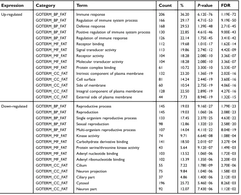
down-regulated DEGs were mainly associated with cilium and neuron projections. GO analysisfindings are shown in Figure 1C and D, and Table 1.
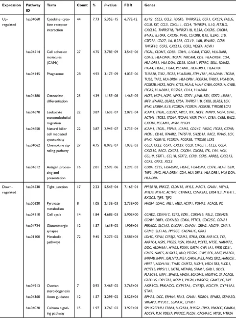
Table 2 and Figure 1E show the results of KEGG analysis. Up-regulated DEGs were mainly enriched in
cytokine–cytokine receptor interactions, cell adhesion
molecules (CAMs), phagosomes, osteoclast
differentia-tion, leukocyte transendothelial migration, natural
killer cell-mediated cytotoxicity, chemokine signaling pathways, and antigen processing and presentation;
down-regulated DEGs were significantly enriched in
tight junctions, pyruvate metabolism, the cell cycle, glutamatergic synapses, metabolic pathways, ovarian steroidogenesis, axon guidance, and calcium signaling pathways.
Identi
fi
cation of hub genes and analysis of
modules from PPI networks
According to the information from STRING, the topfive hub
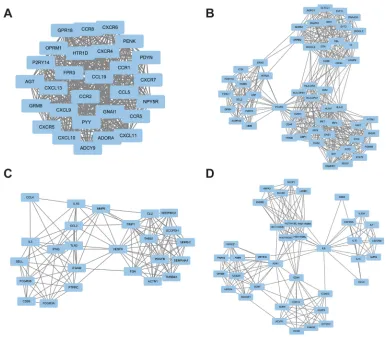
nodes were selected, including interleukin 6 (IL6), vascular endothelial growth factor A (VEGFA), including interleukin 10 (IL10), C-C motif chemokine receptor 5 (CCR5), and C-X-C motif chemokine receptor 4 (CXCR4). Analysis of the relationship between 1,074 nodes and 4,633 edges by plug-in MCODE enabled four modules to be selected, and then functional annotation of the genes from these modules
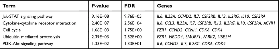
was conducted by DAVID (Figure 2, Tables 3–6). The genes
in these modules were mainly involved in the Janus kinase-signal transducers and activators of transcription (Jak–STAT) signaling pathway, chemokine signaling pathway, endocyto-sis, and cytokine–cytokine receptor interactions.
Table 1Gene ontology analysis of DEGs associated with seminoma
Expression Category Term Count % P-value FDR
Up-regulated GOTERM_BP_FAT Immune response 206 36.20 6.12E−76 1.19E−72
GOTERM_BP_FAT Regulation of immune system process 166 29.17 4.71E−53 9.19E−50
GOTERM_BP_FAT Defense response 168 29.53 1.39E−48 2.71E−45
GOTERM_BP_FAT Positive regulation of immune system process 130 22.85 4.61E−46 9.00E−43
GOTERM_BP_FAT Regulation of immune response 126 22.14 1.75E−45 3.41E−42
GOTERM_MF_FAT Receptor binding 112 19.68 1.01E−17 1.62E−14
GOTERM_MF_FAT Signal transducer activity 113 19.86 2.74E−12 4.42E−09
GOTERM_MF_FAT Receptor activity 104 18.28 2.08E−10 3.36E−07
GOTERM_MF_FAT Molecular transducer activity 104 18.28 2.08E−10 3.36E−07
GOTERM_MF_FAT Protein complex binding 61 10.72 3.30E−10 5.33E−07
GOTERM_CC_FAT Intrinsic component of plasma membrane 132 23.20 1.36E−19 2.02E−16
GOTERM_CC_FAT Cell surface 81 14.24 2.44E−19 3.60E−16
GOTERM_CC_FAT Side of membrane 60 10.54 2.75E−19 4.06E−16
GOTERM_CC_FAT Integral component of plasma membrane 128 22.50 2.89E−19 4.27E−16
GOTERM_CC_FAT External side of plasma membrane 44 7.73 8.94E−19 1.32E−15
Down-regulated GOTERM_BP_FAT Reproductive process 145 19.03 9.16E−27 1.79E−23
GOTERM_BP_FAT Reproduction 145 19.03 1.06E−26 2.08E−23
GOTERM_BP_FAT Single organism reproductive process 133 17.45 2.37E−25 4.63E−22
GOTERM_BP_FAT Sexual reproduction 98 12.86 1.32E−23 2.58E−20
GOTERM_BP_FAT Multi-organism reproductive process 107 14.04 4.11E−22 8.04E−19
GOTERM_MF_FAT Kinase activity 74 9.71 6.64E−08 1.08E−04
GOTERM_MF_FAT Carbohydrate derivative binding 141 18.50 2.01E−07 3.27E−04
GOTERM_MF_FAT Protein serine/threonine kinase activity 43 5.64 9.12E−07 1.49E−03
GOTERM_MF_FAT Adenyl nucleotide binding 103 13.52 1.06E−06 1.72E−03
GOTERM_MF_FAT Adenyl ribonucleotide binding 102 13.39 1.35E−06 2.20E−03
GOTERM_CC_FAT Cilium 55 7.22 1.78E−09 2.70E−06
GOTERM_CC_FAT Neuron projection 75 9.84 1.04E−06 1.58E−03
GOTERM_CC_FAT Ciliary part 37 4.86 1.40E−06 2.12E−03
GOTERM_CC_FAT Cytosol 196 25.72 5.46E−06 8.26E−03
GOTERM_CC_FAT Neuron part 92 12.07 7.43E−06 1.12E−02
OncoTargets and Therapy downloaded from https://www.dovepress.com/ by 118.70.13.36 on 25-Aug-2020
Table 2KEGG pathway analysis of DEGs associated with seminoma
Expression Pathway ID
Term Count % P-value FDR Genes
Up-regulated
hsa04060 Cytokine−
cyto-kine receptor interaction
44 7.73 5.35E−15 6.77E-12 IL1R2,CCL3,CCL2,PDGFB,TNFRSF25,CCR1,CXCL9,FASLG,
CCL8,KIT,CCL5,CXCL11,CCL4,TNFRSF4,IL10,FLT3LG, CXCL10,TNFRSF1B,TNFRSF11B,IL23A,CXCR5,CXCR4,
IFNA5,IL10RA,CXCR6,IFNG,CSF2RB,IL1B,IL2RG,LTB, CSF2RA,CD27,IL6,IL2RB,CCL19,IL6R,IFNAR2,CCR8,
TNFSF10,CCR5,CXCL13,CCR2,VEGFA,ACVR1
hsa04514 Cell adhesion molecules
(CAMs)
27 4.75 2.78E−09 3.54E−06 ITGAL,CLDN7,CD8A,CDH1,L1CAM,ITGB2,HLA-DMB,
CDH3,HLA-DMA,ITGAM,NRCAM,CD2,HLA-DRB4,CD4, HLA-DPB1,HLA-DOA,CD28,ICAM1,PTPRC,SELL,ICAM2,
ITGA4,HLA-E,HLA-F,PECAM1,HLA-DPA1,HLA-DRA
hsa04145 Phagosome 28 4.92 3.17E−09 4.03E−06 TUBB2B,TLR2,ITGB2,HLA-DMB,ATP6V1B1,HLA-DMA,ITGAM,
TUBB,TAP2,HLA-DRB4,HLA-DPB1,FCGR3A,THBS1,HLA-DOA, FCGR3B,NCF2,NCF4,CTSS,HLA-E,HLA-F,CYBA,CORO1A,CYBB,
ITGA5,HLA-DPA1,FCGR2A,CD14,HLA-DRA
hsa04380 Osteoclast
differentiation
25 4.39 1.15E−08 1.46E−05 NCF2,NCF4,ACP5,NFKB2,STAT1,JUNB,BTK,STAT2,LILRB1,
IRF9,IFNAR2,LILRB2,CYBA,TNFRSF11B,CYBB,LILRB3,LCK, IFNG,LILRB4,IL1B,FCGR2A,FCGR3A,FCGR3B,TYROBP,LCP2
hsa04670 Leukocyte
transendothelial migration
22 3.87 1.63E−07 2.07E−04 ICAM1,ITGAL,CLDN7,MYL7,ITK,NCF2,MMP9,NCF4,SIPA1,
ACTN1,ITGB2,ITGA4,ITGAM,VASP,THY1,CYBA,CYBB,RAC2, CXCR4,PECAM1,MSN,RHOH
hsa04650 Natural killer cell mediated
cytotoxicity
22 3.87 2.94E−07 3.73E−04 ICAM1,ITGAL,PTPN6,ICAM2,CD247,FASLG,ITGB2,GZMB, NCR1,CD48,IFNAR2,TNFSF10,SH2D1A,RAC2,IFNA5,LCK,
IFNG,FCER1G,FCGR3A,FCGR3B,TYROBP,LCP2
hsa04062 Chemokine
sig-naling pathway
27 4.75 8.07E−07 1.03E−03 CCL3,CCL2,CCR1,CXCL9,CCL8,CXCL11,CCL5,CCL4,
CXCL10,RAC2,CXCR5,CXCR4,CXCR6,ITK,LYN,HCK,
CCL19,STAT1,CCL18,STAT2,CCR8,CCR5,ARRB2,CXCL13, CCR2,GRK5,XCL2
hsa04612 Antigen
proces-sing and presentation
16 2.81 2.59E−06 3.29E−03 CD8A,CTSS,HLA-DMB,HLA-E,HLA-DMA,CD74,HLA-F,B2M,
TAP2,IFNG,HLA-DRB4,CD4,HLA-DPA1,HLA-DPB1,HLA-DOA, HLA-DRA
Down-regulated
hsa04530 Tight junction 17 2.23 5.54E−04 7.16E−01 PPP2R1B,PRKCZ,CLDN18,MYL5,MAGI1,GNAI1,MYH3, MYLPF,MYH7,ACTN3,CTNNA2,CSNK2A2,EPB41L3,MYH11,
EXOC3,TJP3,TJP2
hsa00620 Pyruvate
metabolism
8 1.05 2.13E−03 2.73E+00 HAGH,LDHC,ME1,ME3,ACYP1,PDHA2,ACACB,PC
hsa04110 Cell cycle 14 1.84 4.68E−03 5.90E+00 CCNE2,CDKN1C,E2F2,FZR1,CDKN1B,RBL2,CDKN2B,
CCNH,DBF4,CDKN2D,CDK6,PTTG1,CDC25C,CCNA1
hsa04724 Glutamatergic
synapse
12 1.57 1.61E−02 1.90E+01 PRKACG,SLC1A2,DLGAP1,GNAO1,GRIA2,ADCY9,GNAI1,
GRM8,SLC1A6,PPP3CC,CACNA1C,GRK3
hsa01100 Metabolic
pathways
72 9.45 2.27E−02 2.58E+01 LDHC,KYNU,CYP2J2,PGAM2,ITPKA,CKB,AKR1C3,TYR,
AKR1C4,AGPS,PTGES,RGN,PDHA2,PCYT2,NT5E,NMNAT2, DDC,ALDH6A1,HYAL3,PDXK,GATM,CYP11A1,PFKP,CDS1,
GMPS,NME5,ALOX15,ADO,PTGDS,CHPF,RFK,ABAT,PLA2G6, INPP4B,INPP1,GALNT3,ME1,CHKA,ME3,MVD,GK2,HMGCS1,
HPRT1,ALDH1A1,TYMS,CKMT2,PLCH1,HSD17B3,PLCD1, PCYT1B,PRPS1L1,UGT8,MTMR6,SPAM1,GAD1,ODC1,
PLA2G16,UAP1,SPHK2,MAOA,BCKDHB,MGAT4C,SI,ACACB, GAPDHS,CYP17A1,ACSM1,PYGM,HMGCS2,GAMT,PC,LIPF
hsa04913 Ovarian steroidogenesis
7 0.92 2.46E−02 2.76E+01 AKR1C3, PRKACG, CYP17A1, CYP2J2, ADCY9, CYP11A1, STAR
hsa04360 Axon guidance 12 1.57 3.29E−02 3.52E+01 EPHA5,DCC,EPHA4,PAK3,GNAI1,ROBO1,EFNB2,SEMA3D, SRGAP3,PPP3CC,SEMA3C,EPHB1
hsa04020 Calcium signal-ing pathway
15 1.97 3.76E−02 3.92E+01 SPHK2EPHB1 ERBB4,SLC25A4,PHKG2,ITPKA,PRKACG,CAMK4, ADCY9,PLN,PDE1A,PPP3CC,PLCD1,CACNA1C,MYLK,HTR2A
OncoTargets and Therapy downloaded from https://www.dovepress.com/ by 118.70.13.36 on 25-Aug-2020
Expression level and survival analysis of
hub genes
Compared with the normal tissues, CCR5 and CXCR4
revealed higher expression levels in seminoma tissues
(P<0.05), but IL6, VEGFA and IL10 not (Figure 3A–E).
Besides, 134 seminoma samples from TCGA database,
grouped by the different expression ofCCR5 andCXCR4,
were used to conduct survival analyses. Table 7 shows the clinicopathological characteristics in patients with seminoma from TCGA cohort. It was found that increased expression
level ofCCR5 (HR 0.72 [0.52–0.98], P=0.047) was
asso-ciated with poor overall survival for seminoma patients, as
well asCXCR4(HR 0.73 [0.59–0.92],P=0.012) (Figure 3F
and G).
Figure 2Top four modules from the PPI network. (A) modules 1; (B) modules 2; (C) modules 3; (D) modules 4.
Table 3The enriched pathways of module 1
Term P-value FDR Genes
Chemokine signaling pathway 4.19E −17
4.35E−14 GNAI1,CCR1,CXCL9,CCL19,CXCL11,CCL5,CXCL10,CCR8,CXCR5,ADCY9,CCR5, CXCR4,CXCL13,CCR2,CXCR6
Cytokine−cytokine receptor interaction
1.36E −12
1.41E−09 CCR1,CXCL9,CCL19,CXCL11,CCL5,CXCL10,CCR8,CCR5,CXCR5,CXCL13,CXCR4, CCR2,CXCR6
Neuroactive ligand-receptor interaction
2.23E −04
2.31E−01 OPRM1,GRM8,P2RY14,FPR3,HTR1D,ADORA1,NPY5R
Toll-like receptor signaling pathway
4.97E −03
5.04E+00 CXCL9,CCL5,CXCL11,CXCL10
Regulation of lipolysis in adipocytes
1.46E −02
1.42E+01 ADCY9,GNAI1,ADORA1
OncoTargets and Therapy downloaded from https://www.dovepress.com/ by 118.70.13.36 on 25-Aug-2020
Discussion
Seminoma accounts for the most part of cases of TGCT, which is the most common malignancy among males between ages 15
and 44 years.1 Understanding its molecular mechanism in
genetic aspects is important for diagnosis and treatment. In the
present study, we analyzed the gene expression profile
GSE8607, containing 40 seminoma samples and three healthy testes samples, by bioinformatics methods, to explore the hub genes which may play crucial roles in tumorigenesis. We
identi-fied 1,636 DEGs, of which 701 were up-regulated and 935 were down-regulated in seminomas compared with control testes.
GO analysis showed that up-regulated DEGs were mainly enriched in defense responses, receptor activity, regulation of immune response processes, and signal transducer activity, while down-regulated DEGs were mainly enriched in reproduc-tive processes, kinase activity, and carbohydrate derivareproduc-tive
binding. Regarding KEGG pathway enrichment analysis, up-regulated DEGs were enriched in CAMs, natural killer cell-mediated cytotoxicity, cytokine–cytokine receptor interactions, and chemokine signaling pathways. Previous studies have sug-gested that cell adhesion plays an important part in the growth, progression, and metastasis of tumors. Moreover, high expres-sion level of CAMs was reported to be associated with poor prognosis in lung and breast cancer, and many other tumor types.17–19In recent years, natural killer T cells were found to be an effective treatment for several cancers, but their efficacy in
seminoma remains unknown.20
We showed that down-regulated DEGs were mainly associated with tight junctions, metabolic pathways, axon guidance, the cell cycle, and calcium signaling pathways. Tight junctions of healthy testes separate the internal and external environment of the testis and protect it from
Table 4The enriched pathways of module 2
Term P-value FDR Genes
Endocytosis 8.30E
−07
8.76E−04 SH3GL3,ADRB2,ARRB2,CBL,DNAJC6,GRK5,HLA-E,BIN1,CLTCL1,SH3GL2, HLA-F
Antigen processing and presentation 3.03E −06
3.19E−03 CD4,HLA-DPA1,HLA-DPB1,HLA-E,HLA-DRA,HLA-F,B2M
Cell adhesion molecules (CAMs) 1.09E −04
1.15E−01 ICAM1,CD4,HLA-DPA1,HLA-DPB1,HLA-E,HLA-DRA,HLA-F
Neuroactive ligand-receptor interaction
6.68E −04
7.03E−01 P2RY6,P2RY10,ADRB2,AVPR1B,F2RL1,NMBR,PTAFR,HTR2A
Phagosome 9.50E
−03
9.58E+00 HLA-DPA1,HLA-DPB1,HLA-E,HLA-DRA,HLA-F
Table 5The enriched pathways of module 3
Term P-value FDR Genes
Cytokine−cytokine receptor interaction 3.34E−05 3.56E−02 IL5,CCL2,PDGFB,VEGFA,IFNG,IL1B,CCL4
Phagosome 8.49E−04 9.01E−01 TLR2,THBS1,FCGR3A,FCGR3B,ITGAM
Osteoclast differentiation 6.00E−03 6.21E+00 IFNG,IL1B,FCGR3A,FCGR3B
Complement and coagulation cascades 1.66E−02 1.64E+01 FGA,SERPING1,SERPINA1
Rap1 signaling pathway 2.15E−02 2.07E+01 PDGFB,VEGFA,THBS1,ITGAM
Table 6The enriched pathways of module 4
Term P-value FDR Genes
Jak-STAT signaling pathway 9.16E−08 9.76E−05 IL6,IL23A,CCND2,IL7,CSF2RB,IL13,IL2RG,IL10,CSF2RA Cytokine-cytokine receptor interaction 2.40E−07 2.56E−04 IL6,CCL3,IL23A,IL7,CSF2RB,IL13,IL2RG,IL10,CSF2RA,ACVR1
Cell cycle 1.66E−03 1.75E+00 FZR1,CCND2,CCNH,CDK6,CDK4
Ubiquitin mediated proteolysis 2.39E−03 2.52E+00 FZR1,NEDD4,SMURF1,PARK2,UBE2H
PI3K-Akt signaling pathway 1.33E−02 1.33E+01 IL6,CCND2,IL7,IL2RG,CDK6,CDK4
OncoTargets and Therapy downloaded from https://www.dovepress.com/ by 118.70.13.36 on 25-Aug-2020
harmful substances. However, it is difficult to maintain a normal structure and function in cancer tissue. Moreover, studies have suggested that the loss of cell cycle regulation leads to genomic instability, and the cell cycle is thought to play an important role in the etiology of
spontaneous cancers.21 Recent evidence indicated that
physiological calcium signaling regulated aerobic metabo-lism, but that pathological calcium overload contributed to
cell death.22 Therefore, monitoring these processes and
pathways may aid the diagnosis or treatment of seminoma.
IL6, VEGFA, IL10, CCR5, andCXCR4were selected as
hub genes because of their high degree of connectivity. Existing evidence suggests that immunologic factors may
affect the development of seminoma, with the
inflammatory cytokines IL6 and IL10 thought to promote
tumor immune evasion through local immunosuppression. Parker et al reported that the extent of lymphocyte infi ltra-tion in seminomas was associated with a reduced risk of
disease recurrence,23while Klein et al documented major
roles for IL6 in shaping the surrounding tumor
microen-vironment by influencing local immune responses.24 IL6
may, therefore, have the potential to become a novel
diag-nostic and immunotherapeutic factor for seminoma.25
However, details about signaling and intercellular interac-tion require further investigainterac-tion. Mohamed reported that
IL10 secreted by tumor-infiltrating
monocytes/macro-phages (CD14+/CD16+) separated from inflammatory
breast cancer patients positively correlated with the
Figure 3Expression levels and survival analyses of DEGs. (A–E) Expression levels of thefive hub genes. *P-value <0.05. (F–G) Kaplan-Meier plot of overall survival for CCR5andCXCR4in seminoma patients.
Abbreviation:HR, hazard ratio.
OncoTargets and Therapy downloaded from https://www.dovepress.com/ by 118.70.13.36 on 25-Aug-2020
expression level of CPB2, which was crucial to
lympho-vascular invasion in inflammatory breast cancer.26
However, further studies are required to clarify the role of IL10 in seminoma.
Angiogenesis is crucial to the progression of many kinds of tumors because of the need for nutrition from blood
vessels.27VEGFA is a subtype of VEGF that functions as
a potent angiogenic factor in blood vessel formation and
regulates the progression of tumors.28,29VEGFA promotes
tumor proliferation and angiogenesis by activating phosphoi-nositide 3-kinase (PI3K)/AKT and
extracellular-signal-regulated kinase (ERK) signaling pathways.30–32 Although
anti-angiogenesis therapies targetingVEGFAinhibit the
pro-gression of many tumor types, their efficacy in seminoma
needs to be investigated. The geneCCR5, encoding one of
the receptors of C-C chemokine ligand 5 (CCL5), promotes
carcinogenesis, stroma genesis, and tumor progression.33
CCL5/CCR5operates via PI3K/AKT, mitogen-activated
pro-tein kinase kinase, and ERK, which in turn activate nuclear factor-κB, leading to activation ofαvβ3 integrin and
contri-buting to cell migration.34–36CCL5/CCR5was found to be
a biomarker of poor prognosis in various cancer types such as pancreas, prostate, breast, ovarian, and renal cancers.37–41 CXCR4 is one of the most commonly overexpressed cyto-kine receptors in malignant tumors, and it accepts the signal of its ligand CXCL12 to mediate cell adhesion, angiogenesis,
proliferation, metastasis, and survival.42 Both CXCR4 and
CXCL12 are important components of the signaling
mechanisms that facilitate the normal migration of primor-dial germ cells from the hindgut to the genital ridges infish
and mammals.43 Although they were reported to play an
important role in the metastasis of seminoma by activating MAP kinase and PI3K pathways, they have a limited role in tumor survival and proliferation.44In the present study, we
found that high expression levels ofCCR5andCXCR4were
associated with poor prognosis for seminoma patients. Maybe they are potential diagnostic biomaker or predictor for prognosis when more studies confirm their values.
Analysis of the four modules selected from the PPI net-work in the present study suggested that the Jak–STAT signal-ing pathway, chemokine signalsignal-ing pathway, endocytosis, and
cytokine–cytokine receptor interactions may be associated
with the occurrence of seminoma. The Jak–STAT pathway is
an evolutionarily conserved signaling cascade which mediates the response to cytokines and growth factors. Cellular responses to the activation of this pathway include differentia-tion, proliferadifferentia-tion, migradifferentia-tion, apoptosis, and cell survival. Moreover, Jak–STAT signaling is integral to homeostatic and developmental processes such as hematopoiesis, immune development, stem cell maintenance, and organismal
growth,45 while its activation by dysregulated chemokines
induces the occurrence and growth of cancer. Palagani et al reported that inhibiting the Jak–STAT pathway might prevent pancreatic cancer progression,46and Li et al showed that its inhibition would promote the antiproliferative effect of meth-otrexate in small cell lung cancer.47However, the relationship
Table 7The clinicopathological characteristics in patients with seminoma from TCGA cohort
Expression of CCR5 P-value Expression of CXCR4 P-value
Low (n=107) High (n=27) Low (n=47) High (n=87)
Age 0.305 0.239
<40 y 88 25 42 71
≥40 y 19 2 5 16
Race 0.442 0.133
White 95 23 45 74
Yellow 7 1 1 7
Black 5 3 1 6
AJCC stage 0.288 0.588
I 76 23 33 68
II 14 2 7 9
III 17 2 7 10
Survival 0.380 0.297
Yes 105 25 47 83
No 2 2 0 4
Note:*p<0.05.
OncoTargets and Therapy downloaded from https://www.dovepress.com/ by 118.70.13.36 on 25-Aug-2020
between seminoma and the Jak–STAT pathway requires
further investigation. Maybe the Jak–STAT pathway is
a promising candidate signaling pathway which affects the tumorigenesis of seminoma.
In conclusion, we used bioinformatics analysis of DEGs to identify key genes and pathways that are closely associated with the occurrence of seminoma. Our work identified several potential targets for biomarkers and for
understanding underlying molecular mechanisms.
However, further study is required to determine the func-tions of these candidates.
Abbreviation list
TCGT, testicular germ cell tumors; AJCC, American Joint Committee on Cancer; DEGs, differentially expressed genes; GO, Gene ontology; KEGG, Kyoto Encyclopedia of Genes and Genomes; STRING, Search Tool for the Retrieval of
Interacting Genes; PPI, protein–protein interaction;
MCODE, Molecular Complex Detection; GEO, Gene Expression Omnibus; BP, biological processes; MF, molecu-lar functions; CC, cellumolecu-lar component; DAVID, Database for Annotation, Visualization and Integrated Discovery; IL 6, interleukin 6; VEGFA, vascular endothelial growth factor A; CCR5, C-C motif chemokine receptor 5; CXCR4,
C-X-C motif chemokine receptor 4; Jak–STAT, the Janus
kinase-signal transducers and activators of transcription sig-naling pathway.
Acknowledgments
We thank Sarah Williams, PhD, from Liwen Bianji, Edanz Group China, for editing the English text of a draft of this manuscript. This study was supported by Foundation of Fujian Provincial Department of Finance (Grant number: 2018B011).
Disclosure
The authors report no conflicts of interest in this work.
References
1. Ghazarian AA, Kelly SP, Altekruse SF, Rosenberg PS, McGlynn KA. Future of testicular germ cell tumor incidence in the United States: forecast through 2026.Cancer. 2017;123:2320–2328. doi:10.1002/cncr.30597 2. Smith ZL, Werntz RP, Eggener SE. Testicular cancer: epidemiology,
diagnosis, and management.Med Clin North Am. 2018;102:251–264. doi:10.1016/j.mcna.2017.10.003
3. Ferlay J, Shin H-R, Bray F, Forman D, Mathers C, Parkin DM. Estimates of worldwide burden of cancer in 2008: GLOBOCAN 2008.Int J Cancer. 2010;127:2893–2917. doi:10.1002/ijc.25516 4. Fankhauser CD, Curioni-Fontecedro A, Allmann V, et al. Frequent
PD-L1 expression in testicular germ cell tumors. Br J Cancer. 2015;113:411–413. doi:10.1038/bjc.2015.244
5. Yamada Y, Takayama K-I, Fujimura T, et al. A novel prognostic factor TRIM44 promotes cell proliferation and migration, and inhi-bits apoptosis in testicular germ cell tumor. Cancer Sci. 2017;108:32–41. doi:10.1111/cas.13105
6. Liu X, Duan H, Zhou S, et al. microRNA-199a-3p functions as tumor suppressor by regulating glucose metabolism in testicular germ cell tumors.Mol Med Rep. 2016;14:2311–2320. doi:10.3892/mmr.2016.5472 7. Davis S, Meltzer PS. GEOquery: a bridge between the Gene Expression Omnibus (GEO) and BioConductor. Bioinformatics. 2007;23:1846–1847. doi:10.1093/bioinformatics/btm254
8. Liang B, Li C, Zhao J. Identification of key pathways and genes in colorectal cancer using bioinformatics analysis. Med Oncol. 2016;33:111. doi:10.1007/s12032-016-0829-6
9. Sun C, Yuan Q, Wu D, Meng X, Wang B. Identification of core genes and outcome in gastric cancer using bioinformatics analysis. Oncotarget. 2017;8:70271–70280. doi:10.18632/oncotarget.20082 10. The Gene Ontology (GO) project in 2006. Nucleic Acids Res.
2006;34:D322–D326. doi:10.1093/nar/gkj021
11. Ashburner M, Ball CA, Blake JA, et al; The Gene Ontology Consortium. Gene ontology: tool for the unification of biology.Nat Genet. 2000;25:25–29. doi:10.1038/75556
12. Kanehisa M, Goto S. KEGG: kyoto encyclopedia of genes and genomes.Nucleic Acids Res. 2000;28:27–30.
13. Huang Da W, Sherman BT, Lempicki RA. Systematic and integrative analysis of large gene lists using DAVID bioinformatics resources. Nat Protoc. 2009;4:44–57. doi:10.1038/nprot.2008.211
14. Szklarczyk D, Franceschini A, Wyder S, et al. STRING v10: protein-protein interaction networks, integrated over the tree of life. Nucleic Acids Res. 2015;43:D447–D452. doi:10.1093/nar/gku1003 15. Shannon P, Markiel A, Ozier O, et al. Cytoscape: a software
envir-onment for integrated models of biomolecular interaction networks. Genome Res. 2003;13:2498–2504. doi:10.1101/gr.1239303 16. Tang Z, Li C, Kang B, et al. GEPIA: a web server for cancer and
normal gene expression profiling and interactive analyses.Nucleic Acids Res. 2017;45:W98–W102. doi:10.1093/nar/gkx247
17. Yang Y, Jiang Y, Xie D, et al. Inhibition of cell-adhesion protein DPYSL3 promotes metastasis of lung cancer. Respir Res. 2018;19:41. doi:10.1186/s12931-018-0740-0
18. Wu J-D, Hong C-Q, Huang W-H, et al. L1 cell adhesion molecule and its soluble form sL1 exhibit poor prognosis in primary breast cancer patients. Clin Breast Cancer. 2018;18:e851–e861. doi:10.1016/j.clbc.2017.12.011
19. Yu H, Gao M, Ma Y, Wang L, Shen Y, Liu X. Inhibition of cell migration by focal adhesion kinase: time-dependent difference in integrin-induced signaling between endothelial and hepatoblastoma cells.Int J Mol Med. 2018;41:2573–2588. doi:10.3892/ijmm.2018.3512
20. Waldowska M, Bojarska-Junak A, Rolinski J. A brief review of clinical trials involving manipulation of invariant NKT cells as a promising approach in future cancer therapies. Cent-Eur J Immunol. 2017;42:181–195. doi:10.5114/ceji.2017.69361 21. Hartwell LH, Kastan MB. Cell cycle control and cancer.Science.
1994;266:1821–1828.
22. Logan CV, Szabadkai G, Sharpe JA, et al. Loss-of-function mutations in MICU1 cause a brain and muscle disorder linked to primary alterations in mitochondrial calcium signaling. Nat Genet. 2014;46:188–193. doi:10.1038/ng.2851
23. Parker C, Milosevic M, Panzarella T, et al. The prognostic significance of the tumour infiltrating lymphocyte count in stage I testicular seminoma managed by surveillance.Eur J Cancer. 2002;38:2014–2019.
24. Klein B, Schuppe H-C, Bergmann M, Hedger MP, Loveland BE, Loveland KL. An in vitro model demonstrates the potential of neoplastic human germ cells to influence the tumour microenvironment.Andrology. 2017;5:763–770. doi:10.1111/andr.12365
25. Klein B, Haggeney T, Fietz D, et al. Specific immune cell and cytokine characteristics of human testicular germ cell neoplasia. Hum Reprod. 2016;31:2192–2202. doi:10.1093/humrep/dew211
OncoTargets and Therapy downloaded from https://www.dovepress.com/ by 118.70.13.36 on 25-Aug-2020
26. Mohamed HT, El-Husseiny N, El-Ghonaimy EA, et al. IL-10 corre-lates with the expression of carboxypeptidase B2 and lymphovascular invasion in inflammatory breast cancer: the potential role of tumor infiltrated macrophages. Curr Probl Cancer. 2018;42:215–230. doi:10.1016/j.currproblcancer.2018.01.009
27. Hicklin DJ, Ellis LM. Role of the vascular endothelial growth factor pathway in tumor growth and angiogenesis. J Clin Oncol. 2005;23:1011–1027. doi:10.1200/jco.2005.06.081
28. London CA, Hannah AL, Zadovoskaya R, et al. Phase I dose-escalating study of SU11654, a small molecule receptor tyrosine kinase inhibitor, in dogs with spontaneous malignancies.Clin Cancer Res. 2003;9:2755–2768.
29. Shibuya M, Claesson-Welsh L. Signal transduction by VEGF recep-tors in regulation of angiogenesis and lymphangiogenesis.Exp Cell Res. 2006;312:549–560. doi:10.1016/j.yexcr.2005.11.012
30. Lichtenberger BM, Tan PK, Niederleithner H, Ferrara N, Petzelbauer P, Sibilia M. Autocrine VEGF signaling synergizes with EGFR in tumor cells to promote epithelial cancer development. Cell. 2010;140:268–279. doi:10.1016/j. cell.2009.12.046
31. Lee T-H, Seng S, Sekine M, et al. Vascular endothelial growth factor mediates intracrine survival in human breast carcinoma cells through internally expressed VEGFR1/FLT1. PLoS Med. 2007;4:e186. doi:10.1371/journal.pmed.0040186
32. Vincent L, Jin DK, Karajannis MA, et al. Fetal stromal-dependent paracrine and intracrine vascular endothelial growth factor-a/vascular endothelial growth factor receptor-1 signaling promotes proliferation and motility of human primary myeloma cells. Cancer Res. 2005;65:3185–3192. doi:10.1158/0008-5472.can-04-3598
33. Cambien B, Richard-Fiardo P, Karimdjee BF, et al. CCL5 neutraliza-tion restricts cancer growth and potentiates the targeting of PDGFRbeta in colorectal carcinoma. PLoS One. 2011;6:e28842. doi:10.1371/journal.pone.0028842
34. Huang C-Y, Fong Y-C, Lee C-Y, et al. CCL5 increases lung cancer migration via PI3K, Akt and NF-kappaB pathways. Biochem Pharmacol. 2009;77:794–803. doi:10.1016/j.bcp.2008.11.014 35. Long H, Xie R, Xiang T, et al. Autocrine CCL5 signaling promotes
invasion and migration of CD133+ ovarian cancer stem-like cells via NF-kappaB-mediated MMP-9 upregulation. Stem Cells. 2012;30:2309–2319. doi:10.1002/stem.1194
36. Kato T, Fujita Y, Nakane K, et al. CCR1/CCL5 interaction promotes invasion of taxane-resistant PC3 prostate cancer cells by increasing secretion of MMPs 2/9 and by activating ERK and Rac signaling. Cytokine. 2013;64:251–257. doi:10.1016/j.cyto.2013.06.313 37. Singh SK, Mishra MK, Eltoum I-EA, Bae S, Lillard JW, Singh R. CCR5/
CCL5 axis interaction promotes migratory and invasiveness of pancreatic cancer cells.Sci Rep. 2018;8:1323. doi:10.1038/s41598-018-19643-0 38. Kondo T, Ito F, Nakazawa H, Horita S, Osaka Y, Toma H. High
expression of chemokine gene as a favorable prognostic factor in renal cell carcinoma.J Urol. 2004;171:2171–2175.
39. Vaday GG, Peehl DM, Kadam PA, Lawrence DM. Expression of CCL5 (RANTES) and CCR5 in prostate cancer. Prostate. 2006;66:124–134. doi:10.1002/pros.20306
40. Khalid A, Wolfram J, Ferrari I, et al. Recent advances in discovering the role of CCL5 in metastatic breast cancer.Mini Rev Med Chem. 2015;15:1063–1072.
41. Tsukishiro S, Suzumori N, Nishikawa H, Arakawa A, Suzumori K. Elevated serum RANTES levels in patients with ovarian cancer correlate with the extent of the disorder. Gynecol Oncol. 2006;102:542–545. doi:10.1016/j.ygyno.2006.01.029
42. Sun X, Cheng G, Hao M, et al. CXCL12/CXCR4/CXCR7 chemokine axis and cancer progression. Cancer Metastasis Rev. 2010;29:709–722. doi:10.1007/s10555-010-9256-x
43. Doitsidou M, Reichman-Fried M, Stebler J, et al. Guidance of pri-mordial germ cell migration by the chemokine SDF-1. Cell. 2002;111:647–659.
44. McIver SC, Loveland KL, Roman SD, Nixon B, Kitazawa R, McLaughlin EA. The chemokine CXCL12 and its receptor CXCR4 are implicated in human seminoma metastasis. Andrology. 2013;1:517–529. doi:10.1111/j.2047-2927.2013.00081.x
45. O’Shea JJ, Plenge R. JAK and STAT signaling molecules in immu-noregulation and immune-mediated disease. Immunity. 2012;36:542–550. doi:10.1016/j.immuni.2012.03.014
46. Palagani V, Bozko P, El Khatib M, et al. Combined inhibition of Notch and JAK/STAT is superior to monotherapies and impairs pancreatic cancer progression. Carcinogenesis. 2014;35:859–866. doi:10.1093/carcin/bgt394
47. Li HX, Zhao W, Shi Y, et al. Retinoic acid amide inhibits JAK/STAT pathway in lung cancer which leads to apoptosis. Tumour Biol. 2015;36:8671–8678. doi:10.1007/s13277-015-3534-8
OncoTargets and Therapy
Dove
press
Publish your work in this journal
OncoTargets and Therapy is an international, peer-reviewed, open access journal focusing on the pathological basis of all cancers, potential targets for therapy and treatment protocols employed to improve the management of cancer patients. The journal also focuses on the impact of management programs and new therapeutic
agents and protocols on patient perspectives such as quality of life, adherence and satisfaction. The manuscript management system is completely online and includes a very quick and fair peer-review system, which is all easy to use. Visit http://www.dovepress.com/ testimonials.php to read real quotes from published authors.
Submit your manuscript here:https://www.dovepress.com/oncotargets-and-therapy-journal
OncoTargets and Therapy downloaded from https://www.dovepress.com/ by 118.70.13.36 on 25-Aug-2020