Original Article
Cloning and pattern of expression of the shiro-uo vasa gene
during embryogenesis and its roles in PGC development
AKIMITSU MIYAKE
1, 2, TAIJU SAITO
1, 3,
TOHRU KASHIWAGI
1, DAISUKE ANDO
1, AKITSUGU YAMAMOTO
4,
TOHRU SUZUKI
5, NORIO NAKATSUJI
6and TAKAKO NAKATSUJI*
,11Department of Marine Biology, Graduate School of Marine Science and Technology, Tokai University, 2 Department of Biological
Science, Graduate School of Science, Tokyo University 3Laboratory of Breeding Science, Graduate School of Fisheries Science, Hokkaido
University 4Cell Biology, Nagahama Institute of Bio-science and Technology 5Laboratory of Bio-industrial Informatics, Graduate School
of Agricultural Science, Tohoku University and 6Institute for Frontier Medical Sciences, Kyoto University, Japan
ABSTRACT The vasa genes are expressed in the germ cell lineage in many organisms, but their expression patterns show large variations. Recent studies suggest that vasa transcripts are involved in germ cell lineage development. In this paper, we isolated the vasa cDNA clone from a teleost, shiro-uo, Leucopsarion petersii and examined its expression pattern during embryogen-esis. Then, we examined the functional significance of vasa mRNA during the formation of primordial germ cells (PGCs). The amino acid sequence of shiro-uo VASA is 61.1% identical to that of zebrafish. In whole-mount in situ hybridization, vasa transcripts appeared at the 4- and 8-cell stages as four spots at both ends of two cleavage planes between the lower tier of blastomeres and the yolk cell mass. At the 16-cell stage, eight spots were observed. After the blastula stage, shiro-uo vasa transcripts showed similar localization as in the zeblafish. Ultrastructural analysis of 4-cell stage embryos revealed the presence of a subcellular organelle that resembled “nuage” in the germ cell lineage observed in the embryos of various organisms. We carried out microma-nipulation of 4- or 8-cell stage embryos to remove the vasa mRNA-containig spots and then measured the number of the vasa-expressing PGCs in the genital ridge of the manipulated embryos. The numbers decreased when all of the four spots were removed, indicating that the vasa-containing spots at early cleavage stages have important functions in the development of PGCs.
KEY WORDS: Shiro-uo, vasa, primordial germ cell (PGC), mRNA localization, teleost
Introduction
The germ cell lineage is an important and specialized group of cells that possesses the ability to produce gametes and offspring. The morphology of PGCs in various fish is similar to that in other organisms (Braat et al., 1999). PGCs are morphologically distinct from somatic cells because of their large cell size, large nuclei and the presence of “nuage” in their cytoplasm (Mohowald, 1962; Czolowska, 1969; Wolf et al., 1983).
Molecular markers used for identification of PGCs have be-come useful research tools. The Drosophila vasa homolog gene has been identified in many other organisms, but its expression patterns during early embryogenesis shows large variations (Start-Gaiano and Lehmann, 2001). During embryonic development in zebrafish, vasa transcrips are localized to the distal parts of the first two cleavage furrows as condensed granules during
cleav-*Address correspondence to: Dr. Takako Nakatsuji. Tokai University School of Marine Science and Technology, Orid 3-20-1, Shimizu-ku, Shizuoka 424-8610, Japan. Fax: +81-543-37-0216. e-mail: [email protected]
Abbreviations used in this paper: PGC, primordial germ cell.
0214-6282/2006/$25.00 © UBC Press
Printed in Spain www.intjdevbiol.com
age and are eventually localized into four cells in the blastula. The number of vasa mRNA-positive cells remains at four until the late blastula stage. Then, zygotic vasa transcription is initiated in PGCs and these vasa mRNA-positive cells migrate to the gonads (Olsen et al., 1997: Yoon et al., 1997: Weidinger et al., 1999; Knaut et al., 2000). In medaka, however, vasa mRNA can be detected uniformly among the blastomeres until late gastrulation. Blastomeres with vasa transcripts move in the embryonic body at the early neurula stage and line up along the anterior-posterior axis on both sides of the embryonic body until the 4-somite stage (Shinomiya et al., 2000). For rainbow trout, vasa mRNA-positive cells are detectable later at the 80-somite stage (Yoshizaki et al., 2000).
B
structures resembling the germ plasm organelle, “nuage”. These expression patterns of vasa RNA are suggestive of its function in PGC development (Knaut et al., 2000). Furthermore, removal of the cytoplasm at the ends of the cleavage planes in zebrafish with the aim of removing vasa and other mRNA components results in a severe reduction in the number of germ cells (Hashimoto et al., 2004).
In this paper, we isolated the vasa cDNA from a teleost, shiro-uo (Family Gobiidae) and studied its expression pattern during embryogenesis while investigating the development of the
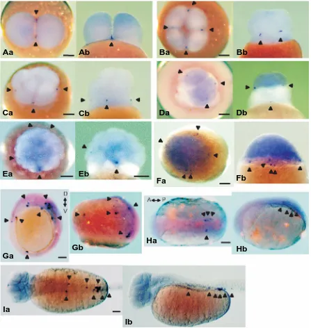
shiro-uo germ cell lineage. We reported that shiro-shiro-uo embryos show a unique cleavage pattern among teleosts (Nakatsuji et al., 1997) and that they are highly transparent and suitable for micromanipu-lation and other experiments. Furthermore, we examined the functional significance of the vasa mRNA-containing spots in the formation of PGCs using micromanipulation. We carried out micromanipulation of 4- or 8-cell stage embryos to remove the vasa mRNA-containig spots and then counted the number of vasa-expressing PGCs in the genital ridges of the manipulated embryos. The numbers decreased significantly when the four Fig. 2. Whole-mount in situ hybridization using vasa probe. (A) 2-cell stage, (B) 4-cell stage, (C) 8-cell stage, (D) 16-cell stage, (E) 32-cell stage,
(F) blastula stage, (G) 90%-epiboly stage, (H) bud stage and (I) 32-somite stage. (Aa, Ba, Ca, Da, Ea, Fa) view from the animal pole. (Ga) view from the vegetal pole. (Ha, Ia) dorsal view. (Ab, Bb, Cb, Db, Eb, Fb, Gb, Hb, Ib) lateral view. D↔V, dorsal↔ventral; A↔P, anterior↔posterior. Arrowheads indicate vasa spots (A, B, C, D, E) and vasa positive cells (F, G, H, I). Scale bars, 100 µm.
Aa
Ba
Ca
Da
Ea
Fa
Ga
Ha
Ia
Ab
Bb
Cb
Db
Eb
Fb
Gb
Hb
B
spots were removed, indicating that the vasa-containing spots at early cleavage stages have important functions in the develop-ment of PGCs.
Results
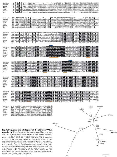
Sequence analysis
We isolated a full-length vasa cDNA clone from shiro-uo, which had an open reading frame of 1938bp and encoded 646 amino acids (Fig. 1A). The amino acid sequence contained eight consensus sequences for the DEAD protein family (Fig.1, under-lined). FASTA analysis indicated that the shiro-uo VASA protein is 69.7% and 61.4% identical to the medaka (Shinomiya et al., 2000) and zebrafish (Olsen et al., 1997; Yoon et al., 1997), respectively. Additionally, the sequence showed 52.1% identity to Xenopus (Komiya et al., 1994), 54.3% identity to Homo sapiens (Castrillon et al., 2000), 55.9% identity to mouse (Fujiwara et al., 1994) and 45.2% identity to Drosophila (Hay et al., 1988; Lasko and Ashburner, 1988). A radial phylogenic tree of VASA proteins produced by the Neighbor-joining method indicated that the deduced shiro-uo VASA protein falls into the fish cluster (Fig. 1B).
Expression pattern during embryogenesis
Whole-mount in situ hybridization was carried out using shiro-uo probes prepared from the 383bp vasa cDNA fragment contain-ing the DEAD box region (Fig. 1A, arrowed region) and the localization of vasa transcripts was investigated from the 2-cell stage to the 32-somite stage (5 day post-fertilization). At the 2-cell
2G). By the bud stage, vasa-positive cells were aligned along both sides of the embryonic body in the trunk region (Fig. 2H) and for the 12-somite stage, the tailbud detached and vasa-positive cells migrated toward the posterior region along the body axis (data not shown). At the 32-somite stage, vasa-positive cells were ob-served as aggregates in the presumptive genital ridge region, although some vasa-positive cells were still localized along both sides of the body axis (Fig. 2I).
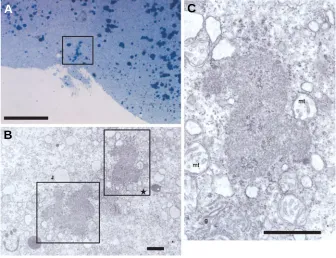
Ultrastructural analysis
Semi-thin sections of 4-cell stage embryos were examined by light microscopy. Distinct aggregated granules were observed at the distal ends of the cleavage furrows, presumptive vasa-con-taining areas (Fig. 3A, square). These granules were distinct from the dark stained round yolk granules. Furthermore, ultrastructural analysis of such granule revealed distinct subcellular structures that resembled nuage, a germ plasm organelle (Fig. 3B, squares). A higher magnification view (square in Fig. 3B) indicated that these nuages were made of fine electron-dense amorphous bodies and were present in close association with mitochondria and the Golgi apparatus (Fig. 3C).
Functional analysis of vasa transcripts
Because of clear morphological criteria, we can easily identify the region in which vasa mRNA may be present. In 4- or 8-stage shiro-uo embryos, the presumed location of the vasa mRNA-containing four spots can be easily identified. To examine the functional significance of the vasa mRNA-containing spots during
Fig. 3. A semi-thin (2 µµµµµm) vertical section (A) and electron micrograph (B,C) of the 4-cell stage of the shiro-uo embryo. (A) Distinct aggregated granules, presumable vasa-containing areas, are indicated in the square. (B) A low magnification view of the electron micrograph of the granules indicated in (A). (C) A higher magnification view of the presumptive germ plasm, “nuage”, indicated by the square labeled with a star in (B). mt, mitochondria; g, Golgi. Scale bars; 50 µm (A); 1 µm (B,C).
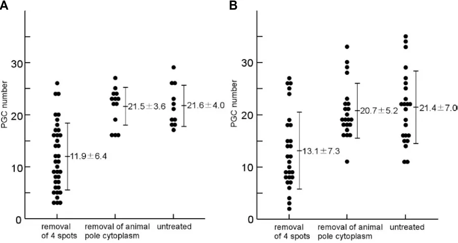
the formation of PGCs, we carried out micromanipulation of 4- or 8-cell stage embryos to remove the cytoplasm at the presumed four sites of the vasa mRNA-containing spots and examined the manipulated embryos at later stages. By whole-mount in situ hybridization, the vasa-positive cells in manipulated and staged embryos were counted. When the four spots were removed, the number of PGCs at the bud stage decreased to 11.9±6.4 (mean±SD, n=36). When those manipulated embryos were ex-amined at the 32-somite stage, the number was 13.1±7.3 (n= 28). As a control experiment, we removed the cytoplasm around the animal pole and the number of PGCs in those control embryos were 21.5±3.6 (n=13) at the bud stage and 20.7±5.2 (n=22) at the 32-somite stage. In the untreated embryos, the number of PGCs were 21.6±4.0 (n=11) at the bud stage and 21.4±7.0 (n=23) at the 32-somite stage. Thus, the number of vasa-positive cells signifi-cantly decreased when all of the four sites, which correspond to the vasa mRNA-containing spots, were removed from the cleav-age stcleav-age embryos (t-test: p<0.01) (Fig.4).
Discussion
In the present study, we cloned and examined the shiro-uo (ice goby), Leucopsarion petersii, vasa gene and identified the eight conserved homology boxes present in all DEAD box proteins. The shiro-uo VASA protein also shares with other VASA-related proteins an N terminus rich in glycine, with multiple repeats of an RGG motif (Fig. 1).
The expression patterns of vasa transcripts during cleavage stages show significant variations among fish. In shiro-uo, upon initiation of the first cleavage, vasa transcript-containing spots were not detectable yet in the whole-mount samples, although there were partial aggregates at the center of the blastomere detectable in histlogical sections of whole- mount samples after in
situ hybridization (data not shown). The four vasa spots became visible at the 4-cell stage. In ukigori and goldfish embryos at the 8-cell stage, vasa transcript accumulation results in eight vasa spots (Otani et al., 2002; Saito et al., 2004). In the shiro-uo 8-cell stage embryos, however, only four vasa spots were observed at both ends of two cleavage planes between the lower tier of the blastomeres and the yolk cell mass, as same as at the 4-cell stage (Fig. 2C). In our previous study on the shiro-uo, involving cell-lineage examination via the injection of a tracer dye, we found that the PGCs mainly originated from lower blastomeres at the 8-cell stage (Saito et al., 2002) and this could be explained by the localization of vasa transcripts at the 8-cell stage. At the 16-cell stage, four new vasa spots appeared between the upper and lower tires of the blastomeres and thus resulted in eight vasa spots (Fig. 2D). These four new spots may be formed by division of the four original vasa spots, or by newly concentrated vasa mRNA. Similarly, in ukigori and goldfish embryos at the16-cell stage, eight vasa spots are present. In contrast, for zebrafish the vasa spots only total four from the 4-cell stage until the 32-cell stage (Olsen et al., 1997; Yoon et al., 1997). After the blastula stages, the expression patterns of vasa transcripts show more similarity among teleost species.
Detection of “nuage-like” subcellular structures using electron microscopy revealed the presence of amorphous fine granules and mitochondria, indicating similarity between the vasa-contain-ing complex and the germ plasm of various groups of organisms. Removal of vasa-containing spots resulted in a significant decrease in PGCs. The number of PGCs was, however, not always lower than that of the controls. This could be due to incomplete removal of containing spots. Also, the vasa-containing complex might not be confined to the spots but also be more diffusely distributed, which might be the origin of the four new spots at the 16-cell stage. In such a case, complete removal
Fig. 4. Removal of the vasa-containg cytoplasm resulted in a decrease in the number of primordial germ cells. (A) The number of PGCs counted at the bud stage. (B) The number of PGCs counted at the 32-somite stage.
Hashimoto et al., 2004; Theusch et al., 2006). Our micromanipu-lation probably removed these complexes at the same time. Taken together, we conclude that the vasa-containing spots at early cleavage stages are functionally important in the develop-ment of PGCs in shiro-uo.
Materials and Methods
Embryos
Shiro-uo embryos were obtained by artificial insemination as de-scribed previously (Arakawa et al., 1999). Fertilized eggs and embryos
were cultured at 19°C in diluted (10%) sterile Hank’s salt solution. The chorion was removed manually using forceps in diluted (50%) sterile Hank’s salt solution supplemented with 5 mM CaCl2.
cDNA Cloning of Shiro-uo vasa
mRNAs were extracted from 30 shiro-uo embryos at the 4- to 5-somite
using a QuickPrep Micro mRNA Purification Kit (Amersham Pharmacia Biotech). First strand cDNAs were synthesized using an oligo (dT) primer and 2nd strand cDNAs were generated using a TimeSever cDNA Synthe-sis Kit (Amersham Pharmacia Biotech). The following PCR primers were designed using a highly conserved region of vasa homologous to that
found in zebrafish (Olsen et al., 1997; Yoon et al., 1997), Xenopus
(Komiya et al., 1994), mouse (Fujiwara et al., 1994), rat (Komiya and
Tanigawa, 1995) and Drosophila (Hay et al., 1988; Lasko and Ashburner,
1988) to amplify a 383bp cDNA fragment of the vasa gene including the
DEAD-box RNA helicase domain;
5’-ATGC(ATGC)TG(CT)GC(ATGC)CA(AG)AC(ATGC)G-3’(upper) and 5’-(AG)AA(ATGC)CCCAT(AG)TC(ATGC)AGCAT-3’(lower). To isolate a full-length coding sequence, 5’- and 3’-RACE were performed after determining the DNA sequence of the 383bp cDNA fragment of the vasa
gene. For 5’-RACE, mRNA was extracted from shiro-uo ovaries and first-strand cDNA was synthesized using a SMART cDNA Library Construc-tion Kit (Clontech). For 3’-RACE, a shiro-uo embryonic cDNA library in the λ zapII vector (Stratagene) was used. The primer sequences used for 5’-and 3’-RACE were as follows.
5’-RACE; 5’-CCTAGCCTCAAGGTGAATCTGGTTG-3’ and 5’-TGCTACGCCGTCTGCCATAAGATGC-3’.
3’-RACE; 5’-GTAAGCCACAGGACACCAGATCAGAG-3’ and 5’-GTATTGGACGAGGCTGACCGAATGC-3’.
The sequence data of the full-length vasa gene are available from
DDBJ under accession number (AB098252).
Whole-mount in situ hybridization
Staged embryos were fixed overnight at 4°C with 4% paraformalde-hyde dissolved in 50% PBS. In situ hybridization was performed as
previously described (Jowett and Lettice, 1994) using Digoxigenin (Dig)-labeled riboprobes, which were prepared from the 383 bp cDNA fragment of shiro-uo vasa containing the DEAD-box region. Antisense RNA probes
were generated using T7 RNA polymerase (Roche).
Electron microscopy
Embryos were fixed with a mixture of 4% paraformaldehyde and 5% glutaraldehyde in 0.1M PBS, rinsed with 0.1M PBS and post-fixed with 1% osmic acid in 0.1M cacodylate buffer. After dehydration through an ethanol series and propylene-oxide, embryos were embedded in Epon, separated from the yolk mass and cut along the animal–vegetal axis.
supplemented with 2% albumen and 5mM CaCl2. Approximately 50 pl of cytoplasm, at the ends of the cleavage furrows in which vasa transcripts
are localized at the 4- or 8-cell stage, was removed from each site with sterile Femtotips II (Eppendorf) and injection equipment (IM-6-2 Microinjector, NARISHIGE). The tip of FemtotipsII was cut to 6 µm diameter for use. We removed all the cytoplasm of the four sites at the ends of the cleavage furrows in which vasa transcripts were presumably
contained. We removed only a limited amount of cytoplasm, ca. 50 pl, from each of the presumed vasa-containing spots for a maximum total of
200 pl from each embryo to avoid interfering with embryonic development and limiting survival. Thirty minutes was allowed to pass to permit the ablated embryos to recover and they were then transferred to dilute (50%) fresh sterile media supplemented with 5 mM CaCl2, antibiotics (100 units/ ml penicillin and streptomycin) and methylene blue. The manipulated embryos were maintained in culture until the bud or 32-somite stage and then fixed with 4% paraformaldehyde. In situ hybridization was then
performed as described and the number of vasa-positive cells was
counted.
Acknowledgements
We thank Dr. H. Takeda for his critical reading of the manuscript. We also thank Mr. and Mrs. T. Matsuura, Mr. and Mrs. K. Toritake, Mr. K. Furukawa and Mr. K. Saito for the supplies of shiro-uo adult fish. This work was supported in part by the Research and Study Program of the Tokai University Educational System General Research Organization.
References
ARAKAWA, T., KANNO,Y., AKIYAMA, N., KITANO, T., NAKATSUJI, N. and NAKATSUJI, T. (1999) Stages of embryonic development of the ice goby (shiro-uo), Leucopsarion petersii. Zool. Sci. 16: 761-773.
BRAAT, A. K., SPEKSNIJDER, J. E. and ZIVKOVIC, D. (1999) Germ line develop-ment in fishes. Int. J. Dev. Biol. 43: 745-760.
CASTRILLON, D. H., QUADE, B. J., WANG, T. Y., QUIGLEY, C. and CRUM, C. P. (2000) The human VASA gene is specifically expressed in the germ cell lineage. Proc. Natl. Acad. Sci. USA 97: 9585-9590.
CZOLOWSKA, R. (1969) Observations on the origin of the “germinal cytoplasm” in Xenopus laevis. J. Embryol. Exp. Morphol. 22: 229-251.
FUJIWARA, Y., KOMIYA, T., KAWABATA, H., SATO, M., FUJIMOTO, H., FURUSAWA, M. and NOCE, T. (1994) Isolation of a DEAD-family protein gene that encodes a murine homolog of Drosophila vasa and its specific expression in germ cell lineage. Proc. Natl. Acad. Sci. USA 91: 12258-12262.
HASHIMOTO, Y., MAEGAWA, S., NAGAI, T., YAMAHA, E., SUZUKI, H., YASUDA, K. and INOUE, K. (2004) Localized maternal factors are required for zebrafish germ cell formation. Dev. Biol. 268: 152-161.
HAY, B., JAN, L. Y. and JAN, Y. N. (1988) A protein component of Drosophila polar granules is encoded by vasa and has extensive sequence similarity to ATP-dependent helicase. Cell 55: 577-587.
JOWETT, T. and LETTICE, L. (1994) Whole mount in situ hybridization on zebrafish embryos using a mixture of digoxigenin and fluorescent probes. Trends Genet. 10: 73.
KNAUT, H., PELEGRI, F., BOHMANN, K., SCHWARZ, H. and NÜSSLEIN-VOLHARD, C. (2000) Zebrafish vasa RNA but not its protein is a component of the germ plasm and segregates asymmetrically before germline specification. J. Cell.Biol.149: 875-888.
KOMIYA, T., ITOH, K., IKENISHI, K. and FURUSAWA, M. (1994) Isolation and characterization of a novel gene of the DEAD box protein family which is specifically expressed in germ cells of Xenopus laevis. Dev. Biol. 162: 354-363.
KÖPRUNNER, M., THISSE, C., THISSE, B. and RAZ, E. (2001) A zebrafish nanos-related gene is essential for the development of primordial germ cells. Genes Dev. 15: 2877-2885.
LASKO, P. F. and ASHBURNER, M. (1988) The product of the Drosophila gene vasa is very similar to eakaryotic initiation factor-4A. Nature 335: 611-617.
MAHOWALD, A. P. (1962) Fine structure of pole cell and polar granules in Drosophila melanogaster. J. Exp. Zool. 151: 201-215.
NAKATSUJI, T., KITANO, T., AKIYAMA, N. and NAKATSUJI, N. (1997) Ice goby (shiro-uo), Leucopsarion petersii, may be a useful material for studying teleo-stean embryogenesis. Zool. Sci. 14: 443-448.
OLSEN, L. C., AASLAND, R. and FJOSE, A. (1997) A vasa-like gene in zebrafish identifies putative primordial germ cells. Mech. Dev. 66: 95-105.
OTANI, S., MAEGAWA, S., INOUE, K., ARAI, K. and YAMAHA, E. (2002) The germ cell lineage identified by vas-mRNA during the embryogenesis in goldfish. Zool. Sci. 19: 519-526.
SAITO, T., OTANI, S., FUJIMOTO, T., SUZUKI, T., NAKATSUJI, T., ARAI, K. and YAMAHA, E. (2004) The germ line lineage in ukigori, Gymnogobius species (Teleostei: Gobiidae) during embryonic development. Int. J. Dev. Biol. 48: 1079-1085.
SAITO, T., OTANI, S., NAGAI, T., NAKATSUJI, T., ARAI, K. and YAMAHA, E. (2002) Germ cell lineage from a single blastomere at 8-cell stage in shiro-uo (ice goby). Zool. Sci. 19: 1027-1032.
SHINOMIYA, A., TANAKA, M., KOBAYASHI, T., NAGAHAMA, Y. and HAMAGUCHI, S. (2000) The vasa-like gene, olvas, identifies the migration path of primordial germ cells during embryonic body formation stage in the medaka, Oryzias latipes. Develop. Growh Differ. 42: 317-326.
STARZ-GAIANO, M. and LEHMANN, R. (2001) Moving towards the next genera-tion. Mech. Dev. 105: 5-18.
THEUSCH, E. V., BROWN K. J. and PELEGRI, F. (2006) Separate pathways of RNA recruitment lead to the compartmentalization of the zebrafish germ plasm. Dev. Biol. 292: 129-141.
WEIDINGER, G., WOLKE, U., KÖPRUNNER, M., KLINGER, M. and RAZ, E. (1999) Identification of tissues and patterning events required for distinct steps in early migration of zebrafish primordial germ cells. Development 126: 5295-5307.
WEIDINGER, G., STEBLER, J., SLANCHEV, K., DUMSTREI, K., WISE, C., LOVELL-BADGE, R., THISSE, C., THISSE, B. and RAZ, E. (2003) dead end, A novel vertebrate germ plasm component, is required for zebrafish primordial germ cell migration and survival. Curr. Biol. 13: 1429-1434.
WOLF, N., PRIESS, J. and HIRSH, D. (1983) Segregation of germline granules in early embryos of Caenorhabditis elegans: an electron microscopic analysis. J. Embryol. Exp. Morphol. 73: 297-306.
YOON, C., KAWAKAMI, K. and HOPKINS, N. (1997) Zebrafish vasa homologue RNA is localized to the cleavage planes of 2- and 4-cell stage embryos and is expressed in the primordial germ cells. Development 124: 3157-3165.
YOSHIZAKI, G., SAKATANI, S., TOMINAGA, H. and TAKEUCHI, T. (2000) Cloning and characterization of a vasa-like gene in rainbow trout and its expression in the germ cell lineage. Mol. Reprod. Develop. 55: 364-371.