Limb muscle development
BODO CHRIST and BEATE BRAND-SABERI*
Anatomisches Institut der Albert-Ludwigs-Universität Freiburg, Germany
ABSTRACT
Skeletal muscle precursors for the limbs originate from the epithelial layer of the
somites, the dermomyotomes. We summarize the steps of limb muscle development from the
specification of precursor cells in the dermomyotome, the directed migration of these cells to and
within the limb buds to muscle growth and differentiation. All steps are controlled by local
signaling between embryonic structures. In dermomyotome development, signals from the
neural tube, the ectoderm and the intermediate and lateral mesoderm result in a medio-lateral
patterning. Only the lateral portions of the dermomyotomes give rise to muscle precursor cells
destined to enter the limb buds. As a prerequisite for migration, precursor cells have to
de-epithelialize as a result of interactions between SF/HGF and its receptor c-met. Precursor cells
adopt a mesenchymal morphology without losing their myogenic specification. This is achieved
by the expression of the transcription factors Pax3, Pax7 and myf5. During migration, premature
differentiation has to be kept at bay to enable motility and proliferation. After having reached their
target sites, the dorsal and ventral myogenic zones, myogenesis is initiated by the activation of
the muscle determination factors MyoD, myogenin and MRF4. Finally, we briefly summarize the
process of muscle hypertrophy and regeneration during which aspects of developmental
pro-cesses are reinitiated.
KEY WORDS:
muscle development, limb bud, hypaxial muscle, migrating precursor cells, signaling
molecules
0214-6282/2002/$25.00
© UBC Press Printed in Spain www.ijdb.ehu.es
*Address correspondence to: Dr. Beate Brand-Saberi. Anatomisches Institut, Lehrstuhl II. Albertstr. 17, D-79104 Freiburg. Fax: +49-761-203-5091. e-mail: [email protected]
Introduction
About thirty years ago, it has been experimentally evidenced
that muscles of the limbs and the ventral body wall originate
from the somites (Christ et al., 1974a, b; Christ et al., 1977;
Chevallier
et al., 1977). At limb levels, the lateral dermomyotomal
edges de-epithelialize and individual mesenchymal muscle
precursor cells migrate into the somatopleural mesoderm of the
limb anlagen where they proliferate, differentiate and
eventu-ally form individual muscles. The muscles become attached to
tendons that have originated from somatopleural cells like the
muscular connective tissue (reviewed by Christ and Ordahl,
1995).
In recent years, many genes and signaling molecules have
been identified that are involved in the process of somite
maturation and compartmentalization, delamination of muscle
precursor cells from the lateral edge of the dermomyotomes,
and control of muscle precursor cell migration, proliferation and
differentiation (reviewed by Brand-Saberi and Christ, 1999). It
is the aim of this review to summarize the current knowledge of
limb myogenesis and to pose questions where problems are
being still unsolved.
Patterning of Dermomyotome and Muscle Precursor
Cell Origin
have been identified to be mediated by Wnt proteins and can be
visualized by the expression of Pax3, Pax7 and myf5 (Goulding et
al., 1994; Kiefer and Hauschka, 2001). Fan et al. (1997) have
shown that cells expressing Wnt1, Wnt3a, Wnt4 and Wnt6 can
induce and maintain the expression of the dermomyotomal
mark-ers Pax3, Pax7, and Sim1 in tissue culture. The expression of
dermomyotomal markers can be enhanced and expanded by an
ectopic expression of Wnt proteins (Capdevila et al., 1998; Wagner
et al., 2000).
In addition to the dorsoventral compartmentalization of the
somite resulting in the formation of dermomyotome and
sclero-tome, mediolateral compartments are formed in the somite. This
results in a patterning of the dermomyotome of which the medial
part gives rise to epaxial muscle and dermis of the back, whereas
the lateral part subsequently yields hypaxial muscle. The border
between the two myogenic lineages divides the epithelial somite
into a medial and a lateral half (Ordahl and Le Douarin, 1992). In
an early thoracic dermomyotome, it is the medial third of this
epithelial layer that consists of epaxial muscle precursors and
prospective dermis cells whereas the lateral two thirds form
hy-paxial muscle (Huang and Christ, 2000). Ehy-paxial muscle precursor
cells express MyoD and muscle proteins and differentiate within
the epaxial myotome, a second epithelial layer to which cells are
added by growth from the dorsomedial dermomyotome lip (Fig. 1.1
and 1.4; Denetclaw et al., 1997; Ordahl et al., 2001).
Like dorso-ventral patterning, the medio-lateral compartments
of the early somite develop as a result of balance a medializing
and lateralizing signals (Ordahl and Le Douarin, 1992; Christ and
Ordahl, 1995). Interestingly, some of the signals involves in
dorso-ventral patterning also play a role in medio-lateral
pattern-ing. Epaxial myogenesis requires signals from the axial
struc-tures. After ablation of both the neural tube and notochord, epaxial
muscle does not develop (Christ, 1970; Rong et al., 1992).
Signals from notochord and neural tube are required to turn on
and maintain MyoD expression in the epaxial myotome of avian
embryos in vivo and in vitro (Pownall et al., 1996; Dietrich et al.,
1997; Münsterberg and Lassar, 1995; Stern et al., 1995; Buffinger
and Stockdale, 1994). The notochord signal required for epaxial
myogenesis has been identified to be Sonic hedgehog (Shh) (Fan
et al., 1995). After ablation of either the entire or the dorsal neural
tube prior to somite formation leaving the notochord in place,
MyoD expression is only transiently expressed in the epaxial
myotome indicating that at least a second signal is required for the
maintenance of epaxial muscle development (Christ et al., 1992;
Spence
et al., 1996; Dietrich et al., 1997; Bober et al., 1994). It has
been shown that Wnt1, Wnt3a and Wnt4 are expressed in the
dorsal half of the neural tube at the time when epaxial myogenesis
is initiated. In mouse embryos lacking both Wnt1 and Wnt3a, the
medial part of the dermomyotome is not formed (Ikeya and
Takada, 1998).
Hypaxial muscle originates from the lateral part of the
dermomyotome (Fig. 1.5, 1.6 and 1.7). At interlimb level, the lateral
dermomyotome develops a hypaxial myotome. The lateral parts of
the dermomyotome and myotome grow as epithelial “muscle buds”
into the somatopleura and give rise to thoracic and abdominal
muscles (Christ et al., 1983). At limb levels, the lateral
dermomyotome desintegrates to release individually migrating
muscle precursor cells that invade the limb buds where they
continue to proliferate and later on differentiate (Fig. 1.5, 1.6 and
1.7; reviewed by Brand-Saberi and Christ, 1999). The mechanisms
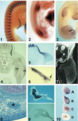
Fig. 1. Development of tissue pattern in the limb. (1)
HH-stage 22 chick
embryo co-stained for expression of Fgf8 (blue) and MyoD (red). Note the
overlapping, but not identical transcript distribution of Fgf8 and MyoD in the
myotomes (Courtesy of Daniel Stolte, Freiburg).
(2)
Lbx1 expression in
HH-stage 21 chick embryo. Dorsal and ventral myogenic zones of the limb buds
are Lbx1-positive. The dorsal myogenic zone has started to downregulate
Lbx-1 expression.
(3)
HH-stage 23 chick embryo; Sonic hedgehog (Shh) in
situ
hybridization.
(4)
Transverse section of a 5-day chick embryo stained for
desmin with an anti-desmin antibody; vm, ventral muscle mass; dm, dorsal
muscle mass.
(5)
Transverse section of a HH-stage 18 chick embryo stained
for the expression of Pax3. Note the Pax3-positive muscle precursor cells
migrating from the lateral dermomyotome into the limb bud.
(6)
of hypaxial muscle cell specification are not well understood to
date. It has been shown that the surface ectoderm is required to
form the hypaxial dermomyotome (Schmidt et al., 2001). Wnt
proteins have been suggested to be the ectodermal signals (Roelink,
1996; Dietrich et al., 1998). Wnt7a and to lesser extent Wnt4 and
Wnt5a can activate myogenesis in mouse paraxial mesoderm
explants (Tajbakhsh et al., 1998). Cauthen et al. (2001) have
recently shown that Wnt6 is uniformely expressed in the surface
ectoderm immediately adjacent to the Pax3 expression domain in
the dorsal somite just at the stage of hypaxial muscle precursor cell
migration.
Another signaling molecule involved in hypaxial muscle
devel-opment is BMP4 which has been shown to induce lateral
charac-teristics of the dermomyotome. Such characcharac-teristics are the
ex-pression of Sim1,(Pourquié et al., 1995, 1996), Lbx1 (Jagla et al.,
1995; Dietrich et al., 1998) and c-met (Bladt et al., 1995). It has
been concluded that Wnt proteins and BMP4 act in concert to
specifiy hypaxial muscle precursor cells (Dietrich et al., 1998).
It is, however, not quite clear if the specification of hypaxial
muscle precursor cells does actually take place within the
dermomyotome because lateral dermomyotomal cells give rise to
different cell types such as endothelial cells of blood vessels and
lymphatics and even cartilage cells forming the scapula blade
(Wilting
et al., 1995; Wilting et al., 1997; Wilting et al., 2000; Huang
et al., 2000). Lateral dermomyotomal cells expressed in addition to
Pax3, Pax7 and myf-5, are specific cellular markers including
VEGF receptor Quek1, the homeobox gene Meis2 and EphA4
(Eichmann et al., 1993; Cecconi et al., 1997; Schmidt et al., 2001).
In any case, muscle precursor cells that have already invaded the
limb buds are found to be determined to form muscle (Mauger and
Kieny, 1980; Wachtler et al., 1981; Wiliams and Ordahl, 2000). To
date it is suggested that signals from the axial structures do not
influence hypaxial myogenesis. Stolte et al. (2002a,b), however,
have been able to show that the expression of Fgf8 even in the
hypaxial myotome depends on Shh produced by the axial
struc-tures meaning that there does exist an influence of notochord and
neural tube on muscle cell development in the hypaxial domain.
Migration of Limb Muscle Precursor Cells
Prerequisite of the migration of muscle precursor cells into the
limb buds is the delamination of cells. It has been shown that
de-epithelialization and subsequent migration of dermomyotomal
cells can be induced ectopically by grafting of proximal limb bud
mesoderm to the flank level (Hayashi and Ozawa, 1995). The
underlying molecular mechanism is an interaction between the
transmembrane tyrosine kinase receptor c-met expressed by the
dermomyotome cells and its ligand scatter factor/hepatocyte growth
factor (SF/HGF) that is produced by somatopleural cells of the limb
buds (Bladt et al., 1995).
Targeting of the genes for either the ligand or the receptor
results in the absence of muscle in the limbs. An ectopic application
of exogenous SF/HGF leads to de-epithelialization of the
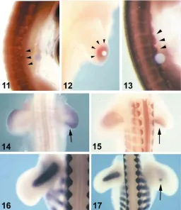
dermomyotomal edges even at interlimb level (Fig. 2.11 and 2.13;
Brand-Saberi et al., 1996; Heymann et al., 1996). The resulting
phenotype of c-met and SF/HGF knockout mice resembles the
phenotype of a naturally occuring mutation in the Pax3 gene called
splotch (Franz et al., 1993; Bober et al., 1994). Pax3 initially
expressed in all the cells of the segmental plate and later on in the
dermomyotome, becomes up-regulated in the lateral part of of the
dermomyotome (Williams and Ordahl, 1994) and has been shown
to regulate the expression of c-met (Epstein et al., 1996; Yang et
al., 1996; Tajbakhsh et al., 1997). The c-met promoter contains a
Pax3 binding site, and Pax3 can drive reporter gene expression
from the c-met promoter in vitro. Therefore, it can be concluded that
Pax3 controls the release of migrating muscle precursors in vivo by
activating c-met. Pax3, Pax7, and myf5 are being expressed in the
migratory muscle precursor cells and in the proliferating cells of
limb premuscular masses (Fig. 2.15; Williams and Ordahl, 1994;
Kiefer and Hauschka, 2001; Swartz et al., 2001).
A gene expressed exclusively in the lateral dermomyotomes at
sites of muscle precursor cell detachment and in migratory
muscle precursors, is the homeobox gene Lbx1 (Fig. 1.2; Jagla et
al., 1995; Dietrich et al., 1998). Lbx1 expression depends on
Pax3, because Lbx1 expression is absent in Splotch mice
(Mennerich et al., 1999; Dietrich et al., 1999). Lbx1 expression is
maintained during muscle precursor cell migration and is
down-regulated shortly after muscle-specific gene expression is
initi-ated in the limb. In mice that lack Lbx1, muscle precursor cells
form and delaminate from the lateral edges of the dermomyotomes
at limb levels but do not move into the limb buds and do not settle
at the sites of future dorsal and ventral muscle masses (Fig. 1.2;
Schäfer and Braun, 1999; Brohmann et al., 2000; Gross et al.,
2000).
Lbx1 is suggested to determine lineage-specific properties
of migrating myogenic precursors and to be essential for the
recognition of cues that guide these cells and maintain their
migratory potential.
It has been shown that the cell adhesion molecule N-cadherin is
involved in the migration and homing of muscle precursor cells as
well as in the differentiation of myoblasts (Brand-Saberi et al., 1996b;
George-Weinstein et al., 1997). The cadherin-mediated
adhesive-ness of cells can be modulated by phosphorylation of intracellular
molecules associated with cadherins, the catenins (reviewed by
Gumbiner 1995; Birchmeier et al., 1996; Huber et al., 1996).
N-cadherin is strongly expressed in the dermomyotome, the migrating
muscle precursors, and more moderately expressed by stationary
somatopleural cells in the myogenic zones and progress zone (Fig.
1.7). After in vivo injection of antibodies and Fab-fragments against
the homophilic binding site of N-cadherin into the wing bud
meso-derm, aggregates of myoblasts are found in the myogenic zone due
to immobilization. It is concluded that the invasion and homing to the
dorsal and ventral myogenic zones where the premuscular masses
are formed depends on homophilic interactions between the
migrat-ing cells and the stationary somatopleural cells by means of
N-cadherin (Brand-Saberi et al., 1996b).
There are further prerequisites for the migration of muscle
precursor cells. Fibronectin has to be available for migration
through the intercellular spaces (Brand-Saberi et al., 1993). The
intercellular spaces have to be large enough, which in vivo is
achieved by differing concentrations of hyaluronic acid (Kosher et
al., 1981; Krenn et al., 1991).
As mentioned earlier, the EphA4 receptor tyrosine kinase is
strongly expressed in the lateral part of the dermomyotomes at the
level where migratory muscle precursors detach (Schmidt et al.,
2001). Recently, it has been suggested that the interaction of
EphA4 that is expressed in the migratory cells and its ligand
ephrin-A5 guides the cells to their appropriate territories in the limb,
disallowing entry into abnormal regions (Swartz et al., 2001).
Muscle precursor cells have to migrate distally in the limbs as
long as the limb anlage grows by distal apposition. They, however,
never reach the most distal region of the growing limb bud, the
so-called progress zone (Brand et al., 1985).
SF/HGF has been found to be continually expressed during of
limb bud outgrowth (Fig. 2.12 and 2.14). Here, it increases the
motility of myogenic precursor cells probably by modulation of
N-cadherin-mediated adhesiveness. At the same time SF/HGF
main-tains their undifferentiated state during migration (Scaal et al.,
1999).
Muscle Growth and Differentiation
Myogenic precursors that have invaded the limb mesenchyme
aggregate and differentiate into dorsal and ventral premuscular
masses (Christ et al., 1977). With the growth of the limb buds the
muscular masses are becoming subdivided in a proximo-distal
direction into stylopodial, zeugopodial and autopodial dorsal and
ventral muscle masses revealing a differentiation gradient from
proximal to distal. Each muscle mass splits up to eventually form
individual, anatomically distinct muscles. Quail-chick chimeras
have given evidence that the muscle pattern is not autonomously
pre-specified within the somitic muscle precursor cells, but
ap-pears to be determined by the somatopleural mesoderm of the limb
bud (Christ and Jacob, 1980; Grim und Wachtler, 1991). Yet it is not
clear which of the specific components of the somatopleural
mesoderm is the source of the patterning information. This is also
true for tendons which develop autonomously and can even be
formed in the absence of muscles (Fig. 1.8; Shellswell and Wolpert,
1977; Jacob and Christ, 1980; Kieny and Chevallier, 1979; Brown
et al., 1999; Kardon, 1999). The later steps of correct
muscle-tendon patterning and the maintenance of muscle-tendons have been
found to depend on reciprocal interactions between muscle and
tendon (Kardon, 1999). Distal tendons differ from more proximal
tendons in their molecular identity. They express the transcription
factors Six1 and Six2 (Oliver et al., 1995) and the receptor EphA4
(Patel et al., 1996) while proximal tendons do not. Other
tendon-specific markers are TGF
β
2,
Eya1 and Eya2 (Xu et al., 1997),
follistatin (D’Sousa and Patel, 1999) and scleraxis (Schweitzer et
al., 2001). Tenascin is first detected in HH-stage 26 wing. It has
been shown that tendon progenitors are induced by ectodermal
signals and that the progenitor cell fate can be repressed by BMP
signaling. The close relationship between ectoderm and early tendon
anlage was earlier shown by Blechschmidt (1961) and Hurle et al.
(1990). The endogenous expression of noggin within the condensing
cartilage is suggested to contribute to the induction of distal tendons
(Schweitzer et al., 2001).Yamamoto et al. (1998) have shown that
Hoxa-11 and Hoxa-13 which in the migrating muscle precursors are
under the control of the limb mesenchyme and polarizing signals are
involved in muscle and tendon patterning in the limb bud.
induces apoptosis and muscle loss (Amthor et al., 1998). The loss
of FGF Receptor 1 (FGFR1) signaling accelerates muscle
differen-tiation resulting in a reduction of muscle size (Itoh et al., 1996;
Flanegan-Steet et al., 2000). It has been shown that FGFs delay
the onset of differentiation of myoblasts obtained from day 4-12
chick wing buds while at the same time there is a subset of
myoblasts derived from HH-stage 23-27 embryos that requires
FGF for myogenic differentiation. It seems that the early population
of limb myoblasts contains discrete subclasses of cells that are
FGF independent. At later stages only FGF independent
myo-blasts persist. This issue of growth factor influence on limb
myogenesis has been most extensively analyzed by the Hauschka
(Clegg et al., 1987; Pirskanen et al., 2000; Seed and Hauschka,
1988; Templeton and Hauschka, 1992) and the Olwin group
(DeHamer et al., 1994; Flanagan-Steet et al., 2000; Hannon et al.,
1996; Olwin et al., 1994a; Olwin et al., 1994b).
More recently myostatin, a member of the TGF
β
superfamily,
has been proposed as a regulator of myogenesis. It is expressed
at early and late stages of myogenesis and regulates the amount
of skeletal muscle cells (McPherron et al., 1997). Myostatin
knock-out mice develop muscles 2-3 times the size of wild-type mice
which is essentially the opposite to what is observed in follistatin
knockout mice (Matzuk et al., 1995). Krüger et al. (2001) have
recently shown that Shh acts as a survival and proliferation factor
for hypaxial muscles corroborating the observation that ectopic
expression of Shh in chicken limb buds induces muscle by
hyper-trophy probably via up-regulation of BMP expression (Figs. 1.3 and
2.17; Duprez et al., 1998; Amthor et al., 1998).
All anatomic muscles of adult vertebrates have their origins in
several waves of muscle fiber formation as development proceeds
(Stockdale, 1997). The primary fibers form during embryonic
development and lay down the anlagen of all future muscles. The
amount of muscle mass formed from the primary myoblasts is
extremely small. Their role may be to define the type, shape and
location of a muscle. Primary fibers traverse the muscle anlage
from tendon to tendon and become innervated at multiple endplates
prior to formation of secondary fibers (Duxson and Usson, 1989;
Duxson and Sheard, 1995). Secondary fibers form and insert on
the surface of primary fibers beginning near the sites of innervation
of primary fibers and initially do not traverse the entire length of a
muscle. The secondary fibers increase rapidly in number and
nucleation and separate from the primary fibers.
All muscle fibers form by fusion of myoblasts with one another
(Stockdale and Holtzer, 1961). The myoblasts can be subdivided
in three categories: embryonic myoblasts, fetal myoblasts and
satellite cells (Stockdale, 1992, 1997). They form fibers which can
be identified by the expression of specific isoforms of myosin heavy
chain (MyHC) and number of nuclei (Miller and Stockdale, 1986;
Ontell
et al., 1993). Embryonic, fetal and adult myoblasts in the limb
have their origins in the migratory population of muscle precursors
(Christ et al., 1977).
Embryonic myoblasts in the limb buds express MyoD and begin
to fuse into small primary fibers, a process that requires surface
molecules which mediate heterophilic and homophilic cell-cell
recognition such as N-CAM, N-cadherin and M-cadherin (reviewed
by Arnold and Braun, 2000). In birds, myoblasts of fetal and adult
characteristics replace the embryonic myoblasts until the end of
the first week. It is still an open question, if the precursors of
embryonic myoblasts are also precursors of fetal and adult
myo-blasts or whether there are separate migratory populations for
each within the somite.
During embryonic development, and before functional
innerva-tion, a highly stereotopic pattern of slow- and fast contracting
primary muscle fibers is established within individual muscles of
the limbs, from distinct populations of myoblasts (Nikovits et al.,
2001). The fibers can be distinguished by distinct morphological
and biochemical properties (Seed and Hauschka, 1984; Stockdale,
1992) and classified by different specific isoforms of the myosin
heavy chain (MyHC) (Miller and Stockdale, 1992; DiMario and
Stockdale, 1997). Nikovits et al. (2001) have concluded that an
intrinsic commitment to either a fast or a slow fiber-type lineage
occurs in myogenic precursors while still within the somite. This is
in accordance with data of fiber type specification in zebrafish
(Blagden
et al., 1997; Currie and Junghans, 1996; Du et al., 1997).
Cann et al., 1999 was able to show that in explant cultures of avian
somites exogenous Shh leads to a marked expansion of the slow
fiber population. It has been suggested that selective amplification
of committed myoblasts to form fast- or slow fibers occurs in
response to proliferative signals that originate in the limb stroma
(Nikovits et al., 2001).
In a recent paper Bren-Mattison and Olwin (2002) have
pre-sented a model according to which Shh represses differentiation of
posterior myoblasts in the ventral muscle mass, allowing mitogens
present in the limb bud mesenchyme to stimulate their proliferation,
while absence of Shh induces precocious differentiation of early
myoblasts fated to express slow MyHC. The precocious
differen-tiation depletes the pool of proliferating myoblasts and ultimately
results in less muscle due to loss of slow MyHC fibers.
Regulator Genes that control Limb Myogenesis
Muscle Hypertrophy and Regeneration
Hypertophy of muscle in the postnatal period in most
verte-brates is under the control of mechanical stress that is transduced
by a number of signaling molecules. It can be identified by an
increase in the number of nuclei in the fibers, increase in the
number of satellite cells, and hypertrophy of the fibers (Kadi and
Thornell, 2000). IGF-I, IGF-II are among the signaling molecules
and are produced by muscle fibers themselves (Gerrard et al.,
1998). Transgenic mice that overexpress IGF-I demonstrate a
marked hypertrophy of muscle fibers and targeted disruption of
IGF-I expression in the mouse leads to a reduction of fiber size and
muscle hypoplasia (Coleman et al., 1995; Fournier and Lewis,
2000). As mentioned earlier, myostatin is involved in the control of
muscle mass. Zhu et al. (2000) have shown that mice with a
disruption in the myostatin gene exhibit a significant increase in
muscle fiber size.
Muscle regeneration requires activation of the mononuclear
satellite cells or myogenic differentiation of bone marrow-derived
hematopoietic stem cells (reviewed by Patel et al., 2002; Parrish,
1996; Bittner et al., 1999; Ferrari et al., 1998). In a reciprocal study,
Jackson et al. (1999) have shown that mononucleated cells from
muscle have the capacity to reconstitute the hematopoietic system
of irradiated mice. The c-met receptor is present on satellite cells
in normal muscle tissue (Tatsumi et al., 1998). Another gene that
has been identified to be expressed in satellite cells is Pax7 (Seale
et al., 2000). A number of growth factors have been found to be
involved in the activation of satellite cell proliferation and MyoD
expression (reviewed by Grounds and Yablonka-Reuveni, 1993)
while HGF/SF inhibits muscle regeneration (Lefaucheur and Sebille,
1995). Floss et al. (1997) have shown that FGF-6 is a critical
component to stimulate, attract, or activate satellite cells for
differ-entiation.
New and unexpected sources of myogenic progenitor cells have
recently been described not only to be the bone marrow
mesen-chyme (Ferrari et al., 1998; Bittner et al., 1999) but also the
limb-bud mesenchyme of muscleless knock-out mice c-met-/- and Pax3
-/- (Bailey et al., 2001) and from the embryonic dorsal aorta (De
Angelis et al., 1999) suggesting that muscle progenitor cells could
be endothelia-derived. In this connection it is interesting to note
that in the somites, myogenic and angioblastic cells exist side by
side and endothelia of the aorta and the limb bud are at least
partially of somitic origin (Wilting et al., 1995; Brand-Saberi et al.,
1995). Finally, a new population of muscle progenitor cells has
been described to reside in adult muscle itself as a so-called
“muscle-derived side population” (Gussoni et al., 1999).
Concluding Remarks
Limb muscle in the adult accounts for more than half of the entire
skeletal muscle mass. This is in marked contrast to the situation in
the early embryo. To understand the process involved in limb
myogenesis we can study the origin of its precursor cells and their
behaviour as they invade and populate the limb bud mesenchyme.
Our insights have become more detailed as we have identified new
regulatory molecules. Correlating the process of muscle
develop-ment with the signals involved in limb bud patterning can be
expected to refine our knowledge further. As in other fields of
research, questions of quantity control will have to be addressed
after having understood some of the questions concerning the
quality control. It is still unknown in how far early and late muscle
populations and satellite cells are connected, or when these
diverge as separate lineages. Answers to these questions can be
expected from clonal studies of myogenic cells in the limb bud. It
can be expected that the study of myogenic stem cells will further
enhance the insights into limb muscle development.
Acknowledgements
We thank the members of our laboratories who have contributed to this
work and Mrs. U. Uhl for typing the manuscript. Work in the laboratory of BC
is supported by the Deutsche Forschungsgemeinschaft (Ch 44/12-1,2,3,
14-1, SFB 592 A1). Work in the laboratory of B.BS is supported by the
Deutsche Forschungsgemeinschaft (Br 957/5-1, 5-2, 5-3; SFB 592 B4).
References
AMTHOR, H., CHRIST, B., WEIL, M. and PATEL, K. (1998). The importance of timing differentiation during limb muscle development. Curr.Biol. 8:642-652.
AMTHOR, H., CHRIST, B. and PATEL, K. (1999). A molecular mechanism enabling continuous embryonic muscle growth - a balance between proliferation and differentiation. Development 126:1041-1053.
ARNOLD, H.H. and BRAUN, T. (2000) Genetics of muscle determination and development. In: Somitogenesis, Part 2. Current Topics in Developmental Biology (ed. C.P. Ordahl) pp.129-164
BAILEY, P., HOLOWACZ, T. and LASSAR, A.B. (2001). The origin of skeletal muscle stem cells in the embryo and the adult. Curr. Opin. Cell Biol. 13:679-689.
BARTON-DAVIS, E., SHOTURNA, D.I., MUSARO, A., ROSENTHAL, N. and SWEENEY, H.L. (1998). Viral mediated expression of insulin-like growth factor blocks the aging-related loss of skeletal muscle function. Proc. Natl. Acad Sci USA 95:15603-15607.
BIRCHMEIER, C., BIRCHMEIER, W. and BRAND-SABERI, B. (1996). Epithelial-mesenchymal transitions in cancer progression. Acta Anat. 156:217-226.
BITTNER, R.E., SCHOFER, C., WEIPOLTSHAMMER, K., IVANOVA, S., STREUBEL, B., HAUSER, E., FREILINGER, M., HOGER, H., ELBE-BURGER, A. and WACHTLER, F. (1999). Recruitment of bone-marrow-derived cells by skeletal and cardiac muscle in adult dystrophic mdx mice. Anat. Embryol. 199:391-396.
BLADT, F., RIETHMACHER, D., ISENMANN, S., AGUZZI, A. and BIRCHMEIER, C. (1995). Essential role for the c-met receptor in the migration of myogenic precursor cells into the limb bud. Nature 376:768-771.
BLAGDEN, C.S., CURRIE, P.D., INGHAM, P.W. and HUGHES, S.M. (1997). Noto-chord induction of zebrafish slow muscle mediated by Sonic hedgehog. Genes. Dev. 11:2163-2175.
BLECHSCHMIDT, B. (1961). The stages of Human Development before Birth. An Introduction to Human Embryology. S. Karger, Basel, London, New York.
BOBER, E., BRAND-SABERI, B., EBENSPERGER, C., WILTING, J., BALLING, R., PATERSON, B.M., ARNOLD, H.H. and CHRIST, B. (1994). Initial steps of myogenesis in somites are independent of influence from axial structure. Devel-opment 120:3073-3082.
BORYCKI, A.G., STRUNK, K.E., SAVARY, R. and EMERSON, C.P.Jr. (1997). Distinct signal/response mechanisms regulate pax1 and QmyoD activation in sclerotomal and myotomal lineages of quail somites. Dev.Biol. 185:185-200.
BORYCKI, A.G. and EMERSON, C.P. (2000). Multiple tissue interactions ans signal transduction pathways control somite myogenesis. Curr. Top Dev. Biol. 165-224.
BRAND-SABERI, B. and CHRIST, B. (1999). Genetic and epigenetic control of muscle development in vertebrates. Review article. Cell Tissue Res. 296:199-212.
BRAND, B., CHRIST, B. and JACOB, H.J. (1985). An experimental analysis of the developmental capabilities of distal parts of avian leg buds. Am. J. Anat. 173:321-340.
BRAND-SABERI, B., EBENSPERGER, C., WILTING, J., BALLING, R. and CHRIST, B. (1993). The ventralizing effect of the notochord on somite differentiation in chick embryos. Anat. Embryol. 188:239-245.
BRAND-SABERI, M., GAMEL, A.J., KRENN, V., MÜLLER, T.S., WILTING, J. and CHRIST, B. (1996b). N-cadherin is involved in myoblast migration and muscle differentiation in the avian limb bud. Dev. Biol. 178:160-173.
BREN-MATTISON, Y. and OLWIN, B.B. (2002). Sonic Hedgehog inhibits the terminal differentiation of limb myoblasts committed to the slow muscle lineage. Dev. Biol. 242:130-148.
BROHMANN, H., JAGLA, K. and BIRCHMEIER, C. (2000). The role of Lbx1 in migration of muscle precursor cells. Development 127:437-445.
BROWN, D., WAGNER, D., LI, X., RICHARDSON, J.A. and OLSON, E.N. (1999). Dual role of the basic helix-loop-helix transcription factor scleraxis in mesoderm formation and chondrogenesis during mouse embryogenesis. Development 126:4317-4329.
BUFFINGER, N. and STOCKDALE, F.E. (1994). Myogenic specification in somites: induction by axial structures. Development 120:1443-1452.
CANN, G.M., LEE, J.W. and STOCKDALE, F.E. (1999). Sonic hedgehog enhances somite cells viability and formation of primary slow muscle fibers in avian segmented mesoderm. Anat. Embryol. 200:239-252.
CAPDEVILA, J., TABIN, C. and JOHNSON, R.L. (1998). Control of dorsoventrakl somite patterning by Wnt-1 and b-catenin. Dev. Biol. 193:182-194.
CAUTHEN, C.A., BERDOUGO, E., SANDLER, J. and BURNS, L.W. (2001). Com-parative analysis of the expression patterns of Wnts and Frizzleds during early myogenesis in chick embryos. Mech. Dev. 104:133-138.
CECCONI, F., PROETZEL, G., ALVAREZ-BOLADO, G., JAY, D. and GRUSS, P. (1997). Expression of Meis2, a knotted-related murine homeoboxgene, indicates a role in the differentiation of the forebrain and the somitic mesoderm. Dev. Dyn. 210:184-190.
CHEVALLIER, A., KIENY, M. and MAUGER, A. (1977). Limb-somite relationship: origin of the limb musculature. J. Embryol. Exp. Morph. 41:245-258.
CHOI, J., COSTA, M.L., MERMELSTEIN, C.S., CHAGAS, C., HOLTZER, S. and HOLTZER, H (1990). MyoD converts primary dermal fibroblasts, chondroblasts, smooth muscle, and retinal pigmented epithelial cells into striated mononucleated myoblasts and multinucleated myotubes. Proc. Natl. Acad. Sci. USA 87:7988-7992.
CHRIST, B. (1970). Experimente zur Lageentwicklung der Somiten. Anat. Anz. Ergänzungs-H. zum Bd. 126:555-564.
CHRIST, B. and JACOB, H.J. (1980). Origin, distribution and determination of chick limb mesenchymal cells. In: “Teratology of the Limbs”, Symposium on Prenatal Dev. Biol, 4th Symp. (H.-J. Merker, H. Nau and D. Neubert, eds.) Pp. 67-77, W. de Gruyter, Berlin
CHRIST, B. and ORDAHL C.P. (1995). Early stages of somite development. Anat. Embryol. 191:381-396.
CHRIST, B., JACOB, H.J. and JACOB, M. (1972). Experimentelle Untersuchungen zur Somitenentstehung beim Hühnerembryo. Z. Anat. Entwickl.-Gesch. 138:82-97.
CHRIST, B., JACOB, H.J. and JACOB, M. (1974a). Über den Ursprung der Flügelmuskulatur. Experientia 30:1446-1448.
CHRIST, B., JACOB, H.J. and JACOB, M. (1974b). Experimentelle Untersuchungen zur Entwicklung der Brustwand beim Hühnerembryo. Experientia (Basel) 30:1449-1451.
CHRIST, B., JACOB, H.J. and JACOB, M. (1977). Experimental analysis of the origin of the wing musculature in avian embryos. Anat. Embryol. 150:171-186.
CHRIST, B., JACOB, H.J. and JACOB. M. (1983). On the origin and development of the ventro-lateral abdominal muscles in the avian embryo. An experimental and ultrastructural study. Anat. Embryol. 166:87-101.
CHRIST, B., BRAND-SABERI, B., GRIM, M. and WILTING, J. (1992). Local signalling in dermomyotomal cell type specification. Anat. Embryol. 186:505-510.
COLEMAN, M.E., DEMAYO, F., YIN, K.C., LEE, H.M., GESKE, R., MONTGOMERY, C and SCHWARTZ, R.J. (1995). Myogenic vector expression of insulin-like growth factor I stimulates muscle cell differentiation and myofiber hypertrophy in transgenic mice. J. Biol. Chem. 270:12109-12116.
CSERJESI, P. and OLSON, E.N. (1991). Myogenin induces the myocyte-specific enhancer binding factor MEF-2 independently of other muscle-specific gene products. Mol. Cell Biol. 11:4854-4862.
CLEGG, C.H., LINKHART, T.A., OLWIN, B.B. and HAUSCHKA, S.D. (1987). Growth factor control of skeletal muscle differentiation: commitment to terminal differen-tiation occurs in G1 phase and is repressed by fibroblast growth factor. J. Cell Biol. 105:949-956.
CURRIE, P.D. and INGHAM, P.W. (1996). Induction of a specific muscle cell type by a hedgehog-like protein in zebrafish. Nature 382:452-455.
DE ANGELIS, L., BERGHELLA, L., COLETTA, M., LATTANZI, L., ZANCHI, M., CUSELLA-DE ANGELIS, M.G., PONZETTO, C. and COSSU, G. (1999). Skel-etal myogenic progenitors originating from embryonic dorsal aorta coexpress endothelial and myogenic markers and contribute to postnatal muscle growth and regeneration. J. Cell Biol. 147:859-878.
DEHAMER, M.K., GUEVARA, J.L., HANNON, K., OLWIN, B.B. and CALOF, A.L. (1994). Genesis of olfactory receptor neurons in vitro: regulation of progenitor cell divisions by fibroblast growth factors. Neuron 13:1083-1097.
DELFINI, M.C., HIRSINGER, E., POURQUIÈ, O. and DUPREZ, D. (2000). Delta1-activated Notch inhibits muscle differentiation without affecting Myf5 and Pax3 expression in chick limb myogenesis. Development 127:5213-5224.
DENETCLAW, W.F., CHRIST, B. and ORDAHL, C.P. (1997). Location and growth of epaxial myotome precursor cells. Development 124:1601-1610.
DEUTSCH, U., DRESSLER, G.R. and GRUSS, P. (1988). Pax1, a member of a paired box homologous murine gene family, is expressed in segment structures during development. Cell 53:617-625.
DIETRICH, S., SCHUBERT, F.R. and LUMSDEN, A. (1997). Control of dorsoventral pattern in the chick paraxial mesoderm. Development 124:3895-3908.
DIETRICH, S., SCHUBERT, F.R., HEALY, C., SHARPE, P.T. and LUMSDEN, A. (1998). Specification of the hypaxial musculature. Development 125:2235-2249.
DIETRICH, S., ABOU-REBYEH, F., BOHMANN, H., BLADT, F., SONNENBERG-RIETHMACHER, E., YAMAAI, T., LUMSDEN, A., BRAND-SABERI, B. and BIRCHMEIER, C. (1999). The role of SF/HGF and c-Met in the development of skeletal muscle. Development 126:1621-1629.
DIMARIO, J.X. and STOCKDALE, F.E. (1997). Both myoblast lineage and innervation determine fiber type and are required for expression of the slow myosin heavy chain 2 gene. Dev. Biol. 188:167-180.
D’SOUZA, D. and PATEL, K. (1999). Involvement of long- and short-range signalling during early tendon development. Anat. Embryol. 200:367-375.
DU, S.J., DEVOTO, S.H., WESTERFIELD, M. and MOON, R.T. (1997). Positive and negative regulation of muscle cell identity by members of the hedgehog and
TGF-β gene families. J. Cell Biol. 139:145-156.
DUPREZ, D., FOURNIER-THIBAULT, C. And LE DOUARIN, N.M. (1998). Sonic hedgehog induces proliferation of commtted skeletal muscle cells in the chick limb. Development 125:495-505.
DUXSON, M.J., USSON, Y. and HARRIS, A.J. (1989). The origin of secondary myotubes in mammalian skeletal muscles: ultrastructural studies. Development 107:743-750.
DUXSON, M.J. and SHEARD, P.W. (1995). Formation of new myotubes occurs exclusively at the multiple nnervation zones of an embryonic large muscle. Dev. Dyn. 204:391-405.
EICHMANN, A., MARCELLE, C., BREANT, C. and LE DOUARIN, N.M. (1993). Two molecules related to the VEGF receptor are expressed in early endothelial cells during avian embryonic development. Mech. Dev. 42:33-48.
EPSTEIN, J.A., SHAPIRO, D.N., CHENG, J., LAM, P.Y. and MAAS, R.L. (1996). Pax3 modulates expression of the c-Met receptor during limb muscle development. Proc. Natl. Acad. Sci. USA 93:4213-4218.
FAN, C-M. and TESSIER-LAVIGNE, M. (1994). Patterning of mammalian somites by surface ectoderm and notochord: evidence for sclerotome induction by a hedgehog homolog. Cell 79:1175-1186.
FAN, C., LEE, C. and TESSIER-LAVIGNE, M. (1997). A role for Wnt proteins in induction of dermomyotome. Dev. Biol. 191:160-165.
FAN, C.M., PORTER, J.A., CHIANG, C., CHANG, D.T., BEACHY, P.A. and TESSIER-LAVIGNE, M. (1995). Long-range sclerotome induction by sonic hedgehog: Direct role of the amino-terminal cleavage product and modulation by the cyclic AMP signaling pathway. Cell 81:457-465.
FERRARI, G., CUSELA-DE ANGELIS, G., COLETTA, M., PAOLUCCI, E., STORNAIUOLO, A., COSSU, G. and MAVILIO, F. (1998). Muscle regeneration by bone marrow-derived myogenic progenitors. Science 279:1529-1530.
FLANAGAN-STEET, H. (2000). Loss of FGF receptor 1 signaling reduces skeletal muscle mass and disrupts myofiber organization in the developing limb. Dev. Biol. 218:21-37.
FOURNIER, M. and LEWIS, M.I. (2000). Influences of IGF-I gene disruption on the cellular profile of the diaphragm. Am. J. Physiol. Endocrinol. Metab. 278:E707-715.
FRANZ, T., KOTHARY, R., SURANI, M.A.H., HALATA, Z. and GRIM, M. (1993). The splotch mutation interferes with muscle development in he limbs. Anat. Embryol. 187:153-160.
FÜCHTBAUER, E.-M. (1995). Expression of M-twist during postimplantation develop-ment of the mouse. Dev. Dyn. 204:316-322.
FÜCHTBAUER, E.M. (2002). Inhibition of skeletal muscle development: less differen-tiation gives more muscle. In: Myogenesis, Results and Problems in Cell Differen-tiation, Vol. 38, B. Brand-Saberi (Ed.): Vertebrate Myogenesis, pp. 143-161, Springer-Verlag Berlin, Heidelberg.
GEORGE-WEINSTEIN, M., GERHART, J., BLITZ, J., SIMAK, E. and KNUDSEN, K.A. (1997). N-cadherin promotes the commitment and differentiation of skeletal muscle precursor cells. Dev. Biol. 185:14-24.
GERRARD, D.E., OKAMURA, C.S., RANALLETTA, M.A: and GRANT, A.L. (1998). Developmental expression and location of IGF-I and IGF-II mRNA and protein in skeletal muscle. J. Anim. Sci. 76:1004-1011.
GOULDING, M.D., LUMSDEN, A. and PAQUETT, A.J. (1994). Regulation of Pax-3 expression in the dermomyotome and its role in muscle development. Development 120:957-971.
GRIM, M. and WACHTLER, F. (1991). Muscle morphogenesis in the absence of myogenic cells. Anat. Embryol. 183:67-70.
GROSS, M.K., MORAN-RIVARD, L., VELASQUEZ, T., NAKATSU, M.N., JAGLA, K., GOULDING, M. (2000). Lbx1 is required for muscle precursor migration along a lateral pathway into the limb. Development 127:413-424.
GROUNDS, M.D. and YABLONKA-REUVENI, Z. (1993). Molecular and cellular biology of muscle regeneration. In: “Molecular and Cellular Biology of Muscular Dystrophy” (T. Partridge, Ed.). pp 210-256. Chapmann and Hall, London.
GUMBINER, B.M. (1995). Signal transduction by β-catenin. Curr. Opin. Cell Biol. 7:634-640.
GUSSONI, E., SONEOKA, Y., STRICKLAND, C.D., BUZNEY, E.A:, KHAN, M.K., FLINT, A.F., KUNKEL, L.M. and MULLIGAN, R.C. (1999). Dystrophin expression in the mdx mouse restored by stem cell transplantation. Nature 401:390-394.
HANNON, K., KUDLA, A.J., MCAVOY, M.J., CLASE, K.L. and OLWIN, B.B. (1996). Differentially expressed fibroblast growth factors regulate skeletal muscle develop-ment through autocrine and paracrine mechanisms. J. Cell Biol. 132:1161-1159.
HAYASHI, K.and OZAWA, E. (1995). Myogenic cell migration from somites is induced by tissue contact with medial region of the presumptive limb msoderm in chick embryos. Development 121:661-660.
HEBROK, M., FÜCHTBAUER, A. and FÜCHTBAUER, E.-M. (1997). Repression of muscle specific gene activation by the murine twist protein. Exp. Cell Res. 232:295-303.
HEYMANN, S., KOUDROVA, M., ARNOLD, H.H., KÖSTER, M. and BRAUN, T. (1996). Regulation and function os SF/HGF during migration of limb muscle precursor cells in chicken. Dev. Biol. 180:655-589.
HUBER, O., BIERKAMP, C. and KEMLER, R. (1996) Cadherins and catenins in development. Curr. Opin. Cell Biol. 8:685-691.
HUANG, R., ZHI, Q., PATEL, K., WILTING, J. and CHRIST, B. (2000). Dual origin and segmental organisation of the avian scapula. Development 127:3789-3794.
HURLE, J.M., ROS, M.A., GANAN, Y., CRITCHLOW, M. and HINCHLIFFE, J.R. (1990). Experimental analysis of the role of ECM in the patterning of the distal tendons of the developing limb bud. Cell Differ. Dev. 30:97-108.
IKEYA, M. and TAKADA, S. (1998). Wnt signaling from the dorsal neural tube is required for the formation of the medial dermomyotome. Development 125:4969-4976.
ITOH, N., MIMA, T. and MIKAWA, T. (1996). Loss of fibroblast growth factors receptor is necessary for terminal differentiation of embryonic limb muscle. Development 122:291-300.
JACKSON, K.A., MI, T. and GOODELL, M.A. (1999). Hematopoietic potential of stem cells isolated from murine skeletal muscle. Proc. Natl. Acad. Sci USA 961:4482-4486.
JAGLA, K., DOLLO, P., MATTEI, M.G., JAGLA, T., SCHUHBAUR, B., DRETZEN, G., BELLARD, F. and BELLARD, M. (1995). Mouse Lbx1 and human LBX1 define a novel mammalian homeobox gene family related to the Drosophila lady bird genes. Mech. Dev. 53:345-356.
JOHNSON, R.L., LAUFER, E., RIDDLE, R.D. and TABIN, C. (1994). Ectopic expression of Sonic hedgehog alters doral-ventral patterning of somites. Cell 79:1165-1173.
KADI, F. and THORNELL, L.E. (2000). Concomitant increases in myonuclear and satellite cell content in female trapezius muscle following strength training. Histochem. Cell Biol. 113:99-103.
KARDON, G. (1999). Muscle and tendon morphogenesis in the avian hind limb. Development 125:4019-4032.
KIEFER, J.C. and HAUSCHKA, S.D. (2001). Myf-5 is transiently expressed in nonmuscle mesoderm and exhibits dynamic regional changes within the presegmented mesoderm and somites I-IV. Dev. Biol. 232:77-90.
KIENY, M. and CHEVALLIER, A. (1979). Autonomy of tendon development in the embryonic chick wing. J. Embryol. Exp. Morphol. 49:153-165.
KOSHER, R.A., SAVAGE, M.P., WALKER, K.H.K. (1981). A gradation of hyaluronate accumulation along the proximodistal axis of the embryonic chick limb bud. J. Embryol. Exp. Morphol. 63:85-98.
KRENN, V., BRAND-SABERI, B. and WACHTLER, F. (1991). Hyaluronic acid influ-ences the migration of myoblasts within the embryonic wing bud. Am. J. Anat. 192:400-406.
KRÜGER, M., MENNERICH, D., FEES, S., SCHÄFER, R., MUNDLOS, S. and BRAUN, T. (2001). Sonic hedgehog is a survival factor for hypaxial muscles during mouse development. Development 128:743-752.
LASSAR, A.B., DAVIS, R.L., WRIGHT, W.E., KADESCH, T., MURRE, C., VORONOVA, A:, BALTIMORE, D. and WEINTRAUB, H. (1991). Functional activity of myogenic HLH proteins requires hetero-oligomerization with E12/E47-like proteins in vivo. Cell 66:305-315.
LEFAUCHEUR, J.P. and SEBILLE, A. (1995). Muscle regeneration following injury can be modified in vivo by immune neutralization of basic fibroblastic growth factor, transforming growth factor beta-1 or insulin-like growth factor I. J. Neuroimmunol. 5:85-91.
MANSOURI, A., STOYKOVA, A., TORRES, M. and GRUSS, P. (1996). Dysgen-esis of cephalic neural crest derivatives in Pax7-/-mutant mice. Development 122:831-838.
MATZUK, M.M., NAIFANG, L., VOGEL, H., SELLHEYER, K., ROOP, D.R. and BRADLEY, A. (1995). Multiple dfects and perinatal death in mice deficient in Follistatin. Nature 374:360-363.
MARCELLE, C., STARK, M. and BRONNER-FRASER, M. (1997). Coordinate actions of BMPs, Wnts, Shh and Noggin mediate patterning of the dorsale somite. Development 124:3955-3963.
MAUGER, A. and KIENY, M. (1980). Migratory and ontogenetic capacities of muscle cells in bird embryos. Wilhelm Roux‘s Arch. Dev. Biol. 189:123-134.
MCMAHON, J.A., TAKADA, S., ZIMMERMANN, L.B., FAN, C.M., HARLAND, R.M. and MCMAHON, A.P. (1998). Noggin-mediated antagonism of BMP signaling is required for growth and patterning of the neural tube and somite. Genes Dev. 12:1438-1452.
MCPHERON, A.C., LAWLER, A.M. and LEE, S.J. (1997). Regulation of skeletal muscle mass in mice by a new TGF-β superfamily member. Nature 387:83-90.
MERINO, R., GANAN, Y., MACIAS, D., ECONOMIDES, A.N., SAMPATH, K. and HURLE, J.M. (1998). Morphogenesis of digits in the avian limb is controlled by FGFs, TGFbetas, and noggin throgh BMP signaling. Dev. Biol. 200:35-45.
MILLER, J.B. and STOCKDALE, FE. (1986). Develomental origins of skeletal muscle fibers: Clonal analysis of myogenic cell lineages based on fast and slow myosin heavy chain expression. Proc. Natl. Acad Sci USA 83:3860-3864.
MENNERICH, D., SCHÄFER, K. and BRAUN, T. (1998). Pax-3 is necessary but not sufficient for lbx1 expression in myogenic precursor cells of the limb. Mech. Dev. 73:147-158.
MOLKENTIN, J.D. and OLSON, E.N. (1996). Defining the reglatory networks for muscle development. Curr. Opin. Gen. Dev. 6:445-453.
MÜNSTERBERG, A.E. and LASSAR, A.B. (1995). Combinatorial signals from the neural tube, floor plate and notochord induce myogenic bHLH gene expression in the somite. Development 121:651-660.
NIKOVITS, W., CANN, G.M., HUANG, R., CHRIST, B. and STOCKDALE, F.E. (2001). Patterning of fast and slow fibers within embryonic muscle is established indepen-dently of signals from the surrounding mesenchyme. Development 128:2537-2544.
OLIVER, G., WEHR, R., JENKINS, N.A., COPELAND, N.G., CHEYETTE, B.N.R., HARTENSTEIN, V., ZIPRSKY, S.L. and GRUSS, P. (1995). Homeobox genes and connective tissue patterning. Development 121:693-705.
OLWIN, B.B., HANNON, K. and KUDLA, A.J. (1994b). Are fibroblast growth factors regulators of myogenesis in vivo? Prog. Growth Factor Res. 5:145-158.
ONTELL, M., ONTELL, M.P., SOPPER, M.M., LYONS, G. and BUCKINGHAM, M. (1993). Contractile protein gene expression in primar myotbes of embryonic mouse hindlimb muscle. Development 117:1435-1444.
ORDAHL, C.P. and LEe DOUARIN, N.M. (1992). Two myogenic lineages within the developing somite. Development 114:339-353.
ORDAHL, C.P., BERDOUGO, E., VENTERS. S.J. and DENETCLAW, W.F. (2001). The dermomyotome dorsomedial lip drives growth and morphogenesis of both primary myotome and dermomyotome epithelium. Development 128:1731-1744.
OTT, M.O., BOBER, E., LYONS, G., ARNOLD, H., BUCKINGHAM, M. (1991). Early expression of the myogenic regulatory gene, myf5, in precursor cells of skeletal muscle in the mouse embryo. Development 111:1097-1107.
PARRISH, E.P., CIFUENTES-DIAZ, C., LI, Z.L., VICART, P., PULIN, D., DREYFUS, P.A., PESCHANSKI, M., HARRIS, A.J. and GARCIA, L. (1996). Targeting wide-spread sites of damage in dystrophic muscle: engrafted macrophages as potential shuttles. Gene Ther. 3:13-20.
PATEL, K., NITTENBERG, R., D’SOUZA, D., IIRVING, C., BURT, D., WILKINSON, D.G. and TICKLE, C. (1996). Expression and regulation of Cek-8, a cell to cell signalling receptor in developing chick limb buds. Development 122:1147-1155.
PATEL, K., CHRIST, B. and STOCKDALE, F.E. (2002). Control of muscle size during embryonic, fetal, and adult life. In. Results and Problems in Cell Differentiation, Vol. 38, B. Brand-Saberi (Ed.): Vertebrate Myogenesis, pp. 163-186. Springer-Verlag Berlin, Heidelberg.
PIRSKANEN, A., KIEFER, J.C. and HAUSCHKA, S.D. (2000). IGFs, insulin, Shh, bFGF, and TGF-beta1 interact synergistically to promote somite myogenesis in vitro. Dev. Biol. 224:189-203.
POURQUIÉ, O., COLTEY, M., BREANT, C. and LE DOUARIN, N.M. (1995). Control of somite patterning by signals from the lateral plate. Proc. Natl. Acad. Sci USA 92:3219-3223.
POURQUIÉ, O., COLTEY, M., TEILLET, M.A., ORDAHL, C.P. and LE DOUARIN, N.M. (1993) Control of dorso-ventral patterning of somite derivatives by notochord and floor plate. Proc. Natl. Acad. Sci USA 90:5242-5346
POURQUIÉ, O., FAN, C.M., COLTEY, M., HIRSINGER, E., WATANABE, Y., BREANT, C., FRANCIS-WEST, P., BRICKELL, P., TESSIER-LAVIGNE, M. and LE DOUARIN, N.M. (1996). Lateral and axial signals involved in avian somite patterning: a role for BMP-4. Cell 84:461-471.
POWNALL, M.E. and Emerson, C.P. (1992). Sequential activation of myogenic regulatory genes during somite morphogenesis in quail embryos. Dev. Biol. 151:67-79.
POWNALL, M., STRUNK, K. and EMERSON, C. (1996). Notochord signals control the transcriptional cascade of myogenic bHLH genes in somites of quail embryos. Development 122:1475-1488.
ROELINK, H. (1996). Tripartite signalling of pattern: interactions between Hedgehogs, BMPs and Wnts in the control of vertebrate development. Curr. Opin. Neurobiol. 6:33-40.
RONG, P.M., TEILLET, M.A., ZILLER, C. and LE DOUARIN, N.M. (1992). The neural-tube notochord complex is necessary for vertebral but not limb and body wall striated muscle differentiation. Development 115:657-672.
RUDNICKI, M.A:, SCHNEGELSBERG, P.N., STEAD, R.H., BRAUN, T., ARNOLD, H.H. and JAENISCH, R. (1993). MyoD or Myf-5 is required for the formation of skeletal muscle. Cell 75:1351-1359.
SASSOON, D.A. (1993). Myogenic regulatory factors: dissecting their role and regula-tion during vertebrate embryogenesis. Dev. Biol. 156:11-23.
SCAAL, M., BONAFEDE, A., DATHE, V., SACHS, M., CANN, G., CHRIST, B. and BRAND-SABERI, B. (1999). SF/HGF is a mediator between limb patterning and muscle development. Development 126:4885-4893.
SCHÄFER, K. and BRAUN, T. (1999). Early specification of limb muscle precursors by the homeobox gene Lbx1h. Nat. Genet. 23:213-216.
SCHMIDT, C., CHRIST, B., PATEL, K. and BRAND-SABERI, B. (1998). Experimental induction of BMP-4 expression leads to apoptosis in the paraxial and lateral plate mesoderm. Dev. Biol. 202:253-263.
SCHMIDT, C., CHRIST, B., MADEN, M., BRAND-SABERI, B. and PATEL, K. (2001). Regulation of EphA4 expression in paraxial and lateral plate mesoderm by ectoderm derived signals. Dev. Dyn. 220:377-386.
SCHWEITZER, R., CYUNG, J.H., MURTAUGH, L.C., BRENT, A.E., ROSEN, V.,
OLSON, E.N., LASSAR, A. and TABIN, C.J. (2001). Analysis of the tendon cell fate using Sceraxis, a specific marker for tendons and ligaments. Development 128:3855-3866.
SEALE, P., SABOURIN, L.A:, GIRGIS-GABARDO, A., MANSOURI, A., GRUSS, P. and RUDNICKI, M.A. (2000). Pax7 is required for the specification of myogenic satellite cells. Cell 102:777-786.
SEED, J. and HAUSCHKA, S.D. (1984). Temporal separation of the migration of distinct myogenic precursor populations into the developing chick wing bud. Dev. Biol. 106:389-393.
SHELLSWELL, G.B. and WOLPERT, L. (1977). The pattern of muscle and tendon development in the chick wing. In Vertebrate Limb and Somite Morphogenesis. (ed. D.A. Ede, J.R. Hinchliffe and M. Balls), pp.71-86. Cambridge University Press.
SPENCE, M.S., YIP, J. and ERICKSON, C.A. (1996). The dorsal neural tube organizes the dermomyotome and induces axial myocytes in the avian embryo. Development 122:231-241.
SPICER, D.B., RHEE, J., CHEUNG, W.L. and LASSAER, A.B. (1996). Inhibition of myogenic bHLH and MEF2 transcription factors by the bHLH protein Twist. Science 272:1476-1480.
STERN, H., BROWN, A. and HAUSCHKA, S. (1995). Myogenesis in paraxial meso-derm: preferential induction by dorsal neural tube and by cells expressing Wnt-1. Developing 121:3675-3686.
STOCKDALE, F.E. (1992). Myogenic cell lineages. Dev. Biol. 154:284-298.
STOCKDALE, F.E. (1997). Mechanisms of formation of muscle fiber types. Cell Struct. Funct. 22:37-43.
STOCKDALE, F.E. and HOLTZER, H. (1961). DNA synhesis and myogenesis. Exp. Cell Res. 24:508-520.
STOETZEL, C., WEBER, B., BOURGEOIS, P., BOLCATO-BELLEMIN, A.L. and PERRIN-SCHMITT, F. (1995). Dorso-ventral and rostral-caudal sequential expres-sion of M-twist in the postimplantation murine embryo. Mech.Dev. 51:251-263.
STOLTE, D., HUANG, R., CANN, G.M., STOCKDALE, F.E. and CHRIST, B. (2002a). Regulation and function of FGF8 in chicken somites (submitted).
STOLTE, D., HUANG, R., KURTZ, H., EHEHALF, F., CANN, G.M., STOCKDALE, F.E., PATEL, K. and CHRIST, B. (2002b). Ventral axis organs regulate expression of myotomal fgf8 that influences rib development. Dev. Biol. (in press).
SWARTZ, M.E., EBERHART, J., PASQUALE, E.B. and KRULL, C.E. (2001). EphA4/ ephrin-A5 interactions in muscle precursor cell migration in the avian forelimb. Development 128:4669-4680.
TAJBAKHSH, S., ROCANCOURT, D., COSSU, G. and BUCKINGHAM, M. (1997). Redefining the genetic hierarchies controlling skeletal myogenesis: Pax-3 and Myf-5 act upstream of MyoD. Cell 89:127-138.
TAJBAKHSH, S., BORELLO, U., VIVARELLI, E., KELLY, R., PAPKOFF, J., DUPREZ, D., BUCKINGHAM, M. and COSSU, G. (1998). Differential activation of Myf5 and MyoD by different Wnts in explants of mouse paraxial mesoderm and the later activation of myogenesis in the absence of Myf5. Development 125:4155-4162.
TATSUMI, R., ANDERSON, J.E., NEVORET, C.J., HALEVY, O. and ALLEN, R.E. (1998). HGF/SF is present in normal adult skeletal muscle and is capable of activating satellite cells. Dev. Biol. 194:114-128.
TEMPLETON, T.J. and HAUSCHKA, S.D. (1992). FGF-mediated aspects of skeletal muscle growth and differentiation are controlled by a high affinity receptor, FGFR1. Dev. Biol. 154:169-181.
WACHTLER, F. CHRIST, B., JACOB, H.J. (1981) On the determination of mesodermal tissues in the avian embryonic wing bud. Anat. Embryol. 161:283-289
WAGNER, J., SCHMIDT, C., NIKOVITS, W. and CHRIST, B. (2000). Compartmental-ization of the somite and myogenesis in chick embryos are influenced by Wnt expression. Dev. Biol. 228:86-94.
WEINTRAUB, H. (1993). The MyoD family and myogenesis: redundancy, networks, and thresholds. Cell 75:1241-1244.
WILLIAMS, B.A. and ORDAHL, C.P. (1994). Pax-3 expression in segmental mesoderm marks early stages in myogenic cell specification. Development 120:785-796.
WILLIAMS, B.A. and ORDAHL, C.P. (2000). Fate restriction in limb muscle precursor cells precedes high-level expression of MyoD family member genes. Development 127:2523-2536.
WILTING, J., PAPOUTSI, M., SCHNEIDER, M. and CHRIST, B. (2000). The lymphatic endothelium of the avian wing is of somitic origin. Dev. Dyn. 217:271-278.
WILTING, J., BRAND-SABERI, B., HUANG, R., ZHI, Q., KÖNTGES, G., ORDAHL, C.P., CHRIST, B. (1995). Angiogenic potential of the avian somite. Dev. Dyn. 202:165-171.
XU, P.X., CHENG, J., EPSTEIN, J.A. and MAAS, R.L. (1997). Mouse Eya genes are expressed during limb tendon development and encode a transcriptional activation function. Proc. Natl. Acad. Sci USA 94:11974-11979.
YAMAMOTO, M., GOTOH, Y., TAMURA, K., TANAKA, M., KAWAKAMI, A., IDE, H. and KUROIWA, A. (1998). Coordinated expression of Hoxa-11 and Hoxa-13 during limb muscle patterning. Development 125:1325-1335.
YANG, X.M., VOGAN, K., GROS, P. and PARK, M. (1996). Expression of the met receptor tyrosine kinase in muscle progenitor cell in somites and limbs in absent in Splotch mice. Development 122:2163-2171.