Computational Design of Self-assembling
Proteins and Protein-DNA Nanowires
Thesis by Yun Mou
In Partial Fulfillment of the Requirements for the
Degree of Doctor of Philosophy
CALIFORNIA INSTITUTE OF TECHNOLOGY Pasadena, California
2014
iv
proposition exam, and this thesis. I would also like to thank Rhonda Digiusto for all her efforts on maintaining the laboratory in a super good shape. I could have failed many of the experiments presented in this thesis if Rhonda did not prepare everything ready and correct for me.
In addition, I would like to thank all Caltech members who kindly gave their time to teach me new techniques, help me troubleshoot problems, and discuss science with me. These generous individuals include Jennifer Keeffe, Matthew Moore, Roberto Chica, Timothy Wannier, Bernardo Araujo, Alexandria Berry, Jan Kostecki, Gene Kym, Toni Lee, Alex Nisthal, Ben Allen, Heidi Privett, Christina Vizcarra, Jost Vielmetter, Jens Kaiser, Pavle Nikolovski, Jie Zhou, Julie Hoy, Mike Anaya, Xin Zhang, Kuang Shen, and Chih-Kai Yang.
Besides my research, I also need to thank many good friends accompanying my personal life these years. I would like to thank Jiun-Yann Yu again, being my best friend at Caltech. With hundreds of movies, beers, street jogging, lunch, and dinner we went through together, you have enriched my Ph.D. life to another level. Joe Yeh as another great alcoholic friend of mine, both your high alcohol tolerance and broad knowledge impressed me very much. I would like to thank Ernie’s for feeding me everyday. I am grateful for the happy time being with Nicole Wu. I won’t forget the encouragement from Yu-Chieh Huang, especially in my most depressing time. I deeply appreciate Chia-Wei Cheng. This journey would never begin and end without your passion and empathy.
v
ABSTRACT
vi
TABLE OF CONTENTS
Acknowledgements
iii
Abstract
v
Table of Contents
vi
Tables and Figures
vii
Abbreviations
x
Chapters
Chapter I
Introduction
1
Chapter II
Using Molecular Dynamics to Predict Domain Swapping of
14
Computationally Designed Protein Variants
Chapter III
Computational Design and Experimental Verification of
42
a Symmetric Homodimer
Chapter IV Computational Design of Self-assembling Protein-DNA Nanowires 77
ix
Figure A-3. Residues I237, Q425, and N426 (green), which changed EPR spectra 138 specifically in the stable complex, are at the conserved motifs (yellow)
that mediate N-G domain rearrangement
Figure A-4. Mutations that disrupt the stable complex did not significantly affect 139 the early intermediate
Figure A-5. Fluorescence decay of donor (DACM)-labeled at SRP (C76) under 140 different experimental conditions
Figure A-6. Distance distributions derived from least-square analyses of the 141 TR-FRET data for each FRET pair in the early intermediate (blue),
stable complex (red), and early intermediate bound with cargo (green)
Figure A-7. Conformational distribution of the early intermediate is broad, and is 142 restricted by formation of the stable complex or the cargo
Figure A-8. Electrostatic interactions between the N-domains of SRP and SR 143 stabilize the early intermediate and accelerate stable complex assembly
Figure A-9. Change complementarity between SRP and SR’s N-domains is 145 essential for the stability of the early intermediate and the kinetics
of stable complex assembly
Figure A-10. The ‘N’ and ‘G’ groups represent possible conformations within the 147 ensemble of the early intermediate
xi
NMR nuclear magnetic resonance NOE nuclear Overhauser EffectNOESY nuclear Overhauser effect spectroscopy
nt nucleotide
PCR polymerase chain reaction PDB protein data bank
RMSD root mean square deviation RMSF root mean square fluctuation
SDS-PAGE sodium dodecyl sulfate polyacrylamide gel electrophoresis Tm melting temperature
1
Chapter 1
10
8. Fleishman SJ, et al. (2011) Computational design of proteins targeting the conserved
stem region of influenza hemagglutinin. Science 332:816-821.
9. Stranges PB, Machius M, Miley MJ, Tripathy A, Kuhlman B (2011) Computational
design of a symmetric homodimer using β-strand assembly. Proc Natl Acad Sci USA
108:20562-20567.
10. Der BS, et al. (2012) Metal-mediated affinity and orientation specificity in a
computationally designed protein homodimer. J Am Chem Soc 134:375-385.
11. Tinberg CE, et al. (2013) Computational design of ligand-binding proteins with high
affinity and selectivity. Nature 501:212-216.
12. Lanci CJ, et al. (2012) Computational design of a protein crystal. PNAS 109:7304-7309.
13. King NP, et al. (2012) Computational design of self-assembling protein nanomaterials
with atomic level accuracy. Science 336:1171-1174.
14. Rothemund PWK (2006) Folding DNA to create nanoscale shapes and patterns. Nature
440:297-302.
15. Sano T, Smith CL, Cantor CR (1992) Immuno-Pcr - Very Sensitive Antigen-Detection by
Means of Specific Antibody-DNA Conjugates. Science 258:120-122.
16. Niemeyer CM, Koehler J, Wuerdemann C (2002) DNA-directed assembly of bienzymic
complexes from in vivo biotinylated NAD(P)H : FMN oxidoreductase and luciferase.
11
17. Fraenkel E, Rould MA, Chambers KA, Pabo CO (1998) Engrailed homeodomain-DNA
complex at 2.2 angstrom resolution: A detailed view of the interface and comparison with
other engrailed structures. J Mol Biol 284:351-361.
18. Marshall SA, Morgan CS, Mayo SL (2002) Electrostatics significantly affect the stability
12
13
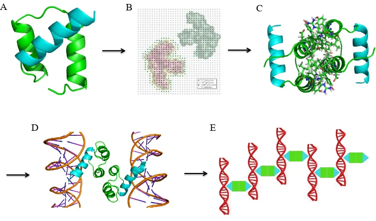
Fig. 1-2. Overall design scheme for a self-assembling protein-DNA nanowire. (A) The protein
scaffold, engrailed homeodomain (ENH), is a monomeric protein with three helices. Helices 1
and 2 (green) were chosen for homodimerization. Helix-3 (cyan) is the DNA-binding domain. (B)
In silico docking was performed to generate a symmetric homodimer configuration using the
structure in (A). (C) The interface of the docked homodimer from (B) was computationally
designed to create protein-protein affinity. (D) Each component of the designed homodimer
specifically binds a dsDNA fragment. The designed homodimer therefore can bring two dsDNA
fragments into close proximity. (E) If each dsDNA fragment has exactly two protein binding sites
located on opposite sides of the DNA helix, the designed homodimer (green and cyan) and the
dsDNA fragment (red) will self-assemble into nanowires.
A B C
14
Chapter 2
Using Molecular Dynamics to Predict Domain Swapping of
15
Abstract
In standard implementations of computational protein design (CPD), a positive-design approach is
used to predict sequences that will stabilize a given backbone structure. Possible competing states
are typically not considered, primarily because appropriate models for them are not available. One
of the competing states, the domain-swapped dimer, is especially compelling, because it is often
nearly identical to its monomeric counterpart, differing by just a few mutations in the hinge region.
Molecular dynamics (MD) simulations provide a computational method to sample different
conformational states of a structure. Here, we tested whether MD could be used as a post-design
screening tool to identify domain-swapped dimers. We hypothesized that a successful
computationally-designed sequence would have backbone dynamics similar to that of the input
structure, and that in contrast, domain-swapped dimers would exhibit increased backbone flexibility
in the hinge region to accommodate the huge conformational change required for domain swapping.
While attempting to engineer a homodimer from the monomeric protein engrailed homeodomain
(ENH), we discovered that we had instead generated a domain-swapped dimer (ENH_DsD). We
ran MD on these proteins and, as expected, observed increased backbone flexibility in the hinge of
the domain-swapped dimer. Two point mutants of ENH_DsD designed to recover the monomeric
fold were then tested with our MD protocol. MD predicted that one of these mutants would adopt
the monomeric structure, and this was confirmed by X-ray crystallography. Similarly,
MD-generated backbone dynamics was found to reflect the domain-swapping tendency of
28
total of three trajectories with different random seeds was run for each sequence and the averaged B
factors ware calculated using the following equation: B = (8π2/3) × (RMSF)2, where the RMSF
units are Å.
References
1. Dahiyat BI, Mayo SL (1997) De novo protein design: fully automated sequence selection.
Science 278:82-87.
2. Pantazes RJ, Grisewood MJ, Maranas CD (2011) Recent advances in computational protein
design. Curr Opin Struc Biol 21:467-472.
3. Röthlisberger D, et al. (2008) Kemp elimination catalysts by computational enzyme design.
Nature 453:190-195.
4. Jiang L, et al. (2008) De novo computational design of retro-aldol enzymes. Science
319:1387-1391.
5. Fleishman SJ, et al. (2011) Computational design of proteins targeting the conserved stem
region of influenza hemagglutinin. Science 332:816-821.
6. King NP, et al. (2012) Computational design of self-assembling protein nanomaterials with
atomic level accuracy. Science 336:1171-1174.
7. Grigoryan G, et al. (2011) Computational design of virus-like protein assemblies on carbon
nanotube surfaces. Science 332:1071-1076.
8. Mandell DJ, Coutsias EA, Kortemme T (2009) Sub-angstrom accuracy in protein loop
29
9. Stranges PB, Machius M, Miley MJ, Tripathy A, Kuhlman B (2011) Computational design
of a symmetric homodimer using β-strand assembly. Proc Natl Acad Sci USA
108:20562-20567.
10. Fleishman SJ, et al. (2011) Community-wide assessment of protein-interface modeling
suggests improvements to design methodology. J Mol Biol 414:289-302.
11. Whitehead TA, Baker D, Fleishman SJ (2013) Computational design of novel protein
binders and experimental affinity maturation. Method Enzymol, ed Amy EK (Academic
Press), Vol Volume 523, pp 1-19.
12. Lim KH, Dyson HJ, Kelly JW, Wright PE (2013) Localized structural fluctuations promote
amyloidogenic conformations in transthyretin. J Mol Biol 425:977-988.
13. Blomberg R, et al. (2013) Precision is essential for efficient catalysis in an evolved Kemp
eliminase. Nature doi:10.1038/nature12623.
14. Malakauskas SM, Mayo SL (1998) Design, structure and stability of a hyperthermophilic
protein variant. Nat Struct Biol 5:470-475.
15. Havranek JJ, Harbury PB (2003) Automated design of specificity in molecular recognition.
Nat Struct Biol 10:45-52.
16. Tinberg CE, et al. (2013) Computational design of ligand-binding proteins with high
affinity and selectivity. Nature 501:212-216.
17. Koder RL, et al. (2009) Design and engineering of an O-2 transport protein. Nature
31
27. Choi EJ, Mayo SL (2006) Generation and analysis of proline mutants in protein G. Protein
Eng Des Sel 19:285-289.
28. O'Neill JW, Kim DE, Baker D, Zhang KYJ (2001) Structures of the B1 domain of protein
L from Peptostreptococcus magnus with a tyrosine to tryptophan substitution. Acta
Crystallogr D 57:480-487.
29. Liang S, et al. (2008) Exploring the molecular design of protein interaction sites with
molecular dynamics simulations and free energy calculations. Biochemistry 48:399-414.
30. Hess B, Kutzner C, van der Spoel D, Lindahl E (2008) GROMACS 4: Algorithms for
highly efficient, load-balanced, and scalable molecular simulation. J Chem Theory Comput
4:435-447.
31. Lindorff-Larsen K, Piana S, Dror RO, Shaw DE (2011) How fast-folding proteins fold.
Science 334:517-520.
32. Emsley P, Cowtan K (2004) Coot: model-building tools for molecular graphics. Acta
Crystallographica Section D-Biological Crystallography 60:2126-2132.
33. Adams PD, et al. (2010) PHENIX: a comprehensive Python-based system for
macromolecular structure solution. Acta Crystallographica Section D-Biological
Crystallography 66:213-221.
34. Shah PS, et al. (2007) Full-sequence computational design and solution structure of a
32
Table 2-1. Sequences of wild-type ENH, ENH_DsD, E23P, and E24P
ENH(WT) TAFSSEQLARLKREFNENRYLTERRRQQLSSELGLNEAQIKIWFQNKRAKI ENH_DsD -E--E--KKA-DLA-YFD-R---EW-RY--QR---E--ER--RR-EQQ- E23P -E--E--KKA-DLA-YFD-R--PEW-RY--QR---E--ER--RR-EQQ- E24P -E--E--KKA-DLA-YFD-R---PW-RY--QR---E--ER--RR-EQQ-
33
Table 2-2. Summary of X-ray crystallography, dimerization assays, CPD calculations, and
MD-derived hinge B factors for each of the proteins tested.
Domain-Swapping
Kd* CPD energy† B-factor‡
ENH (WT) No n/a -127.0 21.1
ENH_DsD Yes 40nM -125.2 70.9
E23P No n/a -123.7 32.7
E24P n/a n/a -120.6 106.6
1HZ5 (WT) No n/a -183.1 78.4
G55A Yes 30uM -166.2 58.2
A52V/D53P/G55A Yes 700pm 249.1 179.6
*Dissociation constant for the domain-swapping dimer.
36
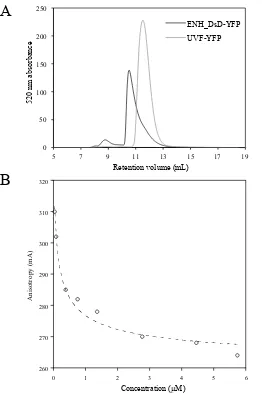
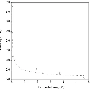
Fig. 2-1. Size exclusion and polarization fluorescence assays indicate that ENH_DsD-YFP is a
high-affinity dimer. (A) Size-exclusion chromatography for ENH_DsD-YFP (black) and a
monomeric control, UVF-YFP (gray). Absorbance at A515 (absorbance peak for YFP) was tracked
for protein elution. UVF is a computationally designed 39-fold mutant of ENH whose NMR
solution structure matches the wild-type (monomeric) fold (34). The retention volumes for
UVF-YFP and ENH_DsD-UVF-YFP are consistent with standards for a monomer and a dimer, respectively. (B)
Polarization fluorescence of ENH_DsD-YFP. The unit of anisotropy (mA) is a thousandth of
anisotropy (A). A Kd of ~40 nM was calculated based on a simple monomer-dimer equilibrium
37
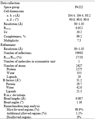
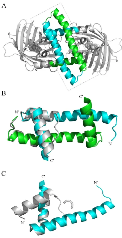
Fig. 2-2. X-ray crystallography reveals that ENH_DsD-YFP is a domain-swapped dimer. (A)
Crystal structure of ENH_DsD-YFP resolved to 1.85 Å. The two chains in ENH_DsD are shown in
green and cyan, respectively, and the YFP sequences are shown in gray. (B) Zoom-in of the dashed
frame in A with the wild-type ENH structure (PDB ID: 1ENH) (gray) superimposed. (C) The
hinge-loop between the first and second helices in the wild-type structure (gray) has flipped over by ~90º
(arrow) and coiled up to form the domain-swapped conformation seen in ENH_DsD-YFP (cyan).
This change leads to a ~180º rearrangement of helix 2 so that helix 1, the hinge, and helix 2 now
38
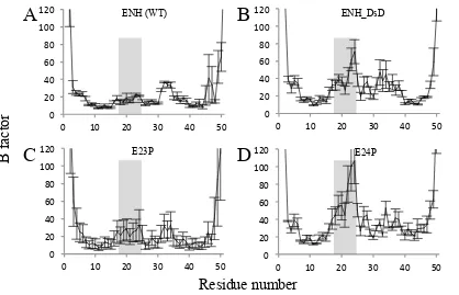
Fig. 2-3. B factor analyses from MD simulations for wild-type ENH (A), ENH_DsD (B), E23P (C),
and E24P (D). 20 ns MD simulations were run on wild-type ENH and on each of the variant
sequences threaded onto the wild-type backbone structure, the RMSF of the Cα atoms was analyzed
using GROMACS for each of three trajectories, and the averaged B factors were calculated. Error
bars: SD for three independent trajectories. Hinge residues (between helices 1 and 2 in wild-type
39
Fig. 2-4. Polarization fluorescence indicates that E23P-YFP is a high-affinity dimer. Polarization
fluorescence of E23P-YFP was measured with a series of diluted samples in buffer containing 100
mM NaCl and 20 mM TrisHCl at pH 8.0. The unit of anisotropy (mA) is a thousandth of anisotropy
(A). The G-factor was determined for each data point. A Kd of ~10 nM was calculated based on a
40
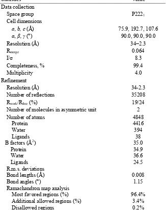
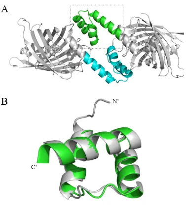
Fig. 2-5. X-ray crystallography of E23P-YFP shows that a single proline mutation in the hinge
recovers the wild-type fold. (A) Crystal structure of E23P-YFP resolved to 2.3 Å. The E23P
sequence is shown in green and cyan (for each of the two chains, respectively) and the YFP
sequences are shown in gray. (B) Zoom-in of the dashed frame in A with the wild-type ENH
structure (gray) superimposed. E23P exhibits the wild-type fold.
41
Fig. 2-6. B factor analyses from MD simulations for the B1 domain of protein L (wild-type) (A),
G55A (B), and the triple mutant A52V/D53P/G55A (C). For G55A and the triple mutant, sequences
were first threaded onto the wild-type monomeric structure. As described above, 20 ns MD
simulations were run, the RMSF of the Cα atoms was analyzed using GROMACS for each of three
trajectories, and the averaged B factors were calculated (see Methods for details). Error bars: SD for
three independent trajectories. Hinge residues are indicated with a gray bar in each panel. The
backbone dynamics of the hinge varies for the different proteins and reflects their domain-swapping
42
Chapter 3
Computational Design and Experimental Verification of
43
Abstract
Homodimers are by far the most common type of protein assembly in nature and have distinct
features compared to heterodimers and higher-order oligomers. Understanding homodimer
interactions at the atomic level is critical both for accurate modeling and for elucidating their
biological mechanisms of action. Computational design of novel protein-protein interfaces can
serve as a bottom-up method to further our understanding of protein interactions. Previous studies
have demonstrated that the de novo design of homodimers can be achieved to atomic-level accuracy
by ß-strand assembly or metal mediation. Here, we report a novel homodimer with C2 symmetry
that is designed via helical interactions. The structure obtained by solution nuclear magnetic
resonance shows that the homodimer exhibits parallel helical packing similar to the designed model.
Because designing for improved functionality often results in decreased thermostability, a stability
design step was introduced. in addition to our standard docking and design procedure, to
compensate for the poor thermostability of the scaffold. This two-step design approach is essential
59
5. Whitehead TA, Baker D, Fleishman SJ (2013) Computational design of novel protein
binders and experimental affinity maturation. Method Enzymol 523:1-19.
6. Halperin I, Ma B, Wolfson H, Nussinov R (2002) Principles of docking: An overview of
search algorithms and a guide to scoring functions. Proteins 47:409-443.
7. Shifman JM, Mayo SL (2003) Exploring the origins of binding specificity through the
computational redesign of calmodulin. Proc Natl Acad Sci USA 100:13274-13279.
8. Havranek JJ, Harbury PB (2003) Automated design of specificity in molecular recognition.
Nat Struct Biol 10:45-52.
9. Sia SK, Kim PS (2003) Protein grafting of an HIV-1-inhibiting epitope. Proc Natl Acad Sci
USA 100:9756-9761.
10. Lewis SM, Kuhlman BA (2011) Anchored design of protein-protein interfaces. Plos One
6:e20872.
11. Fleishman SJ, et al. (2011) Computational design of proteins targeting the conserved stem
region of influenza hemagglutinin. Science 332:816-821.
12. King NP, et al. (2012) Computational design of self-assembling protein nanomaterials with
atomic level accuracy. Science 336:1171-1174.
13. Der BS, et al. (2012) Metal-mediated affinity and orientation specificity in a
computationally designed protein homodimer. J Am Chem Soc 134:375-385.
14. Sammond DW, et al. (2011) Computational design of the sequence and structure of a
60
15. Whitehead TA, et al. (2012) Optimization of affinity, specificity and function of designed
influenza inhibitors using deep sequencing. Nat Biotechnol 30:543-548.
16. Stranges PB, Machius M, Miley MJ, Tripathy A, Kuhlman B (2011) Computational design
of a symmetric homodimer using β-strand assembly. Proc Natl Acad Sci USA
108:20562-20567.
17. Stranges PB, Kuhlman B (2013) A comparison of successful and failed protein interface
designs highlights the challenges of designing buried hydrogen bonds. Protein Sci 22:74-82.
18. Fleishman SJ, et al. (2011) Community-wide assessment of protein-interface modeling
suggests improvements to design methodology. J Mol Biol 414:289-302.
19. Bahadur RP, Chakrabarti P, Rodier F, Janin J (2003) Dissecting subunit interfaces in
homodimeric proteins. Proteins 53:708-719.
20. Harbury PB, Zhang T, Kim PS, Alber T (1993) A switch between 2-stranded, 3-stranded
and 4-stranded coiled coils in Gcn4 leucine-zipper mutants. Science 262:1401-1407.
21. Grigoryan G, Reinke AW, Keating AE (2009) Design of protein-interaction specificity
gives selective bZIP-binding peptides. Nature 458:859-864.
22. Guharoy M, Chakrabarti P (2007) Secondary structure based analysis and classification of
biological interfaces: identification of binding motifs in protein-protein interactions.
Bioinformatics 23:1909-1918.
23. Woolfson DN (2005) The design of coiled-coil structures and assemblies. Adv Protein
61
24. Apgar JR, Gutwin KN, Keating AE (2008) Predicting helix orientation for coiled-coil
dimers. Proteins 72:1048-1065.
25. Karanicolas J, et al. (2011) A de novo protein binding pair by computational design and
directed evolution. Mol Cell 42:250-260.
26. Fraenkel E, Rould MA, Chambers KA, Pabo CO (1998) Engrailed homeodomain-DNA
complex at 2.2 angstrom resolution: A detailed view of the interface and comparison with
other engrailed structures. J Mol Biol 284:351-361.
27. Marshall SA, Mayo SL (2001) Achieving stability and conformational specificity in
designed proteins via binary patterning. J Mol Biol 305:619-631.
28. Shah PS, et al. (2007) Full-sequence computational design and solution structure of a
thermostable protein variant. J Mol Biol 372:1-6.
29. Beck DAC, Daggett V (2004) Methods for molecular dynamics simulations of protein
folding/unfolding in solution. Methods 34:112-120.
30. Privett HK, et al. (2012) Iterative approach to computational enzyme design. Proc Natl
Acad Sci USA 109:3790-3795.
31. Marshall SA, Morgan CS, Mayo SL (2002) Electrostatics significantly affect the stability
of designed homeodomain variants. J Mol Biol 316:189-199.
32. Huang P-S, Love JJ, Mayo SL (2005) Adaptation of a fast Fourier transform-based docking
algorithm for protein design. J Comput Chem 26:1222-1232.
33. Khare SD, Fleishman SJ (2013) Emerging themes in the computational design of novel
62
34. Röthlisberger D, et al. (2008) Kemp elimination catalysts by computational enzyme design.
Nature 453:190-195.
35. Jiang L, et al. (2008) De novo computational design of retro-aldol enzymes. Science
319:1387-1391.
36. Kiss G, Röthlisberger D, Baker D, Houk KN (2010) Evaluation and ranking of enzyme
designs. Protein Sci 19:1760-1773.
37. Oshea EK, Klemm JD, Kim PS, Alber T (1991) X-Ray structure of the Gcn4 leucine zipper,
a 2-stranded, parallel coiled coil. Science 254:539-544.
38. Dahiyat BI, Mayo SL (1996) Protein design automation. Protein Sci 5:895-903.
39. Krissinel E, Henrick K (2007) Inference of macromolecular assemblies from crystalline
state. J Mol Biol 372:774-797.
40. Procko E, et al. (2013) Computational design of a protein-based enzyme inhibitor. J Mol
Biol 425:3563-3575.
41. Murphy PM, Bolduc JM, Gallaher JL, Stoddard BL, Baker D (2009) Alteration of enzyme
specificity by computational loop remodeling and design. Proc Natl Acad Sci USA
106:9215-9220.
42. Mandell DJ, Coutsias EA, Kortemme T (2009) Sub-angstrom accuracy in protein loop
reconstruction by robotics-inspired conformational sampling. Nat Methods 6:551-552.
43. Hu XZ, Wang HC, Ke HM, Kuhlman B (2007) High-resolution design of a protein loop.
63
44. Malakauskas SM, Mayo SL (1998) Design, structure and stability of a hyperthermophilic
protein variant. Nat Struct Biol 5:470-475.
45. Korkegian A, Black ME, Baker D, Stoddard BL (2005) Computational thermostabilization
64
Table 3-1. Library design of the homodimer interface
9 10 13 14 16 17 18 25 28 32
Library K AT L A FY FV DFVY AW FY R
65
Table 3-2. Sequences of wild-type ENH, ENH-c2a, NC3-Ncap, and ENH-c2b
id* Tm† ENH(WT) TAFSSEQLARLKREFNENRYLTERRRQQLSSELGLNEAQIKIWFQNKRAKI - 49 ENH-c2a ---KA-DL--YF---W--Y---R--- 82% n/a NC3-Ncap -E--E--KR--DE--RRD-R---E--RD--QK---E--ER--RR-EQQ- 55% 88 ENH-c2b -E--E--KKA-DLA-YFD-R--PEW-RY--QR---E--ER--RR-EQQ- 47% 62
The three “coils” at the top show the location of the three helices in the ENH wild-type (WT) fold.
*id is the sequence identity compared to ENH.
68
Fig. 3-1. Steps used to design a C2-symmetrical homodimer. The initial scaffold (ENH) used for
docking is shown in silver, and the homodimer model used for all interface designs is shown in
69
Fig. 3-2. SDS-PAGE of purified proteins from soluble fractions. The labels are M: marker, 1: ENH,
2: ENH-c2a, and 3: ENH-c2b. The absence of band in lane 2 indicates that ENH-c2a is not
expressed in soluble fraction.
M 1 2 3
70
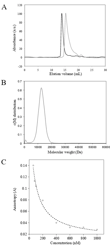
Fig. 3-3. Experimental characterizations of the computational library design. (A) Homo-FRET
assay of the library members. Each dot represents one member in the library. Because of the
homo-FRET effect, members that have lower anisotropic values are likely to have stronger dimer affinities
or higher oligomeric states. Two representative members, shown in orange (named as ENH_DsD)
and magenta were chosen for characterizations in B and C. (B) Size-exclusion chromatography
showed that ENH_DsD (orange curve) is likely a dimer (compared to potein standards) and
magenta variant formed a higher-order oligomer. (C) Sedimentation velocity experiments showed
that ENH_DsD is under a fast equilibrium between monomer and dimer states (orange curve);
magenta variant is a tetramer (magenta curve). The monomer MW is 34.4. kD.
A
73
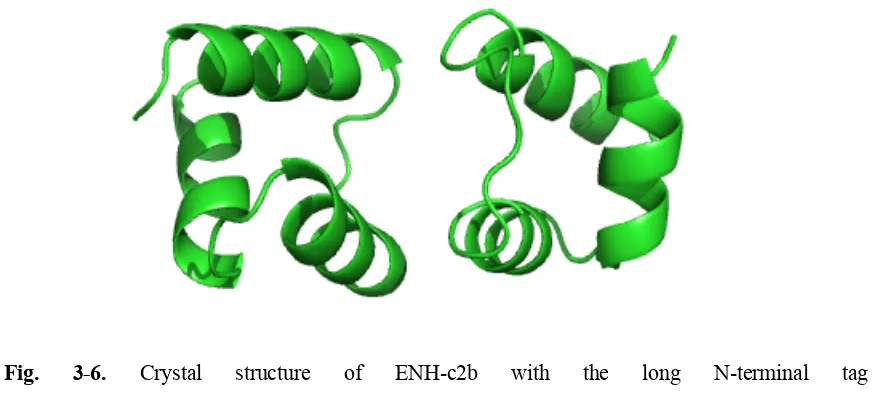
Fig. 3-6. Crystal structure of ENH-c2b with the long N-terminal tag
“MGSSHHHHHHSSGLVPRGSHM” (PDB code: 4NDL). The two chains in proximity make a
crystal contact with helix-1 and helix-2. Inspection evaluated by PISA reveals that this contact
belongs to a crystal packing rather than a biological interface, mainly because of its small area (699
74
Fig. 3-7. Solution NMR structure of ENH-c2b (green) superimposed with design model structure
(gray), viewed from different orientations. (A-B): superposition of just one chain; (C-D):
superposition of the entire dimer structure; (E-F): superposition of left chain showing entire dimer
structure.
A
B
C
D
76
Fig. 3-9. Two-step design strategy for the functional design of homodimers. A single-step design
for functionality often leads to an unstable protein. Stability design prior to functional design can
solve this problem. In this work, single-step functional design of ENH led to an unstable protein
that did not express solubly, referred to as ENH-c2a. However, doing a stability design (NC3-NCap)
prior to functional design led to a stable homodimer, ENH-c2b. Structures shown are computational
77
Chapter 4
Computational Design of Self-assembling
94
34. Dunbrack RL, Jr., Karplus M (1993) Backbone-dependent rotamer library for proteins.
Application to side-chain prediction. J Mol Biol 230(2):543-574.
35. Stemmer WP, Crameri A, Ha KD, Brennan TM, Heyneker HL (1995) Single-step assembly
of a gene and entire plasmid from large numbers of oligodeoxyribonucleotides. Gene
164(1):49-53.
36. Adams PD, et al. (2010) PHENIX: a comprehensive Python-based system for
macromolecular structure solution. Acta Crystallogr D 66:213-221.
37. Emsley P, Cowtan K (2004) Coot: model-building tools for molecular graphics. Acta
95
Table 4-1. Sequences of wild-type ENH and dualENH.
o ooo vv vvv vvv v vv vv oo oo oo ooo o ENH(WT) EKRPRTAFSSEQLARLKREFNENRYLTERRRQQLSSELGLNEAQIKIWFQNKRAKIKKST dualENH ---E--KKA-DLA-YFD----PEW-RY--QR---
The three “coils” at the top show the location of the three helices in the wild-type (WT) fold based
on PDB structure 1ENH. The DNA-binding domain residues are labeled with “o” and the
97
Fig. 4-1. Protein-DNA nanomaterial design strategy. (A) Helix-1 and helix-2 (green) of ENH were
engineered into a homodimerization domain, and helix-3 (blue) is the native DNA-binding domain.
(B) The interface of the docked model was designed for homodimerization. (C) The designed
homodimer, named dualENH, binds two dsDNA fragments on its outward faces. This model was
generated by aligning the homodimer model in b with the ENH-DNA co-crystal structure (PDB
code: 3HDD). (D)Two protein binding sites were engineered onto a dsDNA fragment so that two
dualENH dimers would bind 180º apart along the double-helix. (E) The dualENH protein in c and
the dsDNA fragment in (D) co-assemble into a protein-DNA nanowire. Note that this 2D cartoon is
98
Fig. 4-2. Design model of irregular bulk protein-DNA nanoparticle. (A) Four consecutive ENH
binding sites that each face in a different direction are engineered onto a dsDNA fragment. This
dsDNA building block allows the protein-DNA assembly to occur in all three dimensions. Note that
in this particular design, two neighboring binding sites may not be simultaneously occupied due to
steric hindrance. (B)Cartoon illustrating an irregular shaped nanoparticle formed by co-assembly of
dualENH and the dsDNA shown in (A) The DNA-binding domains of dualENH are shown as blue
99
Fig. 4-3. Circular dichroism (CD) spectroscopy of dualENH. (A) CD spectrum of dualENH at room
temperature. Solid line: before thermal denaturation; dashed line: after thermal denaturation. The
overlapping of the two curves indicates that dualENH folds reversibly. (B) Thermal denaturation
curve measured at 222 nm. Circles: experimental data; line: curve fit with two-state transition model.
The melting temperature of dualENH was determined to be 59 °C.
101
Fig. 4-4. Biophysical characterization of dualENH. (A) Size-exclusion chromatography of dualENH
with three different loading concentrations: solid line: 650 µM; dashed line: 80 µM; dotted line: 5
µM. The highest signals were normalized to 100 for all curves. (B) c(S) model fit from a
sedimentation velocity experiment of 40 µM dualENH. The major peak around S = 1.9 corresponds
to a MW of 18.3 kD, which is about twice that of monomeric dualENH (8.7 kD). The spike at the
left (S < 0.5) may be due to impurities or artifacts from model fitting. (C) Raw data and fitting
residuals for the sedimentation velocity experiment in (B). A total of 378 curves were used for
fitting, but for visual clarity only 1/5th of the curves are shown. The upper graph shows the raw data
(dots) and the fitting curves; the lower figure shows the residuals between the experimental data and
the fit. The square root of variance of the fit is 0.00669. (D)Fluorescence polarization experiment.
Two dsDNA sequences labeled with fluorescein were used as probes to assay dualENH-DNA
binding. Probe-1: 20-nt dsDNA with the binding motif TAATTA; probe-2: same sequence as
probe-1 but with a single-nucleotide mutation to the binding motif (TA[C]TTA). The concentration
of dualENH was varied, while the concentration of the three probes remained constant (25 nM).
Data are shown as mean ± s.e.m. for 3 replicates (E) FRET experiment showing that dualENH
brings two dsDNA fragments within Förster distance. 15-nt dsDNA (TAA)5 were labeled with
either Cy3 or Cy5 to serve as the FRET donor or acceptor. Gray line: 400 nM Cy3-(TAA)5 + 600
nM Cy5-(TAA)5; black line: 400 nM Cy3-(TAA)5 + 600 nM Cy5-(TAA)5 + 4 µM dualENH. (F)
Two control experiments for the FRET experiment in e. Black line: 400 nM Cy3-(TAA)5; Black
dashed line: 400 nM Cy3-(TAA)5 + 4 µM dualENH; Gray line: 600 nM Cy5-(TAA)5; Gray dashed
102
Fig. 4-5. Fluorescence polarization experiments with dualENH and wild-type ENH. (A)
Fluorescence polarization experiment with probe-1, which contains the binding motif TAATTA of
ENH. The concentration of dualENH or wild-type ENH was varied, while the concentration of
probe-1 remained constant (25 nM). Both dualENH and wild-type ENH show saturated binding
when the protein concentration is at or above 100 nM. Data are shown as mean ± s.e.m. for 3
replicates.(B) Same experiments as (A), except that a probe without any TAATNN binding motif
was used. Note that probe-1 used in (A) has a lower fluorescence intensity and polarization than the
probe used in (B) likely due to partial quenching by a guanine nucleotide on the strand opposite the
103
Fig. 4-6. Fluorescence microscopy of protein-DNA nanoobjects. (A) dsDNA (TAA)5 fragments
were labeled with the fluorescent dye Cy3. A fluorescent image was taken of particles formed by
mixing 5 µM dualENH with 2 µM Cy3-(TAA)5 in 20 mM Tris-HCl and 100 mM NaCl at pH 8.0.
The particles formed irregular shapes up to ~5 µm in diameter. (B) Same experiment as in (A)
except that 25-nt dsDNA fragments containing motif-11 (TAATTTAATTT) in the middle
(CGCAGTGTAATTTAATTTCCTCGAC) were used instead of (TAA)5 fragments. All particle
sizes are under the diffraction limit (submicron). The shapes of the particles are slightly oval instead
104
Fig. 4-7. Microscope imaging experiments. (A) The size distribution of the irregular protein-DNA
particles formed by 5 µM dualENH mixed with 2 µM Cy3-(TAA)5. (B) Bright-field microscope
image of 5 µM dualENH mixed with 2 µM Cy3-(TAA)5. A dust particle (upper-left) is evident,
indicating the focal plane is correct. (C) Fluorescence microscope image of 2 µM Cy3-(TAA)5
alone. (D) Fluorescence microscope image of particles formed with 500 nM dualENH mixed with
200 nM Cy3-(TAA)5. (E) Fluorescence microscope image of particles formed with 200 nM
dualENH mixed with 100 nM Cy3-(TAA)5. (F) Fluorescence microscope image of particle
inhibition experiments. A small amount (5 nM) of single-binding-site dsDNA (containing only one
TAATTA motif) was pre-mixed with 500 nM dualENH, then 200 nM Cy3-(TAA)5 was added. The
illumination/camera sensitivity was enhanced to confirm that particle formation is nearly
105
Fig. 4-8. Atomic force microscopy of protein-DNA nanowires. (A) Representative AFM image
obtained after mixing 5 µM dualENH with 2 µM of the two-binding-site dsDNA (25-nt dsDNA
containing motif-11). Nanowire structures ~15 nm wide and up to 300 nm long are clearly visible.
(B) Magnified image of a single nanowire ~250 nm in length. (C) 3D topology display of (B) shows
the height of the nanowire is ~1.0 nm.
106
Fig. 4-9. Co-crystal structure of protein-DNA complex. (A) dualENH forms a symmetric
homodimer using helix-1 and helix-2 (green) as the protein-protein interface. Helix-3 (blue) binds
to the dsDNA in the same way that wild-type ENH does. (B)Two forms of dualENH are present in
the co-crystal structure and occur in a molar ratio of 3:1 (green:cyan). (C), (D) Two forms of
protein-DNA binding are observed in the co-crystal structure and occur in a molar ratio of 1:1. Both
have two dualENH homodimers bound on the designed 11-nt motif TAATTTAATTT. In (C), both
of the dualENH dimers bind in the optimal motif (TAATTT) orientation,whereas in (D), one of the
dualENH dimers (right) binds in the suboptimal orientation (AAATTA, the reverse complementary
sequence of the optimal motif). (E), Slightly kinked nanowire structure found in the co-crystal
107
Fig. 4-10. Co-crystal structure of the protein-DNA complex. (A)structures in the asymmetric unit
cell are shown in color, and the end-to-end packing of neighboring DNA molecules and their bound
proteins are shown in gray. (B), (C) the dualENH homodimer observed in the co-crystal structure
(green or cyan) is superimposed with the design model (gray). The backbone RMSD to the design
model is 3.8 Å (green) and 3.9 Å (cyan), respectively. When only one subunit is aligned between
the more predominant configuration (green) and the design model, the angular displacement
between the other subunits is ~45˚ with about 3 Å translational displacement. The less predominant
configuration has a lower angular displacement ~20˚ but a larger translational displacement ~8 Å.
The calculated energies for the design model and the two crystallographic dimers are -155.2, -140.3
108
Fig. 4-11. dualENH-DNA binding and nanostructure formation are inhibited at high salt
concentrations. (A)Fluorescence polarization experiments of dualENH and probe-1 at various NaCl
concentraions. Probe-1 and dualENH were mixed in buffers with different NaCl concentrations and
fluorescence polarization values were recorded. DualENH-DNA binding dropped significantly from
100 mM to 150 mM NaCl, and was completely absent at 300 mM NaCl. Data are shown as mean ±
s.e.m. for 3 replicates.(B)Fluorescence image of particle experiment at 150 mM salt concentration.
The sample was prepared by mixing 500 nM dualENH and 200 nM Cy3-(TAA)5 in 20 mM
Tris-HCl buffer with 150 mM NaCl. The illumination/camera sensitivity was enhanced to confirm that
109
Appendix
Direct Visualization Reveals Dynamics of a Transient
Intermediate During Protein Assembly
The text of this chapter was adapted from a manuscript coauthored with Xin Zhang, Vinh Q. Lam,
Tetsunari Kimura, Jaeyoon Chung, Sowmya Chandrasekar, Jay R. Winkler, Stephen L. Mayo, and
Shu-ou Shan
Xin Zhang, Vinh Q. Lam*, Yun Mou*, Tetsunari Kimura, Jaeyoon Chung, Sowmya Chandrasekar,
Jay R. Winkler, Stephen L. Mayo, and Shu-ou Shan (2011) Direct visualization reveals dynamics
of a transient intermediate during protein assembly. Proceedings of the National Academy of
Sciences USA 108, 6450-6455. (*these authors contributed equally)
110
AbstractInteractions between proteins underlie numerous biological functions. Theoretical work suggests
that protein interactions initiate with formation of transient intermediates that subsequently relax to
specific, stable complexes. However, the nature and roles of these transient intermediates has
remained elusive. Here, we characterized the global structure, dynamics, and stability of a transient,
on-pathway intermediate during complex assembly between the Signal Recognition Particle (SRP)
and its receptor (SR). We show that this intermediate has overlapping but distinct interaction
interfaces from that of the final complex, and is stabilized by long-range electrostatic interactions. A
wide distribution of conformations is explored by the intermediate; this distribution becomes more
restricted in the final complex and is further regulated by the cargo of SRP. These results suggest a
funnel-shaped energy landscape for protein interactions, and provide a framework for understanding
134
48. Istratov AA, Vyvenko OF (1999) Exponential analysis in physical phenomena. Review of
Scientific Instruments 70:1233-1257.
49. Laurence TA, Kong X, Jager M, Weiss S (2005) Probing structural heterogeneities and
fluctuations of nucleic acids and denatured proteins. Proc Natl Acad Sci U S A
102:17348-17353.
50. Kozakov D, et al. (2010) Achieving reliability and high accuracy in automated protein
docking: ClusPro, PIPER, SDU, and stability analysis in CAPRI rounds 13-19. Proteins
135
Fig. A-1 Mapping the interaction interface of the SRP•SR complexes using EPR spectroscopy. (A)
Left: crystal structure of the SRP•SR NG-domain complex (1JR9). Right: a multi-step mechanism
for SRP-SR complex assembly involving formation of an early intermediate (step 1), and
rearrangement to form the stable complex (step 2). Removal of GTP stalls the complex at the
early intermediate stage. T denotes GTP. (B) Nitroxide spin probes labeled at specific SR residues
change mobility upon formation of the early intermediate (blue), the stable complex (red), or both.
The different classes of spin probe mobility changes are defined in the text. Black denotes the
apo-formed SR. (C) Interaction surface of the stable complex (left) and early intermediate (right)
mapped by EPR. ‘N’ and ‘G’ denote the N- and G-domains of SRP and SR, respectively. The red
136
Fig. A-2 The mobility of spin labels on SR changed upon formation of the early intermediate, stable
complex, or both. (A and B) Spin-labeled SR were screened using the GTPase assay. The activities
137
Supplementary Methods). Two kinetic parameters were assessed: the GTPase rate constants of the
SRP-SR complex (kcat in A) and the association rate constants for stable SRP-SR complex assembly
(as determined by kcat/KminB; see Supplementary Methods). Spin-labeled SR’s that were defective
in either property by a factor of 5 or more were not used for EPR studies (open bars). Spin-labeled
SR’s that were functional in interacting with SRP (grey bars) were used for EPR measurements for
either the early intermediate or the stable complex. (C) Spectra of additional spin probes in SR
changed mobility upon formation of either the early intermediate or the stable complex. Three
different classes were defined in the text based on probe mobility changes. The mobility of spin
label was analyzed from the central line width (ΔH0) and the breadth of the spectra, and summarized
in Figure 1B. Black, blue, and red denote the free protein, the early intermediate, and the stable
complex, respectively. (D) EPR spectra of spin probes in SR that exhibited no significant changes
138
Fig. A-3 Residues I237, Q425, and N426 (green), which changed EPR spectra specifically in the
139
Fig. A-4 Mutations that disrupt the stable complex did not significantly affect the early
intermediate. (A) Positions of SR mutations (cyan and blue) studied herein are shown in the
surface representation of the SR. The three moderately defective mutants are highlighted in blue.
(B) The stability of the early intermediate is insensitive to many mutations that disrupt the stable
complex. (C) The stabilities of the early intermediates formed by mutant SRs were determined by
equilibrium titrations. Nonlinear fits gave Kd values of 4.1 µM for wild-type SR, 13.2 µM for SR
140
Fig. A-5 Fluorescence decay of donor (DACM)-labeled at SRP (C76) under different experimental
conditions. The black, blue, and red curves represent the decay curves for donor-only, the early
intermediate, and the stable complex, respectively. The linear decay of the donor-only sample could
be described by a single decay rate constant. In contrast, the decay curves in both the early
intermediate and stable complex deviated from linearity and were described by multiple decay rate
141
Fig. A-6 Distance distributions derived from least-squares analyses of the TR-FRET data for each
FRET pair in the early intermediate (blue), stable complex (red), and early intermediate bound with
142
Fig. A-7 Conformational distribution of the early intermediate is broad, and is restricted by
formation of the stable complex or the cargo. Left, positions of the G-G (A), NG-NG (B), and
N-N (C) FRET pairs in the stable SRP•SR N-NG-domain complex. Right, FRET distance distributions,
P(r), for each FRET pair in the early intermediate (blue), stable complex (red), and the early
intermediate bound to cargo (green), as derived from maximal entropy analyses of the TR-FRET
143
Fig. A-8 Electrostatic interactions between the N-domains of SRP and SR stabilize the early
intermediate and accelerate stable complex assembly. (A) The SRP and SR N-domains contain
complementarily charged surfaces. (B) Molecular docking simulation generated two groups of
144
shows the structure of the stable complex with the SRP NG-domain aligned in the same
orientation. The nucleotide bound to SRP is shown in CPK coloring. (C) The association rate
constants predicted from Brownian Dynamics calculations for formation of the early intermediate
in the ‘N’ or ‘G’ group, and for the stable complex. The experimentally measured rate constants
are in parentheses. (D) Charge complementarity between the N-domains is critical to the stability
of the early intermediate (black bars) and the kinetics of stable complex assembly (blue bars),
determined using the FRET and GTPase assays, respectively, for the wildtype proteins (WT:WT),
wildtype SRP and mutant SR (WT:RRLRR), mutant SRP and wildtype SR (RK3E:WT), and the
charge reversal SRP and SR mutant pair (RK3E:RRLRR). The kinetic constants were derived
from the data in Figure S5D. FRET efficiency in the early intermediate was recorded at 5 µM
FtsY, at which concentration the FRET value is most sensitive to changes in the stability of the
145
Fig. A-9 Charge complementarity between SRP and SR’s N-domains is essential for the stability
of the early intermediate and the kinetics of stable complex assembly. (A) Sequence alignment of
SRP and SR homologues. The residue numbering is for the E. coli SRP and SR proteins.
Conserved positive and negative residues are denoted in blue and red colors, respectively. (B) The
146
N-domain (left), and the RRLRR mutation in SR generated a moderately positive electrostatic
potential in the SR N-domain (right). (C) The stabilities of the early intermediates formed by
mutant SRP and SR’s were determined by equilibrium titrations. Nonlinear fits of data to eq. 1
(main text) gave Kd values of 4.0 µM for WT:WT (wild-type SRP and SR), 50.1 µM for R2E:WT
[mutant SRP (R2E) and wild-type SR], and 20.1 µM for R2E:RRLRR [mutants SRP (R2E) and
SR (RRLRR)]. (D) The kinetics of stable complex assembly. Nonlinear fits of the data gave
kcat/Km values of 0.72 × 10⁶, 0.056 × 10⁶, 0.080 × 10⁶, and 0.31 × 10⁶ M-1 s-1, respectively, for the
interaction between the wildtype proteins (WT:WT), wildtype SRP and mutant SR (WT:RRLRR),
mutant SRP and wildtype SR (RK3E:WT), and the charge reversal SRP and SR mutants
147
Fig. A-10 The ‘N’ and ‘G’ groups represent possible conformations within the ensemble of the
early intermediate (A-B) Spin probes that changed mobility upon formation of the early
intermediate are close to the interaction surface of either the ‘N’ (magenta residues) or the ‘G’ (red
residues) group.The SRP NG-domain is in gold, and the SR NG-domain is in green. (C-E) Distance
distributions of the three pairs of FRET probes predicted by a combination of the docking structures
148
Fig. A-11 Model of free energy landscapes for the protein assembly process. The conformational
space is broad for the free proteins (grey) and the early intermediate (blue), but becomes more