CSEIT1833105 | Received : 10 March 2018 | Accepted : 18 March 2018 | March-April-2018 [ (3) 3 : 369-374 ]
© 2018 IJSRCSEIT | Volume 3 | Issue 3 | ISSN : 2456-3307
369
A Survey on Screening of Diabetic Retinopathy
Amrita R Krishnan*1, Dr. K S Angel Viji2
1PG Scholar, Department of Computer Science & Engineering, College of Engineering Kidangoor, Kottayam,
India
2Associate Professor, Department of Computer Science & Engineering, College of Engineering Kidangoor,
Kottayam, India
ABSTRACT
Diabetic Retinopathy (DR) is characterized by the progressive deterioration of retina with the appearance of different types of lesions, which includes microaneurysms, hemorrhages, exudates etc. Early detection of lesions plays a significant role in the diagnosis of DR. This paper analyses different methods for the automatic detection of lesions in the screening of DR. Automated lesion detection scheme mainly consists of four steps: vessel extraction and optic disc removal, pre-processing, candidate lesion detection and post processing. The paper gives a survey about the different methods that can be used in the screening of diabetic retinopathy. Keywords : Lesions, Micro aneurysms, Hemorrhages, Exudates.
I.
INTRODUCTION
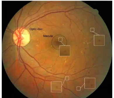
Diabetic Retinopathy (DR) is a progressive micro vascular complication of diabetes that affects the eye. It has been noted as a significant growing public health problem. However, as the disease progresses it leads to distorted and blurred vision, thereby demands early diagnosis to stop sight degradation for blindness prevention. It is characterized by the presence of different lesions including dark lesions such as microaneurysms(MAs), haemorrhages(HEM) and bright lesions such as exudates(EXs). The presence of MAs is the first signs of DR. MA appear reddish, smaller and circular dots. HEMs are caused due to retinal ischemia or the rupture of damaged and abnormally fragile retinal blood vessels. They appear as bright red spots with variable shape and appearances. EXs are yellowish intra-retinal fluid deposits that contain protein, lipid or cellular debris etc. They usually appear as yellowish, bright patches of variable shapes and size [1].
Figure1. An affected retinal image with lesions such as MAs, HEMs and EXs
The following section describes a detailed survey on different detection techniques to detect the lesions separately. Finally it concludes to the most used techniques in the screening of diabetic retinopathy.
II.
LITERATURE SURVEY
This section describes the various methods used in different papers in detecting and classifying different lesions.
A. Algorithms for digital image processing in diabetic retinopathy
This paper proposes the different pre-processing techniques that can be used in the detection of lesions proposed by R.J. Winder et.al [3] .
The main objective of pre-processing techniques is to attenuate image variation by normalizing the original retinal image against a reference model or data set for subsequent viewing, processing or analysis. Variations typically arise within the same image (intra-image variability) as well as between images (inter-image variability) and in order to obtain meaningful information from an image, it is necessary to compensate for this variability. Intraimage variations arise due to differences in light diffusion, the presence of abnormalities, variation in fundus reflectivity and fundus thickness. Inter-image variability is particularly important for longitudinal
studies. Differences between images may be caused by factors including differences in cameras, illumination, acquisition angle and retinal pigmentation. The pre-processing of both monochromatic and colour retinal images may be loosely classified in terms of the correction for non-uniform illumination, contrast enhancement and colour normalization.
A number of general-purpose techniques have been investigated for attenuating this variation and improving the reliability of subsequent operators. Early approaches investigated space-variant filtering schemes supporting locally adaptive contrast enhancements. High-pass filtering and mathematical modeling of the non-uniformity followed by subtraction of this component from the observed image have also been investigated for the correction of non-uniform illumination. However, as noted by Foracchia et al. general-purpose normalization operations typically use metrics derived from the whole image.
B. Detection and classification of retinal lesions for grading of diabetic retinopathy
Digital fundus images normally contain a main region at the center of the image, surrounded by dark background pixels. In the automatic diagnosis of DR, as only the main retinal pixels are required for processing, we therefore separate the background pixels from the foreground in the preprocessing step. Our pre- processing algorithm separates the background using a mean- and variance-based method and it also removes the noise from the image using hue, saturation, and intensity channels. After the background separation, the system extracts the main components, such as the optic disc and the blood vessels, from the retina, something which will help eliminate any spurious and false regions caused by their similarities with bright and dark lesions, respectively.
C. Retinal Microaneurysm Detection Through Local Rotating Cross-Section Profile Analysis
Istvan Lazar and Andras Hajdu propose a method for the automatic detection of microaneurysms (MAs) in color retinal images [4].
Diagnosis of DR is performed by the evaluation of retinal (fundus) images. Manual grading of these images to determine the severity of DR is rather slow and resource demanding. The presence of microaneurysms (MAs) on the retina is the first and most characteristic symptom of this disease. MAs on the retina appear as small, round shaped, red dots. Figure2 shows an example of a fundus image exhibiting signs of DR in terms of the appearance of MAs. The method we propose in this paper realizes the detection of MAs through the analysis of the intensity values along discrete line segments of different directions centered at the candidate pixel.
Figure 2. An example of a fundus image showing signs of DR. MAs are zoomed.
MAs are local intensity maximum structures on the pre-processed retinal image, usually with a Gaussian like intensity distribution. This means that every MA region contains at least one regional maximum also. A local maximum region (LMR), of a gray scale (intensity) image is a connected component of pixels with a given constant intensity value, such that every neighboring pixel of the region has a strictly lower intensity. Therefore, it is sufficient to consider only the LMRs of the pre-processed image as possible MA candidate regions.
D. Automated detection of exudates and macula for grading of diabetic macular edema
Figure3 shows three different fundus images, final image with the appearance of exudates.
Figure 3. Digital image of human retina and different stages of macular edema: (a) healthy retinal image
V.Vijaya kumara and N.Suriyanarayanan compared various optic disc detection methods in the retinal analysis[6]. It facilitates the tracking of various anatomical features and also in the extraction of exudates, drusens etc., present in the retina of human eye. The optic disk is the brightest part in fundus images that can be seen as a pale, round or slightly oval disk. It is the entrance region of blood vessels and also acts as a landmark and reference for the other features in the fundus image. There are several methods for optic disk detection.
In the Principal Component analysis method (PCA), the minimum distance between the original retinal image and its projection onto disk space is located as the center of Optic disk. This detection is accurate but more time consuming. In the next method PCA and active shape model is used, where the shape of the OD is obtained by an active shape method. Here the affine transformation is used to transform the shape from shape space to the image space. This algorithm takes the advantage of top down processing that increases the robustness yet it is time consuming. In lab color morphology, the location of optic disk was by both the automatic initialization of
snake and the application of morphology in color space.
F. Different Methods Used for Extraction of Blood Vessels from Retinal Images
In this paper, Ms. Neha P. Pohankar and Ms. N. R. Wankhade do a survey of different methods of blood vessels segmentation[7]. Blood vessels in retinal images emanate from the Optic Disc (OD). Proper removal of blood vessels and OD is necessary in lesion detection since blood vessels and OD are the significant sources of false positives for dark and bright lesion detection, respectively. Since blood vessels appear as dark elongated structures in retinal images, inaccurate removal of blood vessels would hamper dark lesion detection. There are various methods for automatic segmentation of blood vessels such as thresholding method, tracking method, matched filter method, morphological processing method, region growing method and machine trained classifiers.
having multidirectional structure elements. Then used connect component analysis and an adaptive filter.
G. Blood vessel extraction and optic disc removal using curvelet transform and kernel fuzzy c-means Sudeshna Sil Kar and Santi P. Maity proposes an automatic blood vessel extraction method using matched filtering. translation(location), curvelet is a geometric multiscale transform that is indexed by scale, orientation and location respectively. Vessel detection using matched filtering considers the fact that the gray-level profiles of the cross-section of retinal blood vessels are Gaussian in nature. It is also assumed that the intensity profile is symmetric about the straight line that passes through the center of the vessel. Along with the vessels, pathological images are rich with different non-vascular structures such as bright lesions, exudates, and step and ramp like transient components. For all such images, if the maximum MFR values are directly applied for KFCM based classification, these spurious components are erroneously detected vessels due to their high MFR. To distinguish between the vessel and the non-vessel structures specially for the abnormal images, in addition to MF, negative LoG filter which is the 2nd order derivative of Gaussian is also applied. This justification is due to the asymmetric nature in non-vascular structures and can be modeled as step or ramp functions. LoG filter gives zero response to uniform intensity region. However, when there is a variation in the intensity, it gives positive response on the darker side and negative response on the lighter side. Therefore, at a sharp intensity transition the LoG response contains zero crossing about it's center. This property of the LoG response is utilized
to distinguish the transient like non-vessel structures from the vessels. The LoG filter kernel, which is the second order derivative of the MF kernel.
III.
CONCLUSION
This paper focuses on different techniques or methods for the automatic detection of different lesions mainly for the screening of diabetic retinopathy. It focuses on different pre-processing techniques, methods to extract blood vessels and optic disc removal and different methods used to detect the candidate lesion. The most efficient pre-processing method for dark lesions are curvelet transform and band-pass filter for bright lesions.
IV.
REFERENCES
[1]. Sudeshna Sil Kar and Santi P. Maity, "Automatic Detection of Retinal Lesions for Screening of Diabetic Retinopathy," IEEE Transactions on Biomedical Engineering, 2017 [2]. M. U. Akram, S. Khalid, A. Tariq, S. A. Khan,
and F. Azam, "Detection and classification of retinal lesions for grading of diabetic retinopathy," Computers in Biology and Medicine, vol. 45, pp. 161 - 171, 2014.
[3]. R. Winder, P. Morrow, I. McRitchiea, J. Bailie, and P. Hart, "Algorithms for digital image processing in diabetic retinopathy," Computerized Medical Imaging and Graphics, vol. 33, pp. 608-622, 2009.
[4]. I. Lazar and A. Hajdu, "Retinal microaneurysm detection through local rotating cross-section profile analysis.," IEEE Transactions on Medical Imaging, vol. 32, no. 2, pp. 400-407, 2013.
[5]. M. U. Akram, A. Tariq, S. A. Khan, and M. Y. Javed, "Automated detection of exudates and macula for grading of diabetic macular edema,"
Computer Methods and Programs in
[6]. V.Vijaya Kumari, N.Suriyanarayanan "DETECTION OF OPTIC DISK IN RETINAL IMAGES - A COMPARISON", International Journal on Computer Science and Engineering Vol.1(3), 2009, 192-195.
[7]. Ms. Neha P. Pohankar, Ms. N. R. Wankhade "Different Methods Used for Extraction of Blood Vessels from Retinal Images", 2016 World Conference on Futuristic Trends in Research and Innovation for Social Welfare (WCFTR'16)
[8]. Kuri, Saumitra Kumar, and M. Raju Hossain. "Automated retinal blood vessels extraction using Optimized Gabor filter." Informatics, Electronics & Vision (ICIEV), 2014 International Conference on. IEEE, 2014. [9]. Bauman, Wendall, Di Wu, Ming Zhang,
Jyh-Charn Liu,"On the Adaptive Detection of Blood Vessels in Retinal Images" Computer Science Department Texas A&M University College Station, TX 77843-3112." (2005).
[10]. Shahbeig, Saleh. "Automatic and quick blood