University of Birmingham
Evaluating measurable residual disease in acute
myeloid leukemia
Ravandi, Farhad; Walter, Roland B; Freeman, Sylvie D
DOI:
10.1182/bloodadvances.2018016378
License:
Other (please specify with Rights Statement)
Document Version
Publisher's PDF, also known as Version of record
Citation for published version (Harvard):
Ravandi, F, Walter, RB & Freeman, SD 2018, 'Evaluating measurable residual disease in acute myeloid
leukemia', Blood Advances, vol. 2, no. 11, pp. 1356-1366. https://doi.org/10.1182/bloodadvances.2018016378
Link to publication on Research at Birmingham portal
Publisher Rights Statement: Checked for eligibility: 25/06/2018
This paper was published in the open access journal Blood advances. The final published version can be found at: http://www.bloodadvances.org/content/2/11/1356?sso-checked=true
General rights
Unless a licence is specified above, all rights (including copyright and moral rights) in this document are retained by the authors and/or the copyright holders. The express permission of the copyright holder must be obtained for any use of this material other than for purposes permitted by law.
•Users may freely distribute the URL that is used to identify this publication.
•Users may download and/or print one copy of the publication from the University of Birmingham research portal for the purpose of private study or non-commercial research.
•User may use extracts from the document in line with the concept of ‘fair dealing’ under the Copyright, Designs and Patents Act 1988 (?) •Users may not further distribute the material nor use it for the purposes of commercial gain.
Where a licence is displayed above, please note the terms and conditions of the licence govern your use of this document. When citing, please reference the published version.
Take down policy
While the University of Birmingham exercises care and attention in making items available there are rare occasions when an item has been uploaded in error or has been deemed to be commercially or otherwise sensitive.
If you believe that this is the case for this document, please contact UBIRA@lists.bham.ac.uk providing details and we will remove access to the work immediately and investigate.
REVIEW ARTICLE
Evaluating measurable residual disease in acute myeloid leukemia
Farhad Ravandi,1Roland B. Walter,2,3and Sylvie D. Freeman41Department of Leukemia, The University of Texas MD Anderson Cancer Center, Houston, TX;2Clinical Research Division, Fred Hutchinson Cancer Research Center, Seattle, WA;3Division of Hematology, Department of Medicine, University of Washington, Seattle, WA; and4Department of Clinical Immunology, Institute of Immunology and Immunotherapy, University of Birmingham, Birmingham, United Kingdom
Mounting evidence indicates that the presence of measurable (“minimal”) residual disease (MRD), defined as posttherapy persistence of leukemic cells at levels below morphologic detection, is a strong, independent prognostic marker of increased risk of relapse and shorter survival in patients with acute myeloid leukemia (AML) and can be used to refine risk-stratification and treatment response assessment. Because of the association between MRD and relapse risk, it has been postulated that testing for MRD posttreatment may help guide postremission treatment strategies by identifying high-risk patients who might benefit from preemptive treatment. This strategy, which remains to be formally tested, may be particularly attractive with availability of agents that could be used to specifically eradicate MRD. This review examines current methods of MRD detection, challenges to adopting MRD testing in routine clinical practice, and recent recommendations for MRD testing in AML issued by the European LeukemiaNet MRD Working Party. Inclusion of MRD as an end point in future randomized clinical trials will provide the data needed to move toward
standardizing MRD assays and may provide a more accurate assessment of therapeutic efficacy than current morphologic measures.
Introduction
More than 50% of adult patients with acute myeloid leukemia (AML) relapse after attaining morphologically defined complete remission (CR) with induction chemotherapy.1-3Assessment of posttreatment remission is traditionally based primarily on cytomorphology, with AML relapse conventionally defined as$5% blasts in the bone marrow not attributable to other causes.4-6 Microscopic assessment of bone marrow or peripheral blood morphology relies on examination of a relatively small number of cells (200-500) and its reliability is dependent, in part, on sample quality and the pathologist’s expertise.7As primarily derived from data in younger adults, the risk of relapse following allogeneic hematopoietic stem cell transplantation (allo-HSCT) for consolidation after first CR is 15% to 20% for patients with favorable-risk disease, 20% to 25% for intermediate-risk disease, 30% to 40% for poor-risk disease, and 40% to 50% for very poor-risk disease.8Among older patients (age$60 years) with AML, the respective pooled 2- and 5-year survival estimates following allo-HSCT are 44% and 35% for relapse-free survival (RFS) and 45% and 38% for overall survival (OS).9
Currently, pretreatment factors such as age, cytogenetics, and the presence of certain gene mutations are used to estimate posttreatment risk of relapse based on data from large patient cohorts.5,6,10-12 Table 1 lists prognostic implications of specific mutations in AML as described by the European LeukemiaNet (ELN)10and the National Comprehensive Cancer Network guidelines.6The risk of relapse has been linked to the postchemotherapy persistence of“minimal residual disease”(MRD), which has been defined as leukemic cells at levels below morphologic detection.12Flow cytometric and molecular techniques for assessment of residual leukemia are more sensitive than morphologic assessment, and consensus is growing that MRD might more aptly be called“measurable residual disease,”because the
Submitted 17 January 2018; accepted 23 April 2018. DOI 10.1182/ bloodadvances.2018016378.
presence of any disease detected by these methodologies after treatment is associated with a worse prognosis13,14and detectable leukemia even in morphologic remission may not be“minimal.”MRD monitoring has become part of routine clinical practice in the management of patients with acute lymphoblastic leukemia, acute promyelocytic leukemia (APL), and chronic myeloid leukemia.15-18 Mounting evidence indicates that the presence of MRD is a strong, independent prognostic marker of increased risk of relapse and shorter survival in patients with AML compared with patients with a negative MRD test.4,19-23 This recurrent observation has raised interest in routine MRD testing in AML. To guide the development of a standardized or harmonized approach to MRD testing, the ELN MRD Working Party recently issued consensus recommendations for the measurement and application of MRD in AML (Table 2).24 A prominent strategy to detect MRD is immunophenotypic evaluation by multiparameter flow cytometry (MFC). It is now well established that MRD detected by MFC is an independent prognostic factor for relapse, RFS, and OS.4,19-22In studies of patients ,65 years of age with AML who were fit to receive
cytosine arabinoside plus anthracycline-based induction and consolidation chemotherapy, MRD-negative status as detected by MFC was identified as the most important independent predictor of RFS and OS.25,26Similarly, a retrospective exploratory analysis of data from the Southwest Oncology Group S0106 study showed that MFC-detected MRD after completion of induction chemotherapy could be used to stratify younger patients by risk of AML recurrence, and that MRD status was the single most important predictor of OS and RFS in individual patients.27Data in older patients with AML have also demonstrated the prognostic relevance of MRD monitoring by MFC in patients undergoing traditional cytotoxic chemotherapy induction.28Detection of MRD by MFC during morphological CR before allo-HSCT has also been linked to a substantially higher likelihood of relapse and worse survival.29,30Molecular approaches to detect MRD are equally informative for prognosis. For example, among patients with nucleophosmin 1 (NPM1)–mutated AML who had undergone intensive chemotherapy, the persistence ofNPM1 -mutated transcripts detected using real-time polymerase chain reaction (RT-PCR) was an independent predictor of relapse or
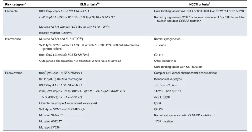
Table 1.Risk status stratification by genetic abnormality per ELN 2017 and NCCN 2017 guidelines
Risk category* ELN criteria10 NCCN criteria6
Favorable t(8;21)(q22;q22.1);RUNX1-RUNX1T1 Core binding factor: inv(16)†,‡or t(16;16)†,‡or t(8;21)†,‡or t(15;17)‡ inv(16)(p13.1;q22) or t(16;16)(p13.1;q22);CBFB-MYH11 Normal cytogenetics:NPM1mutation in absence ofFLT3-ITD or isolated
biallelic (double)CEBPAmutation MutatedNPM1withoutFLT3-ITD or withFLT3-ITDlow§
Biallelic mutatedCEBPA
Intermediate MutatedNPM1andFLT3-ITDhigh§ Normal cytogenetics
Wild-typeNPM1withoutFLT3-ITD or withFLT3-ITDlow§(without adverse-risk genetic lesions)
18 alone
t(9;11)(p21.3;q23.3);MLLT3-KMT2A|| t(9;11)
Cytogenetic abnormalities not classified as favorable or adverse Other nondefined
Core binding factor withKITmutation Poor/adverse t(6;9)(p23;q34.1);DEK-NUP214 Complex ($3 clonal chromosomal abnormalities)
t(v;11q23.3);KMT2Arearranged Monosomal karyotype
t(9;22)(q34.1;q11.2);BCR-ABL1 25, 5q–,–7, 7q–
inv(3)(q21.3q26.2) or t(3;3)(q21.3;q26.2);GATA2,MECOM(EVI1) 11q23–non t(9;11)
25 or del(5q);–7;–17/abn(17p) inv(3), t(3;3)
Complex karyotype,{monosomal karyotype# t(6;9)
Wild-typeNPM1andFLT3-ITDhigh t(9;22)
MutatedRUNX1** Normal cytogenetics: withFLT3-ITD mutation††
MutatedASXL1** TP53mutation
MutatedTP53‡‡
*The prognostic value of a marker is treatment-dependent and may change with new therapies.
†Presence of KIT mutations in patients with t(8:21) and, to a lesser extent, inv(16), confers a high risk of relapse; these patients should be considered intermediate risk and considered for HSCT if available.
‡Other cytogenetic findings in addition to these do not alter risk status.
§Low, low allelic ratio (,0.5); high, high allelic ratio ($0.5); recent studies indicate that AML withNPM1mutation andFLT3-ITD low allelic ratio may also have a more favorable prognosis and patients should not routinely be assigned to allogeneic HCT.75,76
||Presence of t(9;11)(p21.3;q23.3) takes precedence over rare, concurrent adverse-risk gene mutations.
{3 or more chromosomal abnormalities in the absence of 1 of the World Health Organization–designated recurring translocations or inversions: t(8;21), inv(16) or t(16;16), t(9;11), t(v;11) (v;q23.3), t(6;9), inv(3) or t(3;3); AML withBCR-ABL1.
#Defined by the presence of 1 single monosomy (excluding loss of X or Y) with at least 1 additional monosomy or structural chromosome abnormality (excluding core-binding factor AML). **These should not be used as adverse prognostic markers if they occur with favorable-risk AML subtypes.
††FLT3-ITD mutations are considered to confer a significantly poorer outcome in patients with normal karyotype; there is controversy about whetherFLT3-TKD mutations carry equally poor prognosis.
‡‡TP53mutations are significantly associated with AML with complex and monosomal karyotype. NCCN, National Comprehensive Cancer Network.
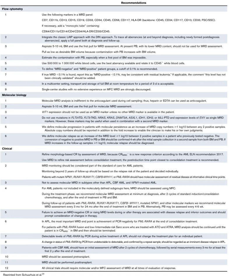
Table 2.ELN recommendations for MRD testing
Recommendations Flow cytometry
1 Use the following markers in a MRD panel:
CD7, CD11b, CD13, CD15, CD19, CD33, CD34, CD45, CD56, CD117, HLA-DR (backbone: CD45, CD34, CD117, CD13, CD33, FSC/SSC). If necessary, add a“monocytic tube”containing:
CD64/CD11b/CD14/CD4/CD34/HLA-DR/CD33/CD45.
2 Integrate the classic LAIP approach with the DfN approach. To trace all aberrancies (at and beyond diagnosis, including newly formed postdiagnosis aberrancies), apply a full panel both at diagnosis and follow-up.
3 Aspirate 5-10 mL BM and use the first pull for MRD assessment. At present PB, with its lower MRD content, should not be used for MRD assessment. Pull as low as desirable BM volume because contamination with PB increases with BM volume.
4 Estimate the contamination with PB, especially when a first pool of BM was impossible.
5 Use 500 000 to 1 000 000 white blood cells, use the best aberrancy available and relate it to CD451white blood cells. 6 To define“MRD-negative”and“MRD-positive”patient groups, a cutoff of 0.1% is recommended.
7 If true MRD,0.1% is found, report this as“MRD-positive,0.1%, may be consistent with residual leukemia.”If applicable, the comment“this level has not been clinically validated”should be added.
8 In a multicenter setting, transport and storage of full BM at room temperature for a period of 3 d is acceptable. 9 Single-center studies with no extensive experience on MFC MRD are strongly discouraged.
Molecular biology
1 Molecular MRD analysis is indifferent to the anticoagulant used during cell sampling; thus, heparin or EDTA can be used as anticoagulant. 2 Aspirate 5-10 mL BM and use the first pull for molecular MRD assessment.
3 WT1expression should not be used as an MRD marker unless no other MRD marker is available in the patient.
4 Do not use mutations inFLT3-ITD,FLT3-TKD,NRAS,KRAS,DNMT3A,ASXL1,IDH1,IDH2, orMLL-PTD and expression levels ofEVI1as single MRD markers. However, these markers may be useful when used in combination with a second MRD marker.
5 We define molecular progression in patients with molecular persistence as an increase of MRD copy numbers$1 log10 between any 2 positive samples. Absolute copy numbers should be reported in addition to the fold increase to enable the clinician to make his or her own judgments.
6 We define molecular relapse as an increase of the MRD level$1 log10 between 2 positive samples in a patient who previously tested negative. The conversion of negative to positive MRD in PB or BM should be confirmed 4 wk after the initial sample collection in a second sample from both BM and PB. If MRD increases in the follow-up samples$1 log10, molecular relapse should be diagnosed.
Clinical
1 Refine morphology-based CR by assessment of MRD, because CRMRD2is a new response criterion according to the AML ELN recommendation 2017. Use MRD to refine risk assessment before consolidation treatment, the postinduction time point closest to consolidation treatment is recommended. 2 MRD monitoring should be considered part of the standard of care for AML patients.
Monitoring beyond 2 years of follow-up should be based on the relapse risk of the patient and decided individually.
Patients with mutantNPM1,RUNX1-RUNX1T1,CBFB-MYH11, orPML-RARAshould have molecular assessment of residual disease at informative clinical time points. 3 Not to assess molecular MRD in subtypes other than APL, CBF AML, andNPM1-mutated AML.
4 For AML patientsnotincluded in the molecularly defined subgroups here, MRD should be assessed using MFC.
During the treatment phase, we recommend molecular MRD assessment at minimum at diagnosis, after 2 cycles of standard induction/consolidation chemotherapy, and after the end of treatment in PB and BM.
During follow-up of patients withPML-RARA,RUNX1-RUNX1T1,CBFB-MYH11, mutatedNPM1, and other molecular markers we recommend molecular MRD assessment every 3 mo for 24 mo after the end of treatment in BM and in PB. Alternatively, PB may be assessed every 4-6 wk.
5 Failure to achieve an MRD-negative CR or rising MRD levels during or after therapy are associated with disease relapse and inferior outcomes and should prompt consideration of changes in therapy.
6 In APL, the most important MRD end point is achievement of PCR-negativity forPML-RARAat the end of consolidation treatment.
For patients withPML-RARAfusion and low-/intermediate-risk Sanz score who are treated with ATO and ATRA, MRD analysis should be continued until the patient is in CRMRD2in BM and then should be terminated.
7 Detectable levels ofPML-RARAby PCR during active treatment of APL should not change the treatment plan for an individual patient.
8 A change in status ofPML-RARAby PCR from undetectable to detectable, and confirmed by a repeat sample, should be regarded as an imminent disease relapse in APL. 9 Patients with CBF AML should have an initial assessment of MRD after 2 cycles of chemotherapy, followed by serial measurements every 3 mo for at least the
first 2 y after the end of treatment. 10 MRD should be assessed pretransplant. 11 MRD should be performed posttransplant.
12 All clinical trials should require molecular and/or MFC assessment of MRD at all times of evaluation of response. Reprinted from Schuurhuis et al.24
death22and of outcomes of allo-HSCT.31,32 Another study using whole-genome or exome sequencing of samples from a cohort of adults with AML found that detection of persistent leukemia-associated mutations in bone marrow cells during remission
;30 days after start of chemotherapy was linked to a significantly increased risk of relapse, shorter event-free survival (EFS), and poorer OS.33
Thus, adding MRD evaluation to other posttreatment assessments (eg, morphologic evaluations) could help guide postremission treatment strategies by identifying patients at high risk of relapse who might benefit from preemptive therapy,34 an appealing treatment concept that will require formal validation. Although not proven to date, the concept of MRD eradication aiding decision-making and improving outcomes of patients with AML is plausible. This is supported by experience with bispecific antibodies such as blinatumomab in ALL.35
This review examines current methods of MRD detection, some challenges in adopting MRD testing in routine clinical practice, and describes some of the recent recommendations from the ELN MRD Working Party consensus statement for the detection of MRD in AML.24
MRD detection methods
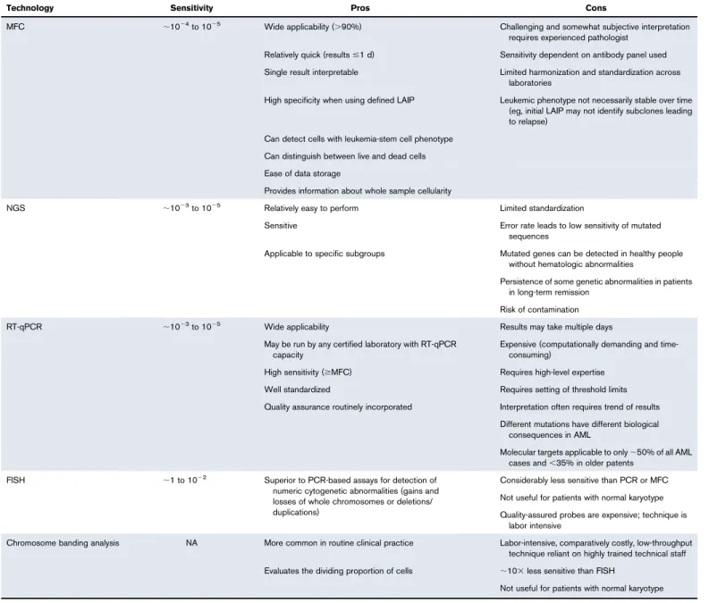
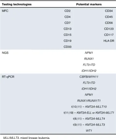
Technologies to measure MRD based on immunophenotype, cytogenetic abnormalities, and molecular mutations each have advantages and disadvantages. The clinical usefulness of MRD detection depends on the choice of MRD marker (eg, specific gene mutation, surface antigen), which in some cases might be a therapeu-tic target.19,36An ideal MRD test should discriminate between cells that would not cause relapse from the smallest clinically significant populations of leukemic cells that hold the potential to cause relapse.37 Current MRD testing methods have not achieved this ideal state, except for PCR testing in APL.38Table 3 summarizes the advantages and limitations of available methods of MRD detection. MFC and RT quantitative PCR (RT-qPCR) are the most commonly used technologies; more recently, next-generation sequencing (NGS) is being used for molecular assessments.6,7,23,37,39,40 Fluorescence in situ hybridization or chromosome banding analysis can detect leukemic cells with cytogenetic abnormalities41 but, because a smaller number of cells are interrogated, are generally less sensitive than MFC or molecular methods. Table 4 lists potential markers for monitoring MRD in AML and the testing technologies used to detect them.15,24,42
MFC
MFC uses panels of fluorochrome-labeled monoclonal antibodies to identify aberrantly expressed antigens located on (or within) leukemic cells.15Instruments that have multiple lasers to detect different fluorochromes, with combinations of multiple monoclonal antibodies, have increased the sensitivity of MFC to detect 1023to 1025 leukemic cells within the white blood cell compartment.43 There are 2 main approaches used to detect MRD by MFC; 1 involves identification of leukemia-associated immunophenotypes (LAIPs) that differ from the majority of normal hematopoietic cells and the other approach entails identification of different-from-normal (DfN) patterns.44LAIP are cells with abnormal patterns of antigens; examples include cross-lineage antigen expression (eg, expression of lymphoid markers in myeloid blasts), asynchronous antigen expression (eg, coexpression of antigens that are not
usually found together during normal cellular differentiation), and over- or underexpression of antigens compared with normal levels.45-47An extensive panel of monoclonal antibodies is required to detect all potentially abnormal LAIP antigen expression patterns in AML, which can number up to 100.26 A standard fixed monoclonal antibody panel is used to identify DfN patterns at all stages of disease/treatment with MFC.44 An advantage of this method is that it does not restrict MRD determination to specific LAIP present at diagnosis and takes immunophenotypic shifts over time into account.44 LAIP detection by MFC is more commonly used than the fixed-antibody method,42but differences between the LAIP and DfN approaches may be minimized if sufficiently large antibody panels ($8 colors) are used for detection.24 Because phenotypes may change over time by gaining or losing specific abnormalities or patterns of abnormalities during disease evolution, a prior MRD target (as defined by a specific LAIP) may be less useful at later time points for the same patient.15,37,48Therefore, the ELN MRD Working Party suggests using a “LAIP-based DfN approach” to monitoring MRD (ie, using the same antibody-fluorochrome combinations with a minimum set of markers during follow-up assessments as those used at diagnosis to track emerging aberrancies) (Table 2).24Researchers continue to work on improving MFC methodology and technology. For example, 1 group has developed a 1-tube assay with a single fluorescence channel that appears to work as well as standard 7-tube antibody panel to accurately quantify CD341CD382leukemic stem cells.49 RT-qPCR
RT-qPCR is used to amplify leukemia-associated genetic abnor-malities. Optimized RT-qPCR assays are more sensitive than MFC, with a detection range of 1024 to 1026.24,43 Additionally, quantitative assays that measure number of leukemic transcripts can be informative of whether transcript levels are rising or falling and can potentially inform further therapy, although benefits of MRD-directed therapy in AML are not yet firmly established.44 Viable targets for molecular MRD monitoring include leukemic fusion genes such as promyelocytic leukemia gene retinoic acid receptor-a (PML-RARA), core-binding factor subunit b myosin heavy chain 11 (CBFB-MYH11), and runt-related transcription factor 1 (RUNX1)/RUNX1 translocated to 1 (RUNX1T1), and mutantNPM1.24Wilms’tumor gene (WT1) should not be used as an MRD marker because of poor sensitivity and specificity unless no other MRD markers are available.24The ELN MRD Working Party recommends against use of Fms-like tyrosine kinase internal tandem duplication (FLT3-ITD), FLT3-TKD, NRAS,KRAS,IDH1,
IDH2,MLL-PTD, and expression levels ofEVI1as single markers of MRD because they are prone to frequent losses or gains; however, these mutations may have prognostic significance if accompanied by other MRD markers.24 Given available molecular targets, RT-qPCR assessment of MRD is thought to be applicable to only
;50% of all AML cases and less than ;35% in older patients (Figure 1), whereas MFC can detect MRD in ;90% of patients when a comprehensive antibody panel is used.19,28,44,46,47,50 Limitations of RT-qPCR–based MRD assays are their depen-dence on specific mutations, requiring individual reference standard curves based on target serial dilutions.51 Digital PCR, a high-throughput technology that generates absolute quantification, can clonally amplify target nucleic acids and does not require a reference standard curve, has greater assay sensitivity than RT-qPCR.52 For example, digital PCR can detect a variety of
NPM1mutation subtypes without the need for multiple plasmid standards.53,54
NGS
Next-generation DNA sequencing technologies, which allow parallel and repeated sequencing of millions of small DNA fragments, can be used to evaluate a few genes or an entire genome.55The ability of NGS to assay large numbers of mutated genes could help trace the evolution of malignant clones, which cannot be done with RT-qPCR.15Studies have demonstrated the feasibility of NGS to monitor mutations for which targeted therapies are available, such as FLT3-ITD56 andIDH1/2,57 and mutations with prognostic relevance, such asCEBPAandNPM1in patients with AML.10A recent study compared a targeted 28-gene NGS
panel for detection of common AML mutations (with variant allele frequency [VAF]$5%) and a 10-color MFC assay of different-from-normal MRD in patients with AML before allo-HSCT.58 Results of the 2 assays were concordant in 71% of patients. For patients in CR or CR with incomplete hematologic recovery (CRi), MRD measured by NGS was much greater than the estimated percentage of aberrant blasts detected by MFC, suggesting that residual mutations persisted in non-blast compartments during remission. Patients found to be MRD-positive with both assays had the highest risk of relapse compared with patients who were negative by both assays and with patients who had discordant assay results.58Similarly, in 340 patients with AML in CR or CR with CRi, there was a 69.1% concordance of MRD detection in the bone marrow using a 54-gene NGS evaluation and an MFC assay;
Table 3.Pros and cons of methods used to detect MRD in AML
Technology Sensitivity Pros Cons
MFC ;1024to 1025 Wide applicability (.90%) Challenging and somewhat subjective interpretation requires experienced pathologist
Relatively quick (results#1 d) Sensitivity dependent on antibody panel used Single result interpretable Limited harmonization and standardization across
laboratories
High specificity when using defined LAIP Leukemic phenotype not necessarily stable over time (eg, initial LAIP may not identify subclones leading to relapse)
Can detect cells with leukemia-stem cell phenotype Can distinguish between live and dead cells Ease of data storage
Provides information about whole sample cellularity
NGS ;1023to 1025 Relatively easy to perform Limited standardization
Sensitive Error rate leads to low sensitivity of mutated sequences
Applicable to specific subgroups Mutated genes can be detected in healthy people without hematologic abnormalities
Persistence of some genetic abnormalities in patients in long-term remission
Risk of contamination
RT-qPCR ;1023to 1025 Wide applicability Results may take multiple days
May be run by any certified laboratory with RT-qPCR capacity
Expensive (computationally demanding and time-consuming)
High sensitivity ($MFC) Requires high-level expertise Well standardized Requires setting of threshold limits Quality assurance routinely incorporated Interpretation often requires trend of results
Different mutations have different biological consequences in AML
Molecular targets applicable to only;50% of all AML cases and,35% in older patents
FISH ;1 to 1022 Superior to PCR-based assays for detection of numeric cytogenetic abnormalities (gains and losses of whole chromosomes or deletions/ duplications)
Considerably less sensitive than PCR or MFC Not useful for patients with normal karyotype Quality-assured probes are expensive; technique is
labor intensive
Chromosome banding analysis NA More common in routine clinical practice Labor-intensive, comparatively costly, low-throughput technique reliant on highly trained technical staff Evaluates the dividing proportion of cells ;103less sensitive than FISH
Not useful for patients with normal karyotype FISH, fluorescence in situ hybridization; NA, not available. QA, quality assurance.
however, persistent mutations were detected by NGS only in 64 patients.23Four-year relapse rate was highest among patients with MRD detected by both methods (73.3%), followed by those with MRD only on NGS (52.3%), those with MRD only on MFC (49.8%), and those who were MRD-negative on both assays (26.7%).23 Factors that complicate the use of NGS to monitor MRD in patients with AML include the genetic clonal heterogeneity at AML diagnosis and during the course of the disease. The predominant leukemic clone at presentation might not be the clone that causes clinical
relapse and mortality.59Moreover, determination of clonality in a given sample can be influenced by the depth of sequencing and the algorithm used to identify mutations.37NGS currently has an intrinsic error rate that limits its sensitivity for most single-nucleotide variants to;1% to 2% of all reads.15NGS typically generates shorter sequence lengths; for example, 1 of the most commonly used technologies, Illumina’s sequencing by synthe-sis, routinely produces read lengths of 75 to 100 base pairs from libraries with insert sizes of 200 to 500 base pairs. Thus, assembly of longer repeats and duplications may suffer from the short read length.60Further, NGS technology is computationally demanding, time-consuming, and still expensive (although it is expected that costs may drop in the future), which might make it difficult to apply in clinical practice. Reflecting the current state of development, the ELN MRD Working Party suggests NGS techniques for MRD measurement are best reserved for clinical trials at this time.24
Challenges to clinical application of
MRD testing
AML is genetically diverse and, currently, there is no uniform approach to detecting the leukemic cells that are biologically capable of and likely to cause relapse.37The genetic heterogeneity of AML and lack of universal antigenic surface markers on leukemic stem cells increase the challenge of standardizing MRD detection protocols. Additional challenges to adopting MRD testing in routine clinical practice for patients with AML have included the absence of interlaboratory standardization or consensus regarding optimal key parameters, including type of specimen, MRD target, timing of MRD assessment, technology (eg, MFC vs RT-qPCR), testing protocols, and lack of established cutoff values,42,61although the ELN MRD Working Party recommendations address several of these issues.24 Variables that affect the ability to detect MRD are assay sensitivity, the skills and expertise of personnel, biologic properties of the leukemic cells, and the quality and number of viable cells used for analyses.15,36,37 The ELN 2017 Recommendations for Diagnosis and Treatment of AML highlight that MRD testing should be performed in experienced, centralized diagnostic laboratories. Because sensitivities vary by type of MRD marker and testing method, reported results should specify the test applied, assay sensitivity, and cutoff values.10Currently, the National Comprehen-sive Cancer Network AML 2018 Clinical Practice Guidelines for
Table 4.Testing technologies and potential markers for monitoring MRD in AML
Testing technologies Potential markers
MFC CD2 CD34 CD4 CD45 CD7 CD56 CD13 CD123 CD15 CD117 CD19 HLA-DR CD33 NGS NPM1 RUNX1 FLT3-ITD IDH1/IDH2 RT-qPCR CBFB/MYH11 FLT3-ITD IDH1/IDH2 NPM1 RUNX1/RUNX1T1 t(10;11)–KMT2A-MLLT10 t(11;19)–KMT2A-ELLorKMT2A-MLLT1 t(6;11)–KMT2A-MLLT4 t(9;11)–KMT2A-MLLT3 WT1 MLL/MLLT3, mixed lineage leukemia.
2% 7% 42% 11% 12% 6% 30% 38% 13% 7% 5% 23% 68% 13% 2% 1% 4% t(15;17)/PML-RARA 0-15 15-60 >60 t(8;21)/RUNX1-RUNX1T1 inv(16)/CBFB-MYH11 11q23/MLL fusions t(6;9)/DEK-CAN NUP98-NSD1 CBFA2T3-GLIS2 NPM1 mutant Other rare fusions
No recurring leukemia-specific marker 1% 1% 1% 1% 1% 1% 2% 4% 4%
AML do not recommend MRD monitoring, but note that ongoing research is moving MRD monitoring to the forefront for all patients with AML.6
Another subject of debate is whether routine clinical sampling for MRD testing should be performed on peripheral blood or bone marrow. The ELN MRD consensus report recommends testing both bone marrow and blood for molecular MRD during treatment.24 Bone marrow sampling generally offers greater sensitivity because MRD levels in peripheral blood are lower than in bone marrow.19 Nevertheless, peripheral blood sampling is less expensive and less painful for patients who may be unwilling to undergo the more frequent bone marrow sampling required to monitor MRD during various courses of treatment.14MRD analysis of peripheral blood requires a minimum of 20 mL of blood; in patients with white blood cell counts,13109/L, more blood may be necessary to improve sensitivity.24The ELN MRD Working Party suggests aspirating 5 to 10 mL of bone marrow using the first pull, noting that contamination from peripheral blood increases as bone marrow sample volume increases.24 Additionally, peripheral blood may be preferable for PCR-based gene expression MRD monitoring because of high background “noise”in bone marrow.14A study of younger adults (ages 18-60 years) with AML found that as detected by RT-qPCR, the presence of mutant NPM1 MRD in peripheral blood in first remission was a strong predictor of relapse, independent of cytogenetics and FLT3-ITDstatus, and might have application in selecting patients who would benefit from allo-HSCT.32
The choice of an MRD target can be confounded by the persistence of genetic abnormalities in patients in long-term remission.62For example, mutated DNMT3A with up to 50% VAF can persist in patients who have been in remission for several years.62Another concern is that some commonly mutated genes in AML, such as
TET2,DNMT3A, andASXL1, can also be mutated in healthy people with no hematologic abnormalities, especially as people age. This phenomenon has been called age-related clonal hematopoiesis of indeterminate potential. Whole exome sequencing of samples from 12 380 people with no hematologic malignancies indicated 10% of persons age.65 years showed evidence of clonal hematopiesis.63 These mutations in older patients with AML may not be the drivers of leukemogenesis.63-65However, the presence of AML-associated mutations may be indicative of early events in the development of hematologic malignancies in some cases because they significantly increase the risk of eventually developing one.65,66
In a recent study, samples from 430 patients with AML in CR or CRi who had at least 1 mutation at diagnosis were obtained between 21 days and 4 months from the start of a second treatment cycle for analysis by targeted NGS and by MFC.23 Mutations in TET2,
DNMT3A, and ASXL1 (DTA) were present during remission in
.50% of patients. Detection of DTA mutations was not associated with a higher 4-year relapse rate than that of patients without these mutations unless they were accompanied by other non-DTA mutations.23The researchers speculated that DTA mutations may have persisted in nonleukemic clones that repopulated the bone marrow after induction chemotherapy.23
A study of patients with de novo AML who had received up to 2 rounds of induction chemotherapy evaluated MRD status at 30 days after treatment using digital sequencing of leukemia-specific mutations in 50 patients in morphologic remission.33Although all patients showed normal morphology at day 30, some patients’
samples showed clearance of all mutations; others showed clearance of only a few of the mutations, which returned at relapse; a third group of patients showed clearance in a subset of the mutations at day 30, but the founding clone mutations persisted in almost every cell.33Patients with EFS durations.12 months were significantly less likely to have persistent disease as indicated by VAF at day 30 than those with EFS #12 months (P5.01); for patients who relapsed, day 30 VAF had increased because cells containing those mutations reexpanded.33These data suggest MRD testing at 30 days posttreatment can provide more important prognostic information than morphologic status, but repeated testing and trends in MRD status over time may be more informative.37The optimum interval duration for sequential MRD testing is unknown and may depend on disease charac-teristics. To avoid false-positive results, some have suggested that a confirmatory MRD test should be performed at 2 to 4 weeks after a positive MRD test before making predictions about impending relapse.37
The optimal time for MRD testing may depend on the type of MRD. Ommen and colleagues showed the kinetics of molecular relapse can differ markedly among leukemias characterized byNPM1, PML-RARA,RUNX1-RUNX1T1, andCBFB-MYH11 AML.67The inves-tigators developed a model to predict the time between molecular relapse and hematologic relapse. They found thatCBFB-MYH11
AML displayed a slower clone regrowth than AML with the other molecular signatures and recommended MRD testing for CBFB-MYH11be performed every 6 months, whereas testing for PML-RARA MRD was recommended for every 2 months.67 There is a continued need to establish the optimal intervals for MRD assessment to predict impending relapse. Currently, the ELN MRD Working Party recommends MRD testing at diagnosis, after 2 cycles of chemotherapy at the closest time point before consoli-dation treatment, and during follow-up of patients withPML-RARA,
RUNX1-RUNX1T1, CBFB-MYH11, mutated NPM1, and other molecular markers. Molecular MRD assessment should be con-ducted in bone marrow and peripheral blood every 3 months for 24 months after the end of treatment, or in peripheral blood every 4 to 6 weeks.24MRD testing should be performed before and after bone marrow transplant.24
More studies are also needed to determine relevant MRD thresholds, which will of necessity vary according to the technology used for assessment and the type of tissue under study. In the study described previously, day 30 samples were assessed for MRD using digital sequencing that included probes covering all exons of 264 recurrently mutated genes in AML.33The VAF threshold was set at 2.5% in bone marrow; investigators noted that because the vast majority of AML-associated somatic mutations are heterozy-gous, a 2.5% VAF threshold suggests that at least 5% of bone marrow cells under examination would contain the mutation(s).33 Indeed, detection of persistent AML-associated mutations in$5% of cells in day 30 remission samples was significantly associated with reduced OS and increased risk of relapse compared with patients who attained mutational clearance.33Studies measuring LAIP by MFC use lower detection thresholds, for example, from 0.01% to 1.0%,26and thresholds may depend on the types of LAIP under study.28The ELN MRD Working Party suggests a threshold of 0.1% to distinguish between MRD positivity and negativity; however, they noted that MRD LAIP levels,0.1% may still signal residual leukemia.24
Remission maintenance therapies
The correlation between MRD status and relapse risk has generated substantial interest in using results of MRD testing to direct therapy decisions for AML patients; for example, therapy might be initiated or intensified when MRD is present, reduced, or discontinued for those who are MRD-negative. Initiating or in-tensifying treatment of patients with MRD may lessen risk of relapse and improve OS, although this remains to be proven.14,37The risks associated with any therapy must be considered when making decisions based on MRD status. At present, there are few published studies that have evaluated MRD stratification and/or therapeutic strategies to eradicate MRD in patients with AML. Azacitidine has been shown to increase expression of epigenetically silenced leukemia antigens and induce a CD81 T-cell response to tumor antigens posttransplant, potentially augmenting a graft-versus-leukemia effect.68-71 At least 2 studies have evaluated preemptive use of azacitidine after SCT based on detection of MRD. The RELAZA phase 2 study evaluated azacitidine after allo-HSCT in 20 patients with CD341AML or myelodysplastic syndromes and signs of MRD, defined as decreases of peripheral blood CD341donor chimerism to,80%, without concomitant signs of hematologic relapse.72After 4 cycles of azacitidine, 10 patients (80%) were MRD-negative; of these, 4 remained MRD-negative at a median follow-up of 347 days. The investigators noted that tracking MRD after allo-HSCT via peripheral blood CD341donor chimerism monitoring allowed preemptive use of azacitidine only when MRD was detected, avoiding unnecessary toxicity in patients in CR at low risk of relapse.72 In another study, 10 patients with mutantNPM1AML and normal karyotype in first or second CR after intensive chemotherapy or autologous or allo-HSCT who showed evidence of molecular relapse (defined as a 1% increase in mutantNPM1transcripts in bone marrow) or persistent MRD in sequential RT-PCR analyses of bone marrow or peripheral blood received azacitidine treatment.73Molecular response was defined as a 1-log reduction in MRD from the pretreatment value. At a median follow-up of 10 months (range, 2-12), patients had received a median of 5 azacitidine treatment cycles. Of the 10 patients, 7 showed a molecular response and remained in CR.73
On a promising note, because of growing acceptance of the prognostic value of MRD, several ongoing AML studies are pro-spectively evaluating the effect of interventions on MRD. Some of these studies require detectable MRD as an eligibility criterion.
Conclusions
The ELN MRD Working Party recommends MRD testing as part of the standard of care for AML patients.24Its recently published guidelines promote widespread adoption of MRD testing for monitoring of therapeutic efficacy and/or the prognoses of patients with AML; however, some questions remain. The MRD Working Party notes that the predictive power of several mutations is low or needs to be clarified. They recommend further study of the clinical implications of detectable flow cytometric MRD levels ,0.1%. Moreover, different MRD thresholds after induction chemotherapy may have variable meaning in differing patient risk groups and the clinical significance of MRD for patients treated with nonintensive therapies such as hypomethylating agents requires more study.24,74Ultimately, the molecular heterogeneity of AML and clonal architecture may prevent a“1-size-fits-all”approach to MRD detection.19 Incorporation of MRD as an end point of ongoing and future randomized clinical trials should provide the data needed to move toward improved understanding of the influence of MRD markers in patients not included in molecularly defined AML subgroups (eg, APL, CBF-AML, AML with BCR-ABL1, AML with NPM1 mutations). These studies will also help determine whether MRD assessment will prove to be a more accurate measure of therapeutic efficacy than current morphologic measures.
Acknowledgments
The authors received editorial support during manuscript devel-opment from Sheila Truten and Kelly Dittmore of Medical Communication Company, Inc. (Wynnewood, PA), who were funded by Celgene Corporation. The authors directed manuscript development and are fully responsible for all content and editorial decisions.
Authorship
Contribution: F.R. prepared the first draft of the manuscript; and all authors contributed equally to revising the manuscript and all ap-proved manuscript content and submission to the journal.
Conflict-of-interest disclosure: The authors declare no competing financial interests.
Correspondence: Farhad Ravandi, Section of Developmental Therapeutics, Department of Leukemia, University of Texas MD Anderson Cancer Center, 1515 Holcombe Blvd, Houston, TX 77030; e-mail: fravandi@mdanderson.org.
References
1. Breems DA, Van Putten WL, Huijgens PC, et al. Prognostic index for adult patients with acute myeloid leukemia in first relapse.J Clin Oncol. 2005;23(9): 1969-1978.
2. Walter RB, Kantarjian HM, Huang X, et al. Effect of complete remission and responses less than complete remission on survival in acute myeloid leukemia: a combined Eastern Cooperative Oncology Group, Southwest Oncology Group, and M. D. Anderson Cancer Center Study.J Clin Oncol. 2010;28(10):1766-1771.
3. Burnett AK, Milligan D, Goldstone A, et al; United Kingdom National Cancer Research Institute Haematological Oncology Study Group. The impact of dose escalation and resistance modulation in older patients with acute myeloid leukaemia and high risk myelodysplastic syndrome: the results of the LRF AML14 trial.Br J Haematol. 2009;145(3):318-332.
4. Chen X, Xie H, Wood BL, et al. Relation of clinical response and minimal residual disease and their prognostic impact on outcome in acute myeloid leukemia.J Clin Oncol. 2015;33(11):1258-1264.
5. Fey MF, Buske C, Group EGW; ESMO Guidelines Working Group. Acute myeloblastic leukaemias in adult patients: ESMO Clinical Practice Guidelines for diagnosis, treatment and follow-up.Ann Oncol. 2013;24(suppl 6):vi138-vi143.
6. National Comprehensive Cancer Network. NCCN Guidelines: acute myeloid leukemia, version 1.2018.
7. DeAngelo DJ, Stein EM, Ravandi F. Evolving therapies in acute myeloid leukemia: progress at last?Am Soc Clin Oncol Educ Book. 2016;35:e302-e312. 8. Cornelissen JJ, Blaise D. Hematopoietic stem cell transplantation for patients with AML in first complete remission.Blood. 2016;127(1):62-70. 9. Rashidi A, Ebadi M, Colditz GA, DiPersio JF. Outcomes of allogeneic stem cell transplantation in elderly patients with acute myeloid leukemia: a
systematic review and meta-analysis.Biol Blood Marrow Transplant. 2016;22(4):651-657.
10. D ¨ohner H, Estey E, Grimwade D, et al. Diagnosis and management of AML in adults: 2017 ELN recommendations from an international expert panel.
Blood. 2017;129(4):424-447.
11. Arber DA, Orazi A, Hasserjian R, et al. The 2016 revision to the World Health Organization classification of myeloid neoplasms and acute leukemia.
Blood. 2016;127(20):2391-2405.
12. Ossenkoppele G, Schuurhuis GJ. MRD in AML: time for redefinition of CR?Blood. 2013;121(12):2166-2168. 13. Goldman JM, Gale RP. What does MRD in leukemia really mean?Leukemia. 2014;28(5):1131.
14. Percival ME, Lai C, Estey E, Hourigan CS. Bone marrow evaluation for diagnosis and monitoring of acute myeloid leukemia.Blood Rev. 2017;31(4): 185-192.
15. Tomlinson B, Lazarus HM. Enhancing acute myeloid leukemia therapy - monitoring response using residual disease testing as a guide to therapeutic decision-making.Expert Rev Hematol. 2017;10(6):563-574.
16. van Dongen JJ, van der Velden VH, Br ¨uggemann M, Orfao A. Minimal residual disease diagnostics in acute lymphoblastic leukemia: need for sensitive, fast, and standardized technologies.Blood. 2015;125(26):3996-4009.
17. De Angelis F, Breccia M. Molecular monitoring as a path to cure acute promyelocytic leukemia.Rare Cancers Ther. 2015;3(1-2):119-132.
18. Paschka P, M ¨uller MC, Merx K, et al. Molecular monitoring of response to imatinib (Glivec) in CML patients pretreated with interferon alpha. Low levels of residual disease are associated with continuous remission.Leukemia. 2003;17(9):1687-1694.
19. Grimwade D, Freeman SD. Defining minimal residual disease in acute myeloid leukemia: which platforms are ready for“prime time”?Blood. 2014; 124(23):3345-3355.
20. San Miguel JF, Vidriales MB, L ´opez-Berges C, et al. Early immunophenotypical evaluation of minimal residual disease in acute myeloid leukemia identifies different patient risk groups and may contribute to postinduction treatment stratification.Blood. 2001;98(6):1746-1751.
21. Buccisano F, Maurillo L, Gattei V, et al. The kinetics of reduction of minimal residual disease impacts on duration of response and survival of patients with acute myeloid leukemia.Leukemia. 2006;20(10):1783-1789.
22. Ivey A, Hills RK, Simpson MA, et al; UK National Cancer Research Institute AML Working Group. Assessment of minimal residual disease in standard-risk AML.N Engl J Med. 2016;374(5):422-433.
23. Jongen-Lavrencic M, Grob T, Hanekamp D, et al. Molecular minimal residual disease in acute myeloid leukemia.N Engl J Med. 2018;378(13):1189-1199. 24. Schuurhuis GJ, Heuser M, Freeman S, et al. Minimal/measurable residual disease in AML: a consensus document from the European LeukemiaNet MRD
Working Party.Blood. 2018;131(12):1275-1291.
25. Ravandi F, Jorgensen J, Borthakur G, et al. Persistence of minimal residual disease assessed by multiparameter flow cytometry is highly prognostic in younger patients with acute myeloid leukemia.Cancer. 2017;123(3):426-435.
26. Terwijn M, van Putten WL, Kelder A, et al. High prognostic impact of flow cytometric minimal residual disease detection in acute myeloid leukemia: data from the HOVON/SAKK AML 42A study.J Clin Oncol. 2013;31(31):3889-3897.
27. Othus M, Wood BL, Stirewalt DL, et al. Effect of measurable (‘minimal’) residual disease (MRD) information on prediction of relapse and survival in adult acute myeloid leukemia.Leukemia. 2016;30(10):2080-2083.
28. Freeman SD, Virgo P, Couzens S, et al. Prognostic relevance of treatment response measured by flow cytometric residual disease detection in older patients with acute myeloid leukemia.J Clin Oncol. 2013;31(32):4123-4131.
29. Walter RB, Buckley SA, Pagel JM, et al. Significance of minimal residual disease before myeloablative allogeneic hematopoietic cell transplantation for AML in first and second complete remission.Blood. 2013;122(10):1813-1821.
30. Walter RB, Gooley TA, Wood BL, et al. Impact of pretransplantation minimal residual disease, as detected by multiparametric flow cytometry, on outcome of myeloablative hematopoietic cell transplantation for acute myeloid leukemia.J Clin Oncol. 2011;29(9):1190-1197.
31. Karas M, Steinerova K, Lysak D, et al. Pre-transplant quantitative determination of NPM1 mutation significantly predicts outcome of allogeneic hematopoietic stem cell transplantation in patients with normal karyotype AML in complete remission.Anticancer Res. 2016;36(10):5487-5498. 32. Balsat M, Renneville A, Thomas X, et al. Postinduction minimal residual disease predicts outcome and benefit from allogeneic stem cell transplantation in
acute myeloid leukemia with NPM1 mutation: a study by the Acute Leukemia French Association Group.J Clin Oncol. 2017;35(2):185-193. 33. Klco JM, Miller CA, Griffith M, et al. Association between mutation clearance after induction therapy and outcomes in acute myeloid leukemia.JAMA.
2015;314(8):811-822.
34. Hourigan CS, Karp JE. Minimal residual disease in acute myeloid leukaemia.Nat Rev Clin Oncol. 2013;10(8):460-471.
35. Topp MS, G ¨okbuget N, Zugmaier G, et al. Long-term follow-up of hematologic relapse-free survival in a phase 2 study of blinatumomab in patients with MRD in B-lineage ALL.Blood. 2012;120(26):5185-5187.
36. Paietta E. Should minimal residual disease guide therapy in AML?Best Pract Res Clin Haematol. 2015;28(2-3):98-105.
37. Hourigan CS, Gale RP, Gormley NJ, Ossenkoppele GJ, Walter RB. Measurable residual disease testing in acute myeloid leukaemia.Leukemia. 2017; 31(7):1482-1490.
38. Grimwade D, Jovanovic JV, Hills RK, et al. Prospective minimal residual disease monitoring to predict relapse of acute promyelocytic leukemia and to direct pre-emptive arsenic trioxide therapy.J Clin Oncol. 2009;27(22):3650-3658.
39. Ravandi F, Jorgensen JL. Monitoring minimal residual disease in acute myeloid leukemia: ready for prime time?J Natl Compr Canc Netw. 2012;10(8): 1029-1036.
40. Chatterjee T, Mallhi RS, Venkatesan S. Minimal residual disease detection using flow cytometry: applications in acute leukemia.Med J Armed Forces India. 2016;72(2):152-156.
41. Wolff DJ, Bagg A, Cooley LD, et al; American College of Medical Genetics Laboratory Quality Assurance Committee. Guidance for fluorescence in situ hybridization testing in hematologic disorders.J Mol Diagn. 2007;9(2):134-143.
42. Mosna F, Capelli D, Gottardi M. Minimal residual disease in acute myeloid leukemia: still a work in progress?J Clin Med. 2017;6(6):E57.
43. Del Principe MI, Buccisano F, Maurillo L, et al. Minimal residual disease in acute myeloid leukemia of adults: determination, prognostic impact and clinical applications.Mediterr J Hematol Infect Dis. 2016;8(1):e2016052.
44. Ossenkoppele G, Schuurhuis GJ. MRD in AML: does it already guide therapy decision-making?Hematology Am Soc Hematol Educ Program. 2016; 2016:356-365.
45. Kern W, Bacher U, Haferlach C, Schnittger S, Haferlach T. The role of multiparameter flow cytometry for disease monitoring in AML.Best Pract Res Clin Haematol. 2010;23(3):379-390.
46. San Miguel JF, Mart´ınez A, Macedo A, et al. Immunophenotyping investigation of minimal residual disease is a useful approach for predicting relapse in acute myeloid leukemia patients.Blood. 1997;90(6):2465-2470.
47. Zeijlemaker W, Schuurhuis GJ. Minimal residual disease and leukemic stem cells in acute myeloid leukemi. InTech. 2013. Available at: http://cdn. intechopen.com/pdfs/39017/InTech-Minimal_residual_disease_and_leukemic_stem_cells_in_acute_myeloid_leukemia.pdf. Accessed 3 May 2018. 48. Zeijlemaker W, Gratama JW, Schuurhuis GJ. Tumor heterogeneity makes AML a“moving target”for detection of residual disease.Cytometry B Clin
Cytom. 2014;86(1):3-14.
49. Zeijlemaker W, Kelder A, Oussoren-Brockhoff YJ, et al. A simple one-tube assay for immunophenotypical quantification of leukemic stem cells in acute myeloid leukemia.Leukemia. 2016;30(2):439-446.
50. Bahia DM, Yamamoto M, Chauffaille ML, et al. Aberrant phenotypes in acute myeloid leukemia: a high frequency and its clinical significance.
Haematologica. 2001;86(8):801-806.
51. Nolan T, Hands RE, Bustin SA. Quantification of mRNA using real-time RT-PCR.Nat Protoc. 2006;1(3):1559-1582.
52. Ommen HB. Monitoring minimal residual disease in acute myeloid leukaemia: a review of the current evolving strategies.Ther Adv Hematol. 2016;7(1): 3-16.
53. Drandi D, Kubiczkova-Besse L, Ferrero S, et al. Minimal residual disease detection by droplet digital PCR in multiple myeloma, mantle cell lymphoma, and follicular lymphoma: a comparison with real-time PCR.J Mol Diagn. 2015;17(6):652-660.
54. Mencia-Trinchant N, Hu Y, Alas MA, et al. Minimal residual disease monitoring of acute myeloid leukemia by massively multiplex digital PCR in patients with NPM1 mutations.J Mol Diagn. 2017;19(4):537-548.
55. Behjati S, Tarpey PS. What is next generation sequencing?Arch Dis Child Educ Pract Ed. 2013;98(6):236-238.
56. Bibault JE, Figeac M, H ´elevaut N, et al. Next-generation sequencing of FLT3 internal tandem duplications for minimal residual disease monitoring in acute myeloid leukemia.Oncotarget. 2015;6(26):22812-22821.
57. Debarri H, Lebon D, Roumier C, et al. IDH1/2 but not DNMT3A mutations are suitable targets for minimal residual disease monitoring in acute myeloid leukemia patients: a study by the Acute Leukemia French Association.Oncotarget. 2015;6(39):42345-42353.
58. Getta BM, Devlin SM, Levine RL, et al. Multicolor flow cytometry and multigene next-generation sequencing are complementary and highly predictive for relapse in acute myeloid leukemia after allogeneic transplantation.Biol Blood Marrow Transplant. 2017;23(7):1064-1071.
59. Ramos NR, Mo CC, Karp JE, Hourigan CS. Current approaches in the treatment of relapsed and refractory acute myeloid leukemia.J Clin Med. 2015; 4(4):665-695.
60. Alkan C, Sajjadian S, Eichler EE. Limitations of next-generation genome sequence assembly.Nat Methods. 2011;8(1):61-65. 61. NCCN. NCCN guidelines, version 1.2017. Acute myeloid leukemia; 2017.
62. Pløen GG, Nederby L, Guldberg P, et al. Persistence of DNMT3A mutations at long-term remission in adult patients with AML.Br J Haematol. 2014; 167(4):478-486.
63. Malcovati L, Cazzola M. The shadowlands of MDS: idiopathic cytopenias of undetermined significance (ICUS) and clonal hematopoiesis of indeterminate potential (CHIP).Hematology Am Soc Hematol Educ Program. 2015;2015:299-307.
64. Jaiswal S, Natarajan P, Silver AJ, et al. Clonal hematopoiesis and risk of atherosclerotic cardiovascular disease.N Engl J Med. 2017;377(2):111-121. 65. Steensma DP, Bejar R, Jaiswal S, et al. Clonal hematopoiesis of indeterminate potential and its distinction from myelodysplastic syndromes.Blood. 2015;
126(1):9-16.
66. Genovese G, K ¨ahler AK, Handsaker RE, et al. Clonal hematopoiesis and blood-cancer risk inferred from blood DNA sequence.N Engl J Med. 2014; 371(26):2477-2487.
67. Ommen HB, Schnittger S, Jovanovic JV, et al. Strikingly different molecular relapse kinetics in NPM1c, PML-RARA, RUNX1-RUNX1T1, and CBFB-MYH11 acute myeloid leukemias.Blood. 2010;115(2):198-205.
68. Goodyear OC, Dennis M, Jilani NY, et al. Azacitidine augments expansion of regulatory T cells after allogeneic stem cell transplantation in patients with acute myeloid leukemia (AML).Blood. 2012;119(14):3361-3369.
69. Goodyear O, Agathanggelou A, Novitzky-Basso I, et al. Induction of a CD81T-cell response to the MAGE cancer testis antigen by combined treatment with azacitidine and sodium valproate in patients with acute myeloid leukemia and myelodysplasia.Blood. 2010;116(11):1908-1918.
70. Jabbour E, Giralt S, Kantarjian H, et al. Low-dose azacitidine after allogeneic stem cell transplantation for acute leukemia.Cancer. 2009;115(9): 1899-1905.
71. Craddock C, Labopin M, Robin M, et al. Clinical activity of azacitidine in patients who relapse after allogeneic stem cell transplantation for acute myeloid leukemia.Haematologica. 2016;101(7):879-883.
72. Platzbecker U, Wermke M, Radke J, et al. Azacitidine for treatment of imminent relapse in MDS or AML patients after allogeneic HSCT: results of the RELAZA trial.Leukemia. 2012;26(3):381-389.
73. Sockel K, Wermke M, Radke J, et al. Minimal residual disease-directed preemptive treatment with azacitidine in patients with NPM1-mutant acute myeloid leukemia and molecular relapse.Haematologica. 2011;96(10):1568-1570.
74. Boddu P, Jorgensen J, Kantarjian H, et al. Achievement of a negative minimal residual disease state after hypomethylating agent therapy in older patients with AML reduces the risk of relapse.Leukemia. 2018;32(1):241-244.
75. Gale RE, Green C, Allen C, et al; Medical Research Council Adult Leukaemia Working Party. The impact of FLT3 internal tandem duplication mutant level, number, size, and interaction with NPM1 mutations in a large cohort of young adult patients with acute myeloid leukemia.Blood. 2008;111(5): 2776-2784.
76. Schlenk RF, Kayser S, Bullinger L, et al; German-Austrian AML Study Group. Differential impact of allelic ratio and insertion site in FLT3-ITD-positive AML with respect to allogeneic transplantation.Blood. 2014;124(23):3441-3449.
77. Kayser S, Schlenk RF, Grimwade D, Yosuico VE, Walter RB. Minimal residual disease-directed therapy in acute myeloid leukemia.Blood. 2015;125(15): 2331-2335.
78. Paietta E. Assessing minimal residual disease (MRD) in leukemia: a changing definition and concept?Bone Marrow Transplant. 2002;29(6):459-465. 79. Salipante SJ, Fromm JR, Shendure J, Wood BL, Wu D. Detection of minimal residual disease in NPM1-mutated acute myeloid leukemia by