en by a small subset (less than 5%) of highly tumorigenic cells with self-renewal properties, analogous to organ-specifi c stem cells, and called cancer stem cells (CSCs)6. According to this model, only the CSCs, but not the re-maining cells in the tumor, can propagate tumorigenesis7. If CSCs retain the features of tissue stem cells, being rare and entering the cell cycle infrequently, they could consti-tute a population that is intrinsically resistant to current therapies designed to kill rapidly dividing cells3. CSCs have the capacity to divide and expand the cancer stem cell pool and to differentiate into the heterogeneous non-tumorigenic cancer cell types8. If this hypothesis is cor-rect, researchers should be refocused their interest on the minority stem cell population that fuels tumor growth. Effective tumor eradication will require obtaining agents
Introduction
Introduction
Ovarian cancer represents the most lethal of all gyne-cologic malignancies. It is an aggressive disease associ-ated with rapid progression to peritoneal metastases, and poor prognosis for patients1,2. Because of lack of early symptoms as well as effective screening, the majority of patients are diagnosed at an advanced stage. Nearly 75% of all patients at stage III or IV disease will achieve a complete clinical remission with primary surgery followed by platinum/taxane based chemotherapy. But 85–90% of patients at advanced stage will have recurrence and die of their disease3–5.
The mechanisms underlying chemoresistance in can-cer are not clear. One hypothesis is that cancan-cers are
driv-Expression of CD133 and CD117 in 64 Serous Ovarian
Expression of CD133 and CD117 in 64 Serous Ovarian
Cancer Cases
Cancer Cases
Snježana Štemberger-Papić
Snježana Štemberger-Papić1, Danijela Vrdoljak-Mozeti, Danijela Vrdoljak-Mozetičč1, Damjana Verša Ostoji, Damjana Verša Ostojićć1, Roberta Rubeša-, Roberta
Rubeša-Mihaljević
Mihaljević1, Ines Krišto, Ines Krištofifićć2, Alemka Brn, Alemka Brnččiićć-Fischer-Fischer2, Maja Kraševi, Maja Kraševićć3 and Senija Eminovi and Senija Eminovićć3
1 University of Rijeka, University Hospital Centre Rijeka, Department of Clinical Cytology, Rijeka, Croatia 2 University of Rijeka, University Hospital Centre Rijeka, Department of Gynaecology, Rijeka, Croatia 3 University of Rijeka, School of Medicine, Department of Pathology, Rijeka, Croatia
A B S T R A C T A B S T R A C T
The cancer stem cells (CSCs) represent a minority of tumor cells that are able to proliferate and self-renew and might be responsible for tumor initiation and maintenance. The CD133 and CD117 are the most commonly used markers for the putative CSCs, especially for the ovarian CSCs, but its clinical signifi cance remains uncertain. The aim of this study was to compare the immunohistochemical expression of CD133 and CD117 in 64 primary ovarian high grade serous carci-noma and peritoneal metastasis, and to examine their potential clinical role. CD133 expression was mainly seen in the apical/endoluminal cell surface of tumor cells and was found in 61% of the carcinoma samples and 41% of the metastasis. The median of CD133 positive cells in tumors was 1 (0.1–7)%, and in metastases was 0.6 (0.1–6) %. CD117 expression appeared as a cytoplasmic and/or membranous stain and was found in 81% of the carcinoma samples and 77% of the metastasis. The median of CD117 positive cells in tumors was 1 (0.1–8)%, and in metastases was 0.1 (0.1–7)%. Multi-variate analysis has shown that patients with high CD133 expression in tumor cells have signifi cantly shorter disease free survival and overall survival (p=0.025 and p=0.014, respectively). Patients with high CD117 expression in tumor cells have signifi cantly shorter disease free survival (p=0.031). Cox´s proportional hazards model identifi ed expression of CD133 protein in tumor as an independent prognostic factor. Our study indicates that the immunohistochemical assessment of CD133 and CD117 expression may have potential clinical value in predicting disease progression and prognosis in the high grade serous ovarian cancer. CD133 proved to be an independent prognostic factor in the high grade serous ovarian cancer patients.
Key words: cancer stem cells, CD117, CD133, immunohistochemistry, epithelial ovarian cancer
that can target cancer stem cells while sparing normal stem cells9.
Putative CSCs can be isolated based on either a surface marker or an intracellular enzyme activity and then as-sessed by a sphere-forming assay in non-adherent culture or by their ability to initiate new tumor growth when xe-notransplanted into immunocompromised mice10. The most commonly used markers for CSCs are: ALDH,Bmi1 ,CD24,CD44,CD90,CD105,CD117,CD133,CD166, EpCAM,SP, but there is no apparent consensus about the „best marker“ for any particular cancer11.
Cancer stem cells were initially isolated from blood cancers12. Cells within solid tumors are less accessible and cell surface markers required to isolate such cells are dif-fi cult to identify6. Over the past decade CSCs have been identifi ed for various epithelial malignancies including cancers of the breast, lung, pancreas, colon and pros-tate7,13–17. Cell surface antigen profi le expression is defi ned by fl ow cytometry and immunohistochemistry techniques. Some studies investigated the clinical role of the immuno-histochemically assessed CSCs; unfavorable prognostic role of high CSCs expression and connection with resis-tance to radiotherapy and chemotherapy have been recog-nized16,18–21. This assumes that high proportion of CSCs signifi es a worse prognosis. A role for CSCs in propagating and maintaining metastasis has been proposed22. It is sup-posed that cell lines in the primary tumors and in the metastasis are derived from the same lineage of cancer stem cells23.
There is still uncertainty relating to the identifi cation of CSCs in ovarian cancer7. The most commonly used markers are: CD44, CD117, CD13311,24–26. Bapat et al. fi rst present the evidence that aggressiveness of human ovar-ian cancer may be a result of transformation and dysfunc-tion of stem cells in the ovary1. The ovarian cancer-initi-ating cells are also isolated from human ovarian primary tumor tissues and showed up-regulation of the CD44 and CD11724,26,27. The CD133 positive cells were detected in ovarian cancer cell lines, in primary cancers and from ascitic fl uid, also in serous and clear cell tumors25,28. Re-cently, Luo et al. demonstrated that human ovarian cancer cells with the CD117+ phenotype possess the unique prop-erties of CSCs (self-renewal, differentiation, tumorigenic potential and chemoresistance)29.
CD133 is the human homologue of mouse prominin-1, a penta-membrane glycoprotein and a cell surface protein originally found on neuroepithelial stem cells in mice. It is a 120kD protein with fi ve transmembrane domains and two large glycosylated extracellular loops30. The function in cancer stem cells has not been established, but it has been found to interact with cholesterol and may be in-volved in maintaining membrane topology and membrane lipid composition. Its expression has been shown to be re-stricted to plasma membrane protrusions in epithelial cells7. CD133+ phenotype was fi rst used to identify and isolate brain tumor stem cells in malignant tumors and now it has been used to defi ne the CSCs populations in diverse malignancies (lung, pancreas, liver, prostate, colorectal, gastric16,20,21,31,32). The CD133 antigen
repre-sents a useful molecule to select and enrich the population of ovarian tumor cells which have a higher clonogenic ef-fi ciency and proliferative potential33. Two splice variants of CD133, CD133/1(akaAC133) and CD133/2(akaAC141) recognize different glycosylated epitopes and most studies use CD133/1.
CD117 (c-kit proto-oncogene) is a 145 kD transmem-brane receptor tyrosine kinase that binds stem cell factor (SCF) and is an important regulator of cell growth. The c-kit/SCF interaction is important for the survival and development of stem cells involved in hematopoiesis, in pancreas development and in melanogenesis, but also motes tumor growth by promoting proliferation and pro-tecting the tumor cell from death14. The c-kit gene product is expressed in a variety of normal human tissue cell types, including breast epithelium, germ cells, melano-cytes, immature myeloid cells, and mast cells. In ovarian cancer samples it was noticed that cells with 117+ pheno-type possess properties of CSCs and chemoresis-tance1,24,29,34. Expression was common in mast cell disease, testicular germ cell tumors, endometrial carcinomas, thy-roid carcinomas, small cell carcinomas, malignant mela-nomas, and ovarian epithelial carcinomas. C-kit positivity is a defi ning feature of gastrointestinal stromal tumors35. Since CD133 and CD117 are the most commonly used markers for putative CSCs, especially for ovarian CSCs, we are decided to analyze immunohistochemically as-sessed expression of these two markers on the samples from high grade serous ovarian carcinoma and their peri-toneal metastasis. Clinical signifi cance of CD133 and CD117 expression is still uncertain, so we analyzed the possible correlation between these two markers and clini-cal factors, especially disease free survival and overall survival. To our knowledge, there are no existing studies that have compared the expression of the immunohisto-chemically assessed markers CD133 and CD117 in pri-mary ovarian carcinomas and their peritoneal metastasis in association with potential clinical role.
Patients and Methods
Patients and Methods
Patients´ data
Patients´ data
The study included 64 patients diagnosed with ovarian high grade serous carcinoma between January 1995 and January 2008 who underwent surgery and treatment in the University Hospital Centre Rijeka, Croatia. The study included a retrospective collection of archival samples and clinical information of the patients. The protocol was ap-proved by Hospital Ethical Commission. All patients had advanced disease and were staged according to FIGO clas-sifi cation (International Federation of Gynecology and Obstetrics) as stage III or IV. All patients underwent pri-mary surgery which included total abdominal hysterec-tomy with bilateral salpingoophorechysterec-tomy, omentechysterec-tomy, pelvic and paraaortic lymphadenectomy and multiple peri-toneal biopsies, followed by chemotherapy. Patients´ re-cords were reviewed and clinical characteristics as well as follow-up data were noted. All histopathological samples
are reclassifi ed according to latest two-tier grading sys-tem of serous ovarian cancer4. The primary ovarian high grade serous cancer samples and their peritoneal metas-tasis samples were analyzed. Response to treatment was grouped into two categories. Complete and partial re-sponse (reduction of the tumor and/or reduction of the CA 125 serum level below 35 U/ml) were grouped together and classifi es as »Yes« (responders), cases with stabilization of disease (weak response and satisfactory general condition) or progression were classifi es as »No« (non-responders).
Immunohistchemistry
Immunohistchemistry
Formalin-fi xed, paraffi n-embedded specimens from 64 patients with serous high grade ovarian carcinoma were analyzed at the Department of Pathology, School of Medi-cine University of Rijeka, Croatia. Tissue sections of pri-mary tumors and their peritoneal metastases were imu-nohistochemically examined for CD133 and CD117 expression.
Immunohistochemical staining for CD133 and CD117 protein was done on 4 μm sections from formalin-fi xed, paraffi n-embedded tissues placed on coated glass slides. After they were deparaffi nised in xylene and rehydrated conventionally, antigen retrieval was performed by incu-bation in a pressurized heating chamber at 125°C for 30 seconds in TrisEDTA buffer (pH9). For CD133 protein slides were incubated with primary antibody (monoclonal-mouse, clone AC133; Milteny Biotec, Germany) diluted 1:50 and incubated overnight at 4°C. For CD117 protein slides were incubated with primary antibody (polyclonal antibody, C-19, c-Kit; Santa Cruz Biotechnology, Santa Cruz, CA) diluted 1:200 and incubated overnight at 4°C. After 30 minutes incubation with secondary antibody (En-vision), 10 minutes incubation with DAB+chromogen was performed.
Immunohistochemical stained slides for CD133 and CD117 expression were independently analyzed by two investigators who were blinded to clinical data. The posi-tive stained cells were counted in ten random and non-overlapping fi elds at high magnifi cation (x200). The re-sults were expressed as the percentage of total number of nuclei counted in the same fi elds28,36. For analysis of pro-gression and survival patients were divided into three groups of CD133 and CD117 status: negative (0% of posi-tive tumor cells), 0.1–0.9% (of posiposi-tive tumor cells), and >0.9% (of positive tumor cells).
Statistical analysis
Statistical analysis
Statistical analysis of data was performed using Sta-tistica for Windows, release 11.0 (StatSoft, Inc., Tulsa, OK). To analyze the distribution of all cases according to clinico-pathological characteristics was used ?2 test or t-test for proportions. ?2 test was used to comparison the difference in distribution of cases CD133 and CD117 ex-pression in tumor and metastasis according to clinico-pathological characteristics. Disease free survival was
calculated from the date of treatment to the date of pro-gression (biochemical or clinical relapse or partial re-sponse) and Overall survival was calculated from the date of treatment to the date of death or the date of last seen. The normality of the distribution of the disease free sur-vival and overall sursur-vival, as the expression of examined markers, were checked by Kolmogorov-Smirnov test. The data were presented by median (5th–95th percentile bound-aries). The Kaplan-Meier method was used to analyze survival (and recidive probability) and the long-rank test was used to estimate differences in survival (and recidive probability). Prognostic factors were examined by Cox’s proportional-hazards regression. All statistical values were considered signifi cant at the p level of < 0.05.
Results
Results
Patient characteristics
Patient characteristics
Median age of patients was 58.2 (40.1–72.1) years, range 31.9–79.9. Clinico-pathological characteristics of the patients are summarized in Table 1. Signifi cantly more patients was under 65 years old (p=0.004). Signifi -cance was noticed in response to treatment and recurrence of the disease (p<0.001). Nearly 69% of all the patients
TABLE 1 TABLE 1
CLINICO-PATHOLOGICAL CHARACTERISTICS OF STUDY SAMPLE Characteristics All (N=64) N (%) Statistics p Age <65 44 (68.8) ≥65 20 (31.2) 0.004* Residual tumor Absent 18 (28.2) <1 cm 23 (35.9) ≥1 cm 23 (35.9) 0.678
Lymph node status
Negative 19 (29.7) Positive 29 (45.3) n.a. 16 (25.0) 0.538 Response to treatment Yes 44 (68.8) No 15 (23.4) n.a. 5 (7.8) <0.001* Recurrence of the disease Yes 57 (89.1) No 7 (10.9) <0.001*
* indicated statistical signifi cance n.a.– not available
had a good response to treatment, but recurrence of the disease was noticed in 89% of them.
CD133 expression protein in high grade serous
CD133 expression protein in high grade serous
ovarian cancer and peritoneal metastasis
ovarian cancer and peritoneal metastasis
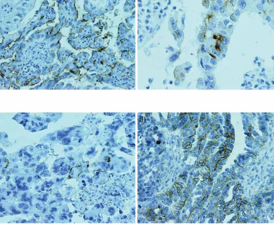
CD133 expression was evaluated in a series of 64 high grade serous ovarian carcinoma and their peritoneal me-tastasis. Expression was mainly seen in the apical/endo-luminal cell surface of tumor cells surrounding a lumen (Figure 1a and c). Cytoplasmic CD133 staining was seen in less than 1% of the solidly arranged tumor cells (but not in all positive cases) (Figure 1b). In some positive cases we noticed strongly stained debris in the lumina of the glands lined by malignant epithelium. Tumor samples were positive for CD133 in thirty nine cases (61%), while metastases were positive in twenty six cases (41%). The median of CD133 positive cells in tumors was 1 (0.1–7)%, range 0.1–9%, and in metastases was 0.6 (0.1–6)%, range 0.1–9%. Although we noticed a more positive cases in tu-mor samples, the difference was not statistically signifi -cant (p>0.05). However, this result has shown a certain trend toward signifi cance.
Comparing all clinico-pathological characteristics and the frequency of CD133 positive tumors and CD133 posi-tive metastases, we don’t reach statistical signifi cance (all p>0.05, Table 2).
CD133 expression in tumor and metastasis was classi-fi ed into three levels: negative (0%), 0.1–0.9%, and more than 0.9%. Distribution of CD133 status is shown in Fig-ure 2. We have noticed that percentage of negative cells are higher in metastasis than in primary tumors, in the group of 0.1–0.9% distribution is similar, and in group of higher positivity we have noticed more positive cells in tumor samples. The difference is signifi cant (?2=10.13, p=0.006).
CD117 expression protein in high grade serous
CD117 expression protein in high grade serous
ovarian cancer and peritoneal metastasis
ovarian cancer and peritoneal metastasis
CD117 positive stain appeared as a cytoplasmic and/or membranous stain (Figure 1d). Nonspecifi c background staining of tumor stroma or adjacent ovarian parenchyma was not encountered. Tumor samples were positive in fi fty two (81%) cases, while metastases were positive in forty nine (77%) cases (Table 2). Although we have noticed
Figure 1. Immunoreaction in the high grade serous ovarian cancer and metastasis. a) High CD133 expression in the high grade serous ovarian cancer (at the apical/endoluminal part of tumor cells). Magnifi cationx200. b) CD133 positive reaction in the cytoplasm of the single tumor cells. Magnifi cationx400. c) Low CD133 expression in the peritoneal metastasis (at the apical/
endoluminal part of tumor cells) Magnifi cationx200. d) CD117 positive expression in the primary serous high grade ovarian cancer
a more positive cases in tumor samples, the difference was not statistically signifi cant (p>0.05). The median of CD117 positive cells in tumor samples was 1 (0.1–8)%, range 0.1– 22%, and in metastases was 0.1 (0.1–7)%, range 0.1–33%. Comparing all clinico-pathological characteristics and the frequency of CD117 positive tumors and CD117 posi-tive metastases, the result did not reach statistical sig-nifi cance (all p>0.05, Table 2).
CD117 expression in tumor and metastasis was classi-fi ed in the same way as CD133. Distribution of CD117 status is shown in Figure 3. We have noticed that distribu-tion of cells are similar considering three groups and loca-tion of positive cells, the signifi cance was not reached (c2=1.23, p=0.541).
Prognostic impact of CD133 and CD117 expression
Prognostic impact of CD133 and CD117 expression
We compare the disease free survival and the overall survival according to three levels of expression of CD133 and CD117 in tumor and in metastasis (Table 3; Figure 4,5,6). The median disease free survival, for all cases, was 11 (1–62) months, range 1–79 months. For CD133 higher positive tumors (>0.9% of positive tumor cells) disease free survival was signifi cantly shorter comparing to CD133 negative tumors and CD133 less positive tumors (0.1– 0.9% of positive tumor cells) (p=0.025). The difference in disease free survival between CD133 positive and CD133 negative metastasis was not statstically significant (p=0.514). For CD117 higher positive cases in tumor, dis-ease free survival was signifi cantly shorter comparing to CD117 negative and CD117 less positive tumor cases (p=0.031). The difference in disease free survival between TABLE 2TABLE 2
COMPARISON OF THE CD133 AND CD117 POSITIVITY IN TUMOR AND IN METASTASIS ACCORDING TO CLINICO-PATHOLOGICAL CHARACTERISTICS
Characteristics Positive CD133 Positive CD117
Expression in tumor (n=39) N (%) Expression in metastasis (n=26) N (%) c2 p Expression in tumor (n=52) N (%) Expression in metastasis (n=49) N (%) c2 p Age <65 26 (66.7) 15 (57.7) 0.22 33 (63.5) 31 (63.3) 0.04 ≥65 13 (33.3) 11 (42.3) 0.637 19 (36.5) 18 (36.7) 0.852 Residual tumor Absent 10 (25.6) 7 (26.9) 13 (25.0) 11 (22.4) <1 cm 13 (33.3) 7 (26.9) 0.31 18 (34.6) 19 (38.8) 0.21 ≥1 cm 16 (41.1) 12 (46.2) 0.855 21 (40.4) 19 (38.8) 0.903 Lymph node status n.a. 11 (28.2) 8 (30.8) 15 (28.9) 13 (26.5) Negative 13 (33.3) 10 (38.4) 0.09 14 (26.9) 11 (22.5) 0.17 Positive 15 (38.5) 8 (30.8) 0.763 23 (44.2) 25 (51.0) 0.683 Response to treatment Yes 22 (56.4) 12 (46.1) 0.32 28 (53.8) 26 (53.1) 0.04 No 17 (43.6) 14 (53.9) 0.574 24 (46.2) 23 (46.9) 0.842 Recurrence Yes 33 (84.6) 22 (84.6) 0.12 46 (88.5) 43 (87.8) 0.01 No 6 (15.4) 4 (15.4) 0.726 6 (11.5) 6 (12.2) 0.927
* indicated statistical signifi cance
Figure 2. Distribution of the CD133 expression in tumor cells and in metastasis, ?2=10.13, p=0.006.
Figure.3. Distribution of the CD117 expression in tumor cells and in metastasis, c2=1.23, p=0.541.
TABLE 3 TABLE 3
COMPARISON OF DISEASE FREE SURVIVAL/OVERALL SURVIVAL ACCORDING TO EXPRESSION OF CD133 AND CD117
IN TUMOR(T) AND IN METASTASIS(M)
Disease free survival /months Overall survival / months N Median (5th–95th) percentile Median (5th–95th) percentile CD133 (T) negative 24 14 (6–73) 34 (10–90) 0.1–0.9% 7 12 (1–25) 27 (1–74) >0.9% 27 9 (1–25) 19 (1–55) p 0.025* 0.014* CD133 (M) negative 35 12 (1–73) 30 (1–74) 0.1–0.9% 12 9 (1–30) 16 (1–65) >0.9% 11 13 (6–63) 23 (9–90) p 0.514 0.190 CD117 (T) negative 11 19 (8–79) 36 (9–96) 0.1–0.9% 21 12 (7–60) 27 (10–73) >0.9% 26 9 (1–30) 19 (1–65) p 0.031* 0.069 CD117 (M) negative 14 13 (7–79) 32 (9–96) 0.1–0.9% 23 10 (6–27) 23 (10–74) >0.9% 21 9 (1–36) 18 (1–64) p 0.140 0.055
*indicated statistical signifi cante difference CD117 positive and CD117 negative metastasis was not
statstically signifi cant (p=0.140).
The median overall survival was 26 (1–73) months, range 1–96 months. For CD133 higher positive tumors the median overall survival was signifi cantly shorter (19 months), comparing to CD133 negative and CD133 less positive cases (34 months and 27 months, respectively) (p=0.014). There were no signifi cant differences in overall survival according to CD133 status in metastases; the signifi cance was not reached (P=0.190). Also, there were no signifi cant differences in overall survival according the CD117 status in tumor samples and in metastases (p=0.069 and p=0.055, respectively).
Table 4. shows the results of Cox´s proportional haz-ards model. Comparing all clinico-pathological character-istics and examined proteins expression, the postoperative survival was signifi cantly related to expression of CD133 in tumor (p=0.024), response to treatment (p< 0.001), and recurrence of the disease (p< 0.001). We can identify ex-pression of CD133 protein in tumor as independent prog-nostic factor calculated by multivariate analysis.
Discussion and Conclusion
Discussion and Conclusion
Our study examined the possible clinical role of the immunohistochemically assessed expression of two poten-tial CSC markers, CD133 and CD117, on samples of high grade serous ovarian cancer and their peritoneal metas-tases. To our knowledge, there is no data available about similar research.
Ovarian cancers are extremely heterogeneous, and this may suggest that different populations of CSCs can be responsible for tumorigenicity in different histological subtypes, although it is possible that several markers are expressed by the same population of ovarian CSCs7,37.So, we analyzed only high grade serous ovarian tumors for better correlation of the results. We can assume that same populations of the putative CSCs can express several dif-ferent markers. Other authors considered that some over-lap of the potential stem cell marker can occur25,28. CD133
Figure 5. Disease free survival curves according to status of CD117 (T) expression, p=0.031.
Figure 6. Overall survival curves according to status of CD133 (T) expression, p=0.014.
TABLE 4 TABLE 4
MULTIVARIATE ANALYSIS ASSESING THE SURVIVAL WITH PROGNOSTIC FACTORS (COX PROPORTIONAL HAZARD REGRESSION)
Variables b SE P HR 95% CI za HR CD133 (T) 0.337 0.106 0.024* 1.33 1.03–1.98 CD133 (M) 0.122 0.132 0.346 0.89 0.53–1.32 CD117 (T) 0.039 0.113 0.716 1.04 0.84–1.27 CD117 (M) 0.034 0.053 0.072 1.02 0.98–1.04 Residual tumor 0.118 0.152 0.292 0.92 0.58–1.27
Lymph node status 0.124 0.386 0.840 1.22 0.56–2.32
Reccurence of dis. –0.055 0.022 <0.001* 0.96 0.92–0.97
Response to treatm. 0.564 0.099 <0.001* 1.82 1.42–2.12
* indicated statistical signifi cance
(b- regression coeffi ccient, SE- Standard Error, HR- Hazard Ratio) seen mainly at the apical/endoluminal surface, and less than 1% of the positive malignant cells showed diffuse cytoplasmic pattern. Immunohistochemically determined CD133 positivity expressed at the apical/endoluminal sur-face was also noticed in samples from pancreatic ductal adenocarcinomas15, and ovarian carcinoma36, while Zhang et al. in ovarian cancer samples observed predominantly membrane/cytoplasmic expression18. CD117 positivity was noticed as a cytoplasmic and/or membranous stain in tu-mor and in metastasis samples. Our observation is consis-tent with data from the literature27,29.
CD133 positivity was seen in the 61% of tumors. Mae-da et al. presents a similar results (60%)21. In other stud-ies authors noticed higher (80%)15,19, or lower number of positive cases (31%)36,18, so the rank of positivity in the literature is quite wide. CD117 positivity was noticed in our study in high number of tumors, 81%, and our results are similar to those obtained in the literature14,35. Some
authors noticed lower number of CD117 positive cases (40% and 52%, respectively)27,29.
The CSCs provide a model of the cancer metastasis in which these cells are able to colonize, expand, and differ-entiate into heterogeneous tumor phenotypes similar to primary tumors38. Both, the primary tumors and the me-tastases would display similar genetic and expression profi les because both populations are supposedly derived from the same lineage of cancer stem cells32. Our results are in the agreement with this hypothesis, because we have found expression of both markers in the samples from tumor and from metastasis. The number of CD133 positive cases was somewhat higher in tumor than in metastasis, but the difference was not statistically signifi cant. The same results we obtanied for CD117. In the literature we did not fi nd the data for the immunohistochemically as-sessed expression of the CD133 and CD117 for metastasis samples.
In our study, the median of CD133 positive cells in tumor specimens was 1 (0.1–7)%, and in metastasis spec-imens was 0.6 (0.1–6) %. The median of CD117 positive cells in tumors was 1 (0.1–8)%, and in metastasis was 0.1 (0.1–6)%. These results indicate that CD133 and CD117 could represent a possible marker for CSCs, because CSCs are only a very small portion, less than 5%, of the total tumor cell population.
The percentage of CD133 expressing cells was signifi -cantly higher in primary ovarian cancer than in perito-neal metastases. Distribution of CD117 positive cells in tumor samples and in metastases is similar to CD133 dis-tribution; we noticed higher percentage of CD117 positive cells in tumor samples, but signifi cance is not reached. This observation may have several reasons. There are studies that suggest that two forms of CSCs exists: stacio-nary CSCs which are incorporated in tumor and persists in all steps of tumor progression; the other form are mobile CSCs that represent smaller pool of tumor cells. They are located at the tumor-host interface and they are capable to disseminate and form metastatic colonies39. Also, we can assume that metastasis in peritoneum cavity has dif-ferent microenvironment than primary tumor in the ova-ry and these different conditions may altered expression of potential CSCs marker. Bignotti et al. detected 156 genes that were expressed differentially between ovarian serous papillary carcinomas and omental metastases40. Our result is with accordance with one found in the lit-erature33. Ferrandina et al. used FACS (fl uorescence acti-vated cell sorting) for determining the percentages of CD133-1 and CD133-2 in ovarian carcinoma samples and in omental metastases. Both the percentages of CD133-1 and CD133-2 expressing cells were signifi cantly lower in omental metastases. Although the two different methods are used for assessment of positive cells (FACS versus immunohistochemistry) similar results were obtained in both studies.
Multivariate analysis showed that tumor expression of both markers has statistical signifi cance according to
dis-ease free survival. For overall survival only CD133 ex-pression in tumor samples reached the signifi cance. The expression of both markers in metastasis samples didn`t show correlation to prognosis.
Our immunohistochemical results show that expres-sion of CD133 in tumor samples best correlate with prog-nosis. Expression of both markers in metastasis samples has no prognostic signifi cance. Similar results for tumor expression were obtained by other authors18,20,21. Ferran-dina et al. found that CD133 expression not provide ad-ditional prognostic information for ovarian cancer patients at all36. But, in the large study (400 ovarian cancer pa-tients) Zang et al. found an association between CD133 status and prognosis18. CD133 is possibly one of the most reliable molecular marker for cancer stem cells in various solid tumors18, and CD117 has only recently been regarded as a possible CSCs surface marker29.
Some studies reported that CSCs phenotype was more resistant to platinum-based therapy, which supports the idea that CSCs may be responsible for chemoresistance of ovarian tumors and serve as chemotherapeutic targets for reducing disease recurrence24,25. It was hypothesized that CD133 and CD117 are putative marker for CSCs and de-fi ning the methods to identify and isolate CD133 and CD117 positive cells in tumor can help adapting the ther-apy to individual cases or possibly developing targeted therapy in the future.
Conclusion
Conclusion
Our study indicates that the immunohistochemical as-sessment of CD133 and CD117 expression may have po-tential clinical value in predicting disease progression or prognosis in high grade serous ovarian cancer patients. According to our results, CD133 proved to be an indepen-dent prognostic factor in high grade serous ovarian cancer patients.
R E F E R E N C E S R E F E R E N C E S
1. BAPAT SA, MALI AM, KOPPIKAR CB, KURREY NK, Cancer Res, 65 (2005) 3025. — 2. SOLJAČIĆ VRANEŠ H, KLARIĆ P, SONICKI Z, GALL V, JUKIĆ M, VUKOVIĆ A, Coll Antropol, 35 (2011) 775. — 3. MENG E, LONG B, SULLIVAN P, MCCLELLAN S, FINAN MA, REED E, SHEVDE L, ROCCONI RP, Clin Exp Metastasis (2012) DOI:10.1007/ s10585-012-9482-4. — 4. SEIDMAN JD, CHO KR, RONNETT BM, KURMAN RJ, Surface epithelial tumors oft he ovary. In: KURMAN JR, HEDRICK ELLENSON L, RONNETT BM (Eds) Blaustein´s pathology of the female genital tract (Springer, New York, 2011). DOI:10.1007/978-1-4419-0489-8. — 5. SOLJAČIĆ VRANEŠ H, KLARIĆ P, KUNA K, KRALJEVIĆ Z, GALL V, JUKIĆ M, Coll Antropol, 36 (2012) 425. — 6. CLARKE MF, DICK JE, DIRKS PB, EAVES CJ, JAMIESON CHM, LEANNE JONES D, VISVADER J, WEISSMAN IL, WAHL GM, Cancer Res, 66 (2006) 9339. — 7. DYALL S, GAYTHER SA, DAFOU D, J Oncol (2010) DOI: 10.1155/2010/105269. — 8. REDŽIĆ A, SMAJILAGIĆ A, ALJIČEVIĆ M, BERBEROVIĆ LJ, Coll Antropol, 34 (2010) 1405. — 9. RAOS M, NEMET D, BOJANIĆ I, SERTIĆ D, BATINIĆ D, DUŠAK V, DUBRAVČIĆ K, MAZIĆ S, SERVENTI-SEIWERTH R, MRSIĆ M, GOLUBIĆ-ĆEPULIĆ B, LABAR B, Coll antropol, 34 (2010) 105. — 10. ALISON MR, ISLAM S, J Pathol, 217 (2009) 144. — 11. ALISON MR,
LIM SML, NICHOLSON LJ, J Pathol, 223 (2011) 147. — 12. DICK JE, BHATIA M, GAN O, KAPP U, WANG JCY, Stem Cells, 15 (1997) 199. — 13. LOPEZ JI, CAMENISCH TD, STEVENS MV, SANDS BJ, MC-DONALD J, SCHROEDER A, Cancer Res, 65 (2005) 6755. — 14. BUT-NOR KJ, BRUCHETTE JL, SPORN TA, HAMMAR SP, ROGGLI VL, Arch Pathol Lab Med, 128 (2004) 538. — 15. IMMERVOLL H, HOEM D, SAKARIASSEN PO, STEFFENSEN OJ, MOLVEN A, BMC Cancer, (2008) DOI:10.1186/1471-2407-8-48. — 16. LUGLI A, IEZZI G, HOSTET-TLER I, MURARO MG, MELE V, TORNILLO L, CARAFA V, SPAG-NOLI G, TERRACCIANO L, ZLOBEC I, Br J Cancer, 103 (2010) 382. — 17. ICKOWSKI KA, Am J Transl Res, 3 (2011) 1. — 18. ZHANG J, GUO X, YOUNG CHANG D, ROSEN DG, MERCADO-URIBE I, LIU J, Modern Pathol, 25 (2012). — 19. LEELAVAT K, THONGTAWEE, NARONG S, SUBWONGCHAROEN S, TREEPONGKARUNA S, World J Gastroenterol, 17 (2011) 1192. — 20. ZEPPERNICK F, AHMADI R, CAMPOS B, DICTUS C, HELMKE BM, BECKER N, LICHTER P, UN-TERBERG A, RADLWIMMER B, HEROLD-MENDE CC, Clin Cancer Res, 14 (2008 123. — 21. MAEDA S, SHINCHI, KURAHARA H, MAT-AKI Y, MAEMURA K, SATO M, NATSUGOE S, AIKOU T, TAKAO S, Br J Cancer, 98 (2008) 1389. — 22. MIKI J, FURUSATO B, LI H, GU Y,
TAKAHASHI H, EGAWA S, SESTERHENN IA, MCLEOD DG, SRIV-ASTAVA S, RHIM JS, Cancer Res, 67 (2007) 3153. — 23. LEVINA V, MARRANGONI AM, DEMARCO R, GORELIK E, LOKSHIN AE, PloS One, (2008) DOI:10.1371/journal.pone.0003077. — 24. ZHANG S, BALCH C, CHAN MW, Cancer Res, 68 (2008) 4311. — 25. BABA T, CONVERY PA, MATSUMARA N, WHITAKER RS, KONDOH E, PER-RY T, HUANG Z, BENTLEY RC, MORI S, FUJII S, MARKS JR, BER-CHUCK A, MURPHY SK, Oncogene, 28 (2009) 209-218. — 26. STEF-FENSEN KD, ALVERO AB, YANG Y, WALDSTROM M, HUI P, HOLMBERG JC, SILASI DA, JAKOBSEN A, RUTHERFORD T, MOR G, J Oncol (2011) DOI:10.1155/2011/620523. — 27. RASPOLLINI MR, AMUNNI G, VILLANUCCI A, BARONI G, TADDEI A, TADDEI GL, Annals of Oncology, 15 (2004) 594. — 28. CURLEY M, THERRIEN VA, CUMMINGS CL, Stem Cells, 27 (2009) 2975. — 29. LUO L, ZENG J, LIANG B, ZHAO Z, SUN L, CAO D, YANG J, SHEN K, Exp Mol Pathol, 91 (2011) 598. — 30. MIZRAK D, BRITTAN M, ALISON MR, J Pathol, 214 (2008) 3. — 31. HU Y, FU L, Am J Cancer Res, 2 (2012) 330. — 32.
KUSUMBE AP, MALI AM, BAPAT SA, Stem Cells, 27 (2009) 498. — 33. FERRANDINA G, BONANNO G, PIERELLI L, PERILLO A, PRO-COLI A, MARIOTTI A, CORALLO M, MARTINELLI E, RUTELLA S, PAGLIA A, ZANNONI G, MANCUSO S, SCAMBIO G, Int J Gynecol Cancer, 18 (2008) 506. — 34. CHOI YP, SHIM HS, GAO MQ, KANG S, CHO NH, Cancer Lett, 307 (2011) 62. — 35. ARBER DA, TAMAYO R, WEISS LM, Hum Pathol, 29 (1998) 498. — 36. FERRANDINA G, MAR-TINELLI E, PETRILLO M, PRISCO MG, ZANNONI G, SIOLETIC S, SCAMBIA G, BMC Cancer, (2009) CD133 DOI:10.1186/1471-2407-9-221. — 37. FOSTER R, BUCHANOVICH RJ, RUEDA BR, Cancer Lett, (2012), Available from:http://dx.doi.org/10.1016/j.canlet.2012.10.023. — 38. PONNUSAMY MP, BATRA SK, J Ovarian Res, 12 (2008) 1. — 39. BRABLETZ T, JUNG A, SPADERNA S, HLUBEK F, KIRCHNER T, Nat Rev Cancer 5 (2005) 744. — 40. BIGNOTTI E, TASSI RA, CALZA S, RAVAGGI A, BANDIERA E, ROSSI E, DONZELLI C, PASINETTI B, PECORELLI S, SANTIN AD, Am J Obstet Gynecol, 196 (2007) 245. DOI:10.1016/j.ajog.2006.10.874.
S. Štemberger-Papić
University of Rijeka, University Hospital Centre Rijeka, Department of Clinical Cytology, Cambierijeva 17, 51000 Rijeka, Croatia
e-mail: snjezana.stemberger@ri.t-com.hr
IZRAŽAJ BILJEGA CD133 I CD117 U 64 BOLESNICE SA SEROZNIM RAKOM JAJNIKA IZRAŽAJ BILJEGA CD133 I CD117 U 64 BOLESNICE SA SEROZNIM RAKOM JAJNIKA S A Ž E T A K
S A Ž E T A K
Tumorske matične stanice (cancer stem cells-CSCs) predstavljaju manji dio stanica u tumoru koje se mogu samoob-navljati i proliferirati, te su vjerojatno odgovorne za početak i održavanje tumorskog rasta. CD133 i CD117 su najčešće korišteni biljezi za potencijalne CSCs, osobito za CSCs jajnika, no njihov klinički značaj još uvijek je nedovoljno istražen. Cilj ovog istraživanja bio je usporediti imunohistokemijski izražaj biljega CD133 i CD117 kod 64 slučaja primarnog se-roznog raka jajnika visokog gradusa i pripadajućih peritonealnih metastaza, te ispitati njihovu potencijalnu kliničku ulogu. Izražaj biljega CD133 uočen je u apikalnim/ endoluminalnim dijelovima tumorskih stanica, u 61% tumora i 41% metastaza. Medijan CD133 pozitivnih stanica u tumorima bio je 1 (0,1–7)%, a u metastazama 0,6 (0,1–6)%. Izražaj biljega CD117 uočen je kao citoplazmatsko i/ili membransko bojenje, a pronađeno je u 81% tumora i 77% metastaza. Medijan CD117 pozitivnih stanica u tumorima bio je 1 (0,1–8)%, a u metastazama 0,1 (0,1–6)%. Multivarijatna analiza pokazala je da bolesnice s jačim izražajem biljega CD133 u tumoru imaju statistički značajno kraće vrijeme do pojave recidiva i vrijeme preživljenja (p=0,025 odnosno p=0,014). Bolsnice s jačim izražajem biljega CD117 u tumoru imaju statistički značajno kraće vrijeme do pojave recidiva bolesti (P = 0,031). Cox-ovom regresijskom metodom izražaj biljega CD133 u tumoru uočen je kao nezavisni prognostički čimbenik. Naša studija pokazuje da imunohistokemijski izražaj biljega CD133 i CD117 može imati potencijalnu kliničku vrijednost u predviđanju progresije bolesti i prognoze kod sero-znog raka jajnika visokog gradusa. Izražaj biljega CD133 je nezavisni prognostički čimbenik kod pacijenata sa seroznim rakom jajnika visokog gradusa.