Original Citation:
Minimally invasive implantation of continuous flow left ventricular assist devices: the evolution of surgical
techniques in a single centre experience
Publisher:
Published version: DOI:
Terms of use: Open Access
(Article begins on next page)
This article is made available under terms and conditions applicable to Open Access Guidelines, as described at http://www.unipd.it/download/file/fid/55401 (Italian only)
Availability:
This version is available at: 11577/3275355 since: 2018-09-24T09:46:47Z
10.1111/aor.13339
Università degli Studi di Padova
Accepted
Article
This article has been accepted for publication and undergone full peer review but has not
been through the copyediting, typesetting, pagination and proofreading process, which may
lead to differences between this version and the Version of Record. Please cite this article as
doi: 10.1111/aor.13339
DR. MASSIMILIANO CARROZZINI (Orcid ID : 0000-0001-5506-0201) DR. JONIDA BEJKO (Orcid ID : 0000-0001-5015-4823)
Article type : Thoughts & Progress
Title
Minimally invasive implantation of continuous flow left ventricular assist devices: the evolution of surgical techniques in a single centre experience
Authors
Massimiliano Carrozzini, MD1*; Jonida Bejko, MD1*; Alvise Guariento, MD1; Maurizio Rubino, MD1; Roberto Bianco, MD1; Vincenzo Tarzia, MD1; Dario Gregori, PhD2; Tomaso Bottio, MD1; Gino Gerosa, MD1;
*These authors equally contributed to this work
Affiliations
1 Cardiac Surgery Unit, Department of Cardiac, Thoracic and Vascular Sciences, University of Padova, Padova, Italy
2 Unit of Biostatistics, Epidemiology and Public Health, Department of Cardiac, Thoracic and Vascular Sciences, University of Padova, Italy
Accepted
Article
Corresponding author Massimiliano Carrozzini
Cardiac Surgery Unit, Department of Cardiac, Thoracic and Vascular Sciences, University of Padova. Via Giustiniani 2, 35100 Padova, Italy.
Tel: +39-049-8212428; fax: +39-049-8212409 E-mail: [email protected]
Author contributions
- Carrozzini M: concept/design; data collection; data analysis; data interpretation; drafting article; critical revision of article; approval of article
- Bejko J: concept/design; data collection; data interpretation; drafting article; critical revision of article; approval of article
- Guariento A: drafting article; approval of article - Rubino M: approval of article
- Bianco R: approval of article - Tarzia V: approval of article
- Gregori D: data analysis; statistics; critical revision of article; approval of article
- Bottio T: concept/design; data interpretation; drafting article; critical revision of article; approval of article
- Gerosa G: critical revision of article; approval of article Received: April 5, 2018
Revised: July 20, 2018
Abstract Objective
To evaluate the evolution of our surgical experience with the implantation of a continuous flow left ventricular assist device (LVAD), from the original full sternotomy approach to less invasive surgical strategies including mini-sternotomy and/or mini-thoracotomies.
Accepted
Article
Methods
We reviewed all consecutive patients implanted with a continuous flow LVAD at our Institute. To exclude the possible bias related to the device used, out of 91 collected LVADs implants, we selected only those patients (n=42) who received, between 2012 and 2015, the HeartWare HVAD. The analysis focused on the surgical approach used for the LVAD implant. Most of the patients (95%) were affected by dilated or ischemic cardiomyopathy, with an INTERMACS class I-II in the majority of cases (86%).
Results
The LVAD implant was performed through a full sternotomy in 10 patients (24%); the remaining 32 cases (76%) were managed with minimally invasive procedures. These were left mini-thoracotomy with upper mini-sternotomy (20 patients, 62%), right and left mini-thoracotomy (7 patients, 22%) and a recently developed left mini-thoracotomy with outflow graft anastomosis to the left axillary artery (5 patients, 16%). The most common adverse event on device was right heart failure (26%). Eighteen patients (43%) were transplanted. Overall estimated 24 months survival (on device or after transplant) was 68±7%. The causal analysis, adjusted by propensity score weighting baseline data and sample size, showed that left mini-thoracotomy with outflow anastomosis to the left axillary artery resulted in a significantly reduced rate of post implant right heart failure (p<0.01), and mechanical ventilation time (p=0.049).
Conclusions
In our series, by applying mini-invasive implant techniques in the majority of cases, mid-term survival of continuous flow LVADs in severely compromised patients was satisfactory. In the adjusted analysis, the left anterior mini-thoracotomy with outflow anastomosis to the left axillary artery showed the most favourable results.
Keywords: Left Ventricular Assist Device; HeartWare; HVAD; minimally invasive; implant; surgical technique
Accepted
Article
Main text Introduction
During the last decades, left ventricular assist devices (LVADs) have become a viable therapeutic option for patients with end-stage heart failure1–4. First generation LVADs were bulky and space-demanding devices; thus, in the early era, the standard surgical approach for LVADs implantation was limited to the full median sternotomy5. Subsequent improvements in pump technology and design, with a progressive miniaturization of the devices, enhanced their performance and reliability and allowed the development of less invasive implantation strategies. Several minimally invasive techniques have been, so far, proposed, such as left thoracotomy combined with an upper mini-sternotomy, or combination of right and left mini-thoracotomies6–8. Since this is a novel and evolving field, the results of minimally invasive procedure for LVAD implant are still under investigation. Here we describe these techniques and focus on the latest developed surgical approach, consisting in a left mini-thoracotomy for exposure of the left ventricular apex combined with the left axillary artery surgical isolation, where the outflow graft anastomosis is performed. The aim was to analyse and compare the results of the standard sternotomy approach and of the different minimally invasive implant techniques.
Materials and Methods
Study design and patient population
This is a single-centre retrospective study on prospectively collected data. We reviewed all consecutive patients who received a continuous flow LVAD at our Institution from 2008 to 2015. During this period, we implanted 91 patients: 49 (54%) received the Jarvik 2000 (Jarvik Heart, Inc., Manhattan, NY, USA), and 42 (46%) the HeartWare HVAD (HeartWare Inc., Framingham, MA). To exclude a possible bias related to the device used, we selected for the analysis only the HeartWare HVAD patients. These were implanted between 2012 and 2015, when our expertise in the management of mechanical circulatory supports was already established. The study was approved by the Institutional Review Board; an individual patient consent was waived.
Accepted
Article
Adverse events on device were classified according to the INTERMACS definitions1.
Surgical technique
Here we systematically describe the possible surgical approaches for LVADs implantation, focusing on tricks and traps of minimally invasive techniques. These consist in the left anterior mini-thoracotomy (LMT) for left ventricle apex exposure, combined with one of three possibilities: 1) the upper mini-sternotomy (UMS); 2) the right anterior mini-thoracotomy (RMT); 3) the left axillary artery isolation (LAA). The first two options entail performing the outflow graft anastomosis at the ascending aorta, whereas in the last case the outflow is connected to the left axillary artery (Figure 1). In our experience, the first 10 cases (24%) of HVAD implantation were performed through a full median sternotomy. Thereafter, the path evolved towards less invasive approaches and the LMT was combined with UMS in 20 patients (48%), with RMT in 7 patients (17%), and with LAA in 5 patients (12%).
When a minimally invasive approach is employed, the first surgical step is the execution of the LMT with left ventricle apex exposure and positioning of the sewing ring. Subsequently, the UMS or the RMT is performed, the LVAD inflow is inserted into the left ventricle and the outflow graft is tunnelled through the pericardium and anastomosed to the ascending aorta. Those cases managed with left axillary artery isolation enable a different surgical plan, which will be described later in the dedicated part.
Left anterior mini-thoracotomy
The patient is placed in supine position and a pillow or a gel pad is used to elevate the chest at the interscapular level, as for a sternotomy access. The skin incision is performed just below the areola in men, and at the inframammary groove in women. The intercostal space to access is chosen depending on the position of the apex (usually it corresponds to the fifth intercostal space), which can be identified by palpation or with the aid of echocardiography. Once the pericardium is exposed and opened, the left ventricular apex is available without any further heart manipulation. The correct
Accepted
Article
insertion site of the LVAD is localized on the left ventricular wall by echocardiographic assessment, using a finger pushed on the apex to simulate the inflow cannula. A dermographic pen can be used to mark this site. The sewing ring is, then, secured with interrupted pledgetted 2-0 polypropylene sutures.
Upper mini-sternotomy
An inverted T-shape mini-sternotomy is performed by sawing the sternal manubrium only or together with a part of the sternal body, up to the third-forth intercostal space. According to the extension of the mini-sternotomy, a different section of the ascending aorta is exposed. When the sternum is divided up to the fourth intercostal space, the anastomosis of the outflow graft will correspond to the anterior face of the aorta, near the sino-tubular junction. In the case the sternum is split up to the third intercostal space, the anastomosis will be performed at the front face of the ascending aorta in an intermediate position. In cases of isolated sternal manubrium division, the anastomosis will be performed in the front position of the aorta, but very close to the emergence of the brachiocephalic artery. The outflow vascular graft is tunnelled underneath the pericardium from the LMT to the UMS and is accurately arranged and measured in order to cut at the right length, without kinking or overstretching it.
Right anterior mini-thoracotomy
The LVAD implantation through a RMTstarts with an incision at the second right intercostal space. The incision, of almost 4-5 cm, is performed proximally to the body of the sternum with a lateral direction, along the correspondent intercostal space. Once opened the pericardium longitudinally, and suspended to the skin in its proximal (next to the first operator) edge, in the foreground appears the ascending aorta, on a lower level lies the superior vena cava, while the right atrial appendage extends to the most proximal part of the aorta. The pulmonary artery, instead, runs much deeper and medial in the surgical field. When the implantation of a right ventricular assist device (RVAD) is required, the only way to expose it is to release the proximal pericardial suspension and to pull on the distal
Accepted
Article
pericardial edge until a slight twisting of great vessels axis is achieved, to get the pulmonary artery in a higher plane respect to the aorta.
After great vessels exposure, the outflow graft is tunnelled through the LMT underneath the pericardium, using a blunt-tip instrument, and is stretched to the RMT. The vascular graft is anastomosed, then, to the lateral face of the ascending aorta, in its intermediate portion.
Left axillary artery outflow graft anastomosis
So far, this implant technique exclusively applies to the HVAD, due to its peculiar outflow graft size (10 mm of diameter). Other currently available devices provide a larger vascular prosthesis, which are incompatible with the usual size of the axillary artery.
This approach enables a different surgical plan: after LMT, the axillary artery isolation and the outflow graft anastomosis can be performed first, followed by the connection to the LVAD and its insertion into the left ventricular apex in a second step.
There are two possible surgical incision: sub-clavicular incision and delto-pectoral incision.
The sub-clavicular incisionis performed through a 6-8 cm incision below and parallel to the lateral two-thirds of the clavicle. The axillary artery, in its proximal portion, is located just below the vein, and, once circled, it can easily be mobilized and externalized.
A further approach to the axillary artery is the so-called delto-pectoral incision, which allows access to the second (median) portion of the vessel. This more peripheral surgical approach is preferred in the presence of a defibrillator in the sub-clavicular position. The skin incision of nearly 5 cm is performed in the delto-pectoral groove, along the line that connects the midclavicular line with the point of intersection between the anterior margin of the deltoid and the side edge of the biceps muscle. In this position, the median nerve crosses the artery, while the vein remains anteromedial. The axillary
Accepted
Article
artery is exposed by inferiorly mobilizing the nerve. Particular attention must be paid, to respect the nerve structures during both artery dissection and vascular clamp placement.
Generally, the calibre of the axillary artery is less than 8 mm, while the diameter of HVAD vascular prosthesis is 10 mm. According to the dimension of the artery, we apply different surgical procedures to perform the outflow graft anastomosis (Figure 2), as follows:
A. Axillary artery diameter >8 mm
After placing the vascular clamp proximally and distally, the artery is cut crosswise. The proximal portion of the axillary artery is end-to-end anastomosed to the HVAD vascular prosthesis with a 5-0 polypropylene suture. Thereafter, the distal portion of the axillary artery is end-to-side anastomosed to the vascular prosthesis (Figure 2 – A).
B. Axillary artery diameter <8 mm
This is the more common case, which we manage by interposing a 2-2.5 cm length of vascular prosthesis of 8 mm in diameter to the axillary artery, to create a prosthetic bridge along its course. After clamping, the axillary artery is transversely interrupted, and the vascular prosthesis is end-to-end anastomosed proximally and distally, using 5-0 polypropylene sutures. An incision is, then, performed on the interposition graft, along the long axis of the vessel and the LVAD outflow graft is end-to-side connected with it (Figure 2 – B).
The distal portion of the axillary artery is banded, to avoid a preferential flow to the left arm. Once the LVAD outflow is anastomosed, it has to be tunnelled within the chest. In both types of incision, we choose the first intercostal space as entry point. Through the same access used for the graft anastomosis, with the aid of a retractor, the muscle pectoralis major is gently mobilized to expose the first intercostal space. An umbilical tape is introduced from the LMT through this space. At this point, an armed Tex vascular prosthesis is tunnelled through the thorax following the tape. The Gore-Tex prosthesis stands for protection of the outflow graft along the intrathoracic course, and in the way-out of the chest through the intercostal space. The outflow graft is tunnelled inside the Gore-Tex prosthesis and, then, connected to the LVAD.
Accepted
Article
Statistical analysis
Continuous data are presented as median and interquartile range (IQR); discrete variables are expressed as number and percentages. Categorical variables were compared with the chi-square test or the Fisher’s exact test, when appropriate. Continuous variables were compared with the t test or by one-way analysis of variance (to compare multiple groups). Survival curves were drawn by competing risk analysis. Statistical significance was set at a p-value <0.05.
A causal analysis was conducted to estimate the effect of surgical access on several outcomes. Propensity score analysis was targeted to the average treatment effect on the population (ATE), which answers the question of how, on average, the outcome of interest would change if everyone in the population had been assigned to a particular treatment rather than all receiving another single treatment. Propensity weights have been estimated by Generalized Boosted Models (GBM)9 using sex, age at implant, INTERMACS profile, primary cardiac diagnosis, glomerular filtration rate (GFR), serum bilirubin, tricuspid annular plane systolic excursion (TAPSE) at echocardiogram, pre-operative ECMO implantation and follow-up time as covariates. GBM has been implemented as a tree-based regression model built in an iterative fashion (10000 iterations each). Kolmogorov-Smirnov (KS) statistics was used as stopping rule and balance measure and its maximum for summarizing across balance metrics. Causal effect of surgical access on outcomes was estimated by propensity score-adjusted Generalized Linear Models. Quasi-binomial models have been used for right heart failure (severe-acute and severe), major bleeding, 30-days mortality and presence of cerebral fatal and non-fatal events (ischemic and haemorrhagic). For talking skewness in the data distribution, GLM for mechanical ventilation time, ICU stay and in-hospital stay was built using a Gamma with log-link function.
The statistical analysis was performed using the “R” system10.
Accepted
Article
Results
Between 2012 and 2015, 42 patients underwent HVAD implantation at our Institution. Median age at implant was 52 years (IQR: 40-58). Baseline patient’s characteristics are summarized in Table 1. Most of the cases were in INTERMACS profiles I-II (86%). Overall, 8 (19%) patients required preoperative support with intra-aortic balloon pump (IABP), 16 (38%) were on extracorporeal membrane oxygenation (ECMO) and 4 (10%) on para-corporeal LVAD. Patients implanted through different surgical approaches had comparable baseline characteristics (Table 1). Fourteen patients (44%) received off-pump and 12 (38%) on-ECMO implantation of LVAD in the mini-invasive group. On the contrary, all full sternotomy cases were managed with the aid of cardiopulmonary bypass, except for one case of on-ECMO implantation.
Post implant outcomes overall and by surgical approach adopted are detailed in Table 2. Right heart failure (severe-acute and/or severe) occurred in 11 cases (26%) and was the most common adverse event on device, considering both at implant and surveillance periods. Among these, 8 (19%) patients experienced sever-acute RHF, requiring RVAD implant. Thirty-day mortality was 17 % (n=7); other six deaths occurred during a median follow-up of 22 months (IQR 7-32), thus, the overall observed mortality was 31% (n=13). Eighteen patients (43%) underwent heart transplant. Estimations of overall survival at 12 and 24 months were 71±7% (95% Confidence Interval – 95% CI: 68-74) and 68±7% (95% CI: 64-72); whereas the probabilities to undergo heart transplant were 37±10% (95% CI: 31-43) and 45±10% (95% CI: 29-61), respectively (Figure 3). Unadjusted univariate analysis of outcomes showed comparable results among patients managed with standard (full sternotomy) or minimally invasive surgical approach (Table 2).
To evaluate the impact of each technique on results, we performed a causal analysis adjusted by propensity score weighting baseline data, follow up time and sample size. Effective Sample Sizes (ESS) were 9.56 for full sternotomy, 18.67 for UMS, 6.99 for RMT and 4.99 for LMT-LAA. Effects of surgical access on outcomes, both crude and after propensity score adjustment, are presented in Table 4. Left anterior mini-thoracotomy combined to left axillary artery isolation showed
Accepted
Article
a significantly reduced rate of post implant right heart failure (severe-acute and/or severe) (p<0.01), and mechanical ventilation time (p=0.049), in the adjusted analysis.
Discussion
In the current era, where the number of available donor hearts is insufficient to meet the needs of patients waiting for heart transplant, the LVAD therapy has increasingly gained relevance worldwide1–3,11. Minimally invasive procedures in cardiac surgery are known to be associated with many favourable effects, such as reduction of blood loss and rate of infections, leading to improved outcomes12. With the reduced profile and dimension of the newest generation devices, the use of minimally invasive techniques is, nowadays, possible even for LVADs implantation6,13–16. Thus, the emerging question is whether these surgical approaches should be always pursued or not17.
Although the full median sternotomy provides the best and easiest access to the heart and the adjacent structures, it has many detrimental effects. Median sternotomy decreases lung volumes and reduces thoracic motion with significantly lowered functional residual capacity and total lung capacity months later. This, combined with a higher postoperative pain, can lead to respiratory failure or a prolonged mechanical ventilation time18. Moreover, the increased surgical trauma exposes the patient to a greater risk of bleeding and infection12. The typical LVAD candidate is physically compromised, fragile, often with impaired pulmonary and renal function19–22 and may not have sufficient resources to tolerate major surgical insults. Therefore, device implant through smaller, less traumatic incisions is a desirable goal, which could help achieving a faster postoperative recovery. Bedridden patients inevitably develop muscle atrophy, which compromises respiratory mechanics and prolong wound healing, while an early implementation of exercise training represent a key therapeutic intervention in frail patients23. Nonetheless, the favourable effects of minimally invasive LVAD implantation can go far beyond a fastened physical recover: some authors showed a decrease in the rate of right heart failure compared to the standard surgical approach24,25. This has been linked to the avoidance of pericardial opening, which could allow for a preservation of right ventricle geometry and, therefore, function26. Another important advantage of mini-invasive techniques is the possibility to implant the
Accepted
Article
device without using the cardiopulmonary bypass27. In fact, the thoracotomy approach to the left ventricle apex allows for the LVAD insertion without heart manipulations, which could cause haemodynamic decline. The avoidance of cardiopulmonary bypass enables a reduction in the heparin dose administered, decreasing the risk of bleeding, and in the inflammatory response, improving the postoperative course24. On the other hand, the minimally invasive techniques for LVADs implantation are complex procedure and, thus, can be applied only in experienced centre, by well-trained teams.
In this study, we reviewed our surgical experience with HeartWare HVAD and described the available techniques for mini-invasive implantation. We first evolved from the full sternotomy to the left anterior mini-thoracotomy combined with the upper mini-sternotomy. This approach allows for an optimal great vessels exposure and enables the possibility of off-pump or on-ECMO implantation. Nevertheless, it imposes a partial sternotomy and do not permit the complete preservation of thoracic physiology and mechanics. For this reason, we moved to the double (right and left) mini-thoracotomy, which has all the advantages of a real sternal-sparing technique. However, in this procedure, the outflow anastomosis to the ascending aorta is more demanding due to the narrowed surgical field; moreover, the graft tunnelling underneath the pericardium can be particularly complicated in presence of a full loaded heart, often preventing the off-pump implantation. Finally, this technique is not able to avoid the re-entry risk of a subsequent heart transplant, due to the outflow graft that crosses the mediastinum. In the last development, we combined the left anterior mini-thoracotomy with the outflow anastomosis to the left axillary artery. This procedure deserves specific considerations. It allows an almost complete preservation of mediastinal structures; this unique feature makes it preferable in re-do patients or in case of calcified ascending aorta, besides the obvious advantage at the heart transplant time. The connection to the left axillary artery presents some technical issues: the calibre of the vessel, usually, does not match with that of the graft; the outflow need to be tunnelled in the thorax and trough an intercostal space, being subject to kinking or compression; the left arm is at risk of overflow. To overcome the first issue, we propose a surgical approach that is dependent on the calibre of the LAA. Rare cases with a vessel diameter >8 mm are managed with end-to-end graft anastomosis and end-to-side connection of distal LAA (Figure 2 – A). Whereas, when the calibre is
Accepted
Article
<8 mm (which is the usual condition), we first interpose a prosthetic bridge along the LAA course, then, we perform an to-side graft anastomosis (Figure 2 – B). Other authors reported a direct end-to-side anastomosis28; we prefer using the prosthetic bridge to expand the lumen of the LAA (up to 8 mm) and avoid a potential narrowing at the anastomosis site. To simplify the procedure and reduce the operative time, the outflow anastomosis can be performed as a free graft, with subsequent tunnelling in the thorax and connection to the LVAD in a second step, though the strain-relief placement through the LMT could result troublesome. The use of a double-lumen endotracheal tube for mechanical ventilation can facilitate this part, allowing for a temporary exclusion of the left lung. To avoid kinking or compression of the outflow graft in its course within the chest and the intercostal space, we cover it with an armed Gore-Tex prosthesis. Finally, to prevent preferential left arm flow, which could cause reduced systemic perfusion and arm swelling, we perform a bending of the LAA distal to the graft anastomosis, guided by a continuous pressure monitoring of both radial arteries. The distal flow in the left arm is assessed after pump start and the bend is tightened until the pressure in both arms is equalized. Another advantage of LAA use is the possibility to fine-tune the deairing phase. After performing the anastomosis of the graft and its connection with the LVAD, a first device deairing is performed retrogradely. Subsequently, the LVAD is inserted in the left ventricle and the system is deaired, by low-speed pump activation, through a fine needle placed distally in the outflow graft. This main, antegrade, deairing is performed keeping a clamp on the LAA proximal to the anastomosis, in order to prevent potential air embolism to the cerebral vessels. Despite the numerous presented pros, the LAA use has also significant cons. First, cases of severe-acute right heart failure, requiring RVAD placement, have not a straightforward management. In these cases, to expose the pulmonary artery, which is not directly accessible, an additional surgical access is required. We encountered this situation in one patient, which we managed by direct cannulation of the pulmonary artery through a small left mini-thoracotomy in the second intercostal space. This approach is relatively simple and guarantees a good exposure of the target vessel, avoiding more invasive procedures; emergent situations can be initially stabilized by peripheral ECMO. Moreover, there are concerns regarding possible LVAD flow alterations due to the anastomosis with the LAA, that could be subject to narrowing when the left arm is extensively elevated or could be at increased risk of graft
Accepted
Article
thrombosis28. No data, so far, support these hypotheses. In our series, we observed an initial restriction of pump flow (to 2.5-3 L/min) that progressively increased to normal values within the first week; no significant arm swelling or neurapraxia was noticed. There were no significant mean arterial pressure differences among the arms after implant or during follow-up. We educated the patients not to overextend (above the head) the left arm and unexpected flow losses were not experienced. This was a minor movement restriction, which did not significantly affect patient’s activities. On the other hand, INTERMACS-defined suspected pump thrombus was detected in one out of five patients and could solve after increasing the anticoagulant and antiplatelet therapy, suggesting that a higher antithrombotic level is advisable in these patients.
Our experience bases on severely compromised patients with an INTERMACS profile I-II in 86% of cases (85% in the minimally invasive group). This makes our series unique and precludes a possible comparison with the present reports in the literature7,13,24,25,28. The number of severe adverse events observed was relatively high. However, it should be considered again the critical status characterizing most of the patients at baseline. Moreover, we reported a cumulative rate of complications that included both at implant and surveillance periods. Anyhow, by applying mini-invasive techniques in the majority of cases (76%) the overall mid-term survival was satisfactory. RHF was the most frequent adverse event and 19% of cases required temporary RVAD implant. Pre-implant right ventricular (RV) function was comparable among patients undergoing different surgical approaches. As a matter of fact, in this study, the implant technique was applied independently of RV function. We investigated the effect on outcomes of the different techniques used by means of propensity weighted causal analysis. In this adjusted comparison, the LMT with outflow anastomosis to LAA resulted in a significant reduction of the rate of right heart failure (INTERMACS-defined, severe-acute and severe) and of mechanical ventilation time. Thus, in our series, the last developed technique was the most effective in improving post implant outcomes. Of course, this result should be taken cautiously, as the small number of cases managed with each procedure limited our analysis, despite the advanced statistical method applied. We believe that, expanding the number of treated patients,
Accepted
Article
the favourable effect of mini-invasive approaches will definitely be highlighted, as seen in other fields of cardiac surgery.
Conclusions
In our series, by applying mini-invasive implant techniques in the majority of cases, mid-term survival of continuous flow LVADs in severely compromised patients was satisfactory. In the adjusted analysis, the left anterior mini-thoracotomy with outflow anastomosis to the left axillary artery showed the most favourable results. We believe that, expanding the experience, mini-invasive procedures will establish as the standard approaches for LVAD implant.
References
1 Kirklin JK, Pagani FD, Kormos RL, Stevenson LW, Blume ED, Myers SL, et al. Eighth annual INTERMACS report: Special focus on framing the impact of adverse events. J Heart Lung Transplant 2017;36:1080–6. https://doi.org/10.1016/j.healun.2017.07.005.
2 Kirklin JK, Cantor R, Mohacsi P, Gummert J, De By T, Hannan MM, et al. First Annual IMACS Report: A global International Society for Heart and Lung Transplantation Registry for Mechanical Circulatory Support. J Heart Lung Transplant Off Publ Int Soc Heart Transplant 2016;35:407–12. https://doi.org/10.1016/j.healun.2016.01.002.
3 de By TMMH, Mohacsi P, Gahl B, Zittermann A, Krabatsch T, Gustafsson F, et al. The European Registry for Patients with Mechanical Circulatory Support (EUROMACS) of the European Association for Cardio-Thoracic Surgery (EACTS): second report. Eur J Cardiothorac Surg 2017. https://doi.org/10.1093/ejcts/ezx320.
4 Aaronson KD, Slaughter MS, Miller LW, McGee EC, Cotts WG, Acker MA, et al. Use of an intrapericardial, continuous-flow, centrifugal pump in patients awaiting heart transplantation.
Circulation 2012;125:3191–200. https://doi.org/10.1161/CIRCULATIONAHA.111.058412. 5 Slaughter MS. Implantation of the HeartWare Left Ventricular Assist Device. Semin Thorac
Accepted
Article
6 Ricklefs M, Hanke JS, Dogan G, Napp LC, Feldmann C, Haverich A, et al. Less Invasive Surgical Approaches for LVAD Implantation -State of the Art-. Semin Thorac Cardiovasc Surg 2018. https://doi.org/10.1053/j.semtcvs.2018.01.002.
7 Haberl T, Riebandt J, Mahr S, Laufer G, Rajek A, Schima H, et al. Viennese approach to minimize the invasiveness of ventricular assist device implantation. Eur J Cardiothorac Surg 2014;46:991– 6. https://doi.org/10.1093/ejcts/ezu051.
8 Bottio T, Bejko J, Guariento A, Tarzia V, Pittarello D, Gerosa G. Bilateral mini-thoracotomy off-pump Jarvik 2000 implantation in regional asymmetric paravertebral analgesia: J Cardiovasc Med
2016;17:160–4. https://doi.org/10.2459/JCM.0000000000000191.
9 McCaffrey DF, Griffin BA, Almirall D, Slaughter ME, Ramchand R, Burgette LF. A tutorial on propensity score estimation for multiple treatments using generalized boosted models. Stat Med
2013;32:3388–414. https://doi.org/10.1002/sim.5753.
10 R Development Core Team. R: a language and evironment for statistical computing. n.d.
11 Lund LH, Edwards LB, Dipchand AI, Goldfarb S, Kucheryavaya AY, Levvey BJ, et al. The Registry of the International Society for Heart and Lung Transplantation: Thirty-third Adult Heart Transplantation Report-2016; Focus Theme: Primary Diagnostic Indications for Transplant. J Heart Lung Transplant Off Publ Int Soc Heart Transplant 2016;35:1158–69.
12 Schmitto JD, Mokashi SA, Cohn LH. Minimally-Invasive Valve Surgery. J Am Coll Cardiol
2010;56:455–62. https://doi.org/10.1016/j.jacc.2010.03.053.
13 Maltais S, Anwer LA, Tchantchaleishvili V, Haglund NA, Dunlay SM, Aaronson KD, et al. Left Lateral Thoracotomy for Centrifugal Continuous-Flow Left Ventricular Assist Device Placement: An Analysis from the Mechanical Circulatory Support Research Network. ASAIO J 2017:1. https://doi.org/10.1097/MAT.0000000000000714.
14 Schmitto JD, Molitoris U, Haverich A, Strueber M. Implantation of a centrifugal pump as a left ventricular assist device through a novel, minimized approach: Upper hemisternotomy combined with anterolateral thoracotomy. J Thorac Cardiovasc Surg 2012;143:511–3. https://doi.org/10.1016/j.jtcvs.2011.07.046.
Accepted
Article
15 Bottio T, Bejko J, Falasco G, Bortolussi G, Gallo M, Tarzia V, et al. Less-invasive off-pump ventricular assist device implantation in regional paravertebral analgesia. J Artif Organs Off J Jpn Soc Artif Organs 2014;17:275–7. https://doi.org/10.1007/s10047-014-0764-2.
16 Bejko J, Pittarello D, Falasco G, Di Gregorio G, Tarzia V, Rizzoli G, et al. A pilot study on the efficacy and safety of a minimally invasive surgical and anesthetic approach for ventricular assist device implantation. Int J Artif Organs 2018;41:28–36. https://doi.org/10.5301/ijao.5000647. 17 Schumer EM, Slaughter MS. The Minimally Invasive Approach to Left Ventricular Assist Device
Implantation: Is Smaller Better? Rev Esp Cardiol Engl Ed 2018;71:2–3. https://doi.org/10.1016/j.rec.2017.08.003.
18 Boezaart AP, Lucas SD, Elliott CE. Paravertebral block: cervical, thoracic, lumbar, and sacral:
Curr Opin Anaesthesiol 2009;22:637–43. https://doi.org/10.1097/ACO.0b013e32832f3277.
19 von Haehling S, Anker SD, Doehner W, Morley JE, Vellas B. Frailty and heart disease. Int J Cardiol 2013;168:1745–7. https://doi.org/10.1016/j.ijcard.2013.07.068.
20 Fulster S, Tacke M, Sandek A, Ebner N, Tschope C, Doehner W, et al. Muscle wasting in patients with chronic heart failure: results from the studies investigating co-morbidities aggravating heart failure (SICA-HF). Eur Heart J 2013;34:512–9. https://doi.org/10.1093/eurheartj/ehs381.
21 Tarzia V, Bortolussi G, Bianco R, Buratto E, Bejko J, Carrozzini M, et al. Extracorporeal life support in cardiogenic shock: Impact of acute versus chronic etiology on outcome. J Thorac Cardiovasc Surg 2015;150:333–40. https://doi.org/10.1016/j.jtcvs.2015.02.043.
22 Carrozzini M, Toto F, Gerosa G, Bottio T. Irreversible cardiac failure with intraventricular thrombosis: A novel technique of paracorporeal biventricular assist device implantation with ventricles excision. J Thorac Cardiovasc Surg 2017. https://doi.org/10.1016/j.jtcvs.2017.11.085. 23 Kato A. Muscle wasting is associated with reduced exercise capacity and advanced disease in
patients with chronic heart failure. Future Cardiol 2013;9:767–70. https://doi.org/10.2217/fca.13.74.
24 Sileshi B, Haglund NA, Davis ME, Tricarico NM, Stulak JM, Khalpey Z, et al. In-hospital outcomes of a minimally invasive off-pump left thoracotomy approach using a centrifugal
Accepted
Article
continuous-flow left ventricular assist device. J Heart Lung Transplant 2015;34:107–12. https://doi.org/10.1016/j.healun.2014.09.023.
25 Strueber M, Meyer AL, Feussner M, Ender J, Correia J-C, Mohr F-W. A minimally invasive off-pump implantation technique for continuous-flow left ventricular assist devices: Early experience.
J Heart Lung Transplant 2014;33:851–6. https://doi.org/10.1016/j.healun.2014.05.016.
26 Unsworth B, Casula RP, Kyriacou AA, Yadav H, Chukwuemeka A, Cherian A, et al. The right ventricular annular velocity reduction caused by coronary artery bypass graft surgery occurs at the moment of pericardial incision. Am Heart J 2010;159:314–22. https://doi.org/10.1016/j.ahj.2009.11.013.
27 Cheung A, Lamarche Y, Kaan A, Munt B, Doyle A, Bashir J, et al. Off-Pump Implantation of the HeartWare HVAD Left Ventricular Assist Device Through Minimally Invasive Incisions. Ann Thorac Surg 2011;91:1294–6. https://doi.org/10.1016/j.athoracsur.2010.08.031.
28 Riebandt J, Haberl T, Mahr S, Rajek A, Laufer G, Schima H, et al. Off-Pump HeartWare Ventricular Assist Device Implantation With Outflow Graft Anastomosis to the Left Subclavian Artery. Ann Thorac Surg 2014;97:2214–6. https://doi.org/10.1016/j.athoracsur.2013.10.054.
Accepted
Article
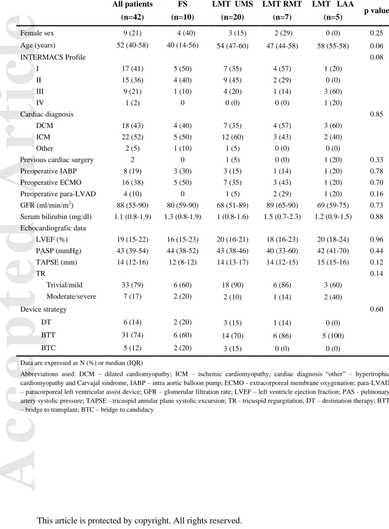
Table 1 - Baseline patient’s characteristics All patients (n=42) FS (n=10) LMT UMS (n=20) LMT RMT (n=7) LMT LAA (n=5) p value Female sex 9 (21) 4 (40) 3 (15) 2 (29) 0 (0) 0.25 Age (years) 52 (40-58) 40 (14-56) 54 (47-60) 47 (44-58) 58 (55-58) 0.06 INTERMACS Profile 0.08 I 17 (41) 5 (50) 7 (35) 4 (57) 1 (20) II 15 (36) 4 (40) 9 (45) 2 (29) 0 (0) III 9 (21) 1 (10) 4 (20) 1 (14) 3 (60) IV 1 (2) 0 0 (0) 0 (0) 1 (20) Cardiac diagnosis 0.85 DCM 18 (43) 4 (40) 7 (35) 4 (57) 3 (60) ICM 22 (52) 5 (50) 12 (60) 3 (43) 2 (40) Other 2 (5) 1 (10) 1 (5) 0 (0) 0 (0)
Previous cardiac surgery 2 0 1 (5) 0 (0) 1 (20) 0.33
Preoperative IABP 8 (19) 3 (30) 3 (15) 1 (14) 1 (20) 0.78 Preoperative ECMO 16 (38) 5 (50) 7 (35) 3 (43) 1 (20) 0.70 Preoperative para-LVAD 4 (10) 0 1 (5) 2 (29) 1 (20) 0.16 GFR (ml/min/m2) 88 (55-90) 80 (59-90) 68 (51-89) 89 (65-90) 69 (59-75) 0.73 Serum bilirubin (mg/dl) 1.1 (0.8-1.9) 1.3 (0.8-1.9) 1 (0.8-1.6) 1.5 (0.7-2.3) 1.2 (0.9-1.5) 0.88 Echocardiografic data LVEF (%) 19 (15-22) 16 (15-23) 20 (16-21) 18 (16-23) 20 (18-24) 0.96 PASP (mmHg) 43 (39-54) 44 (38-52) 43 (38-46) 40 (33-60) 42 (41-70) 0.44 TAPSE (mm) 14 (12-16) 12 (8-12) 14 (13-17) 14 (12-15) 15 (15-16) 0.12 TR 0.14 Trivial/mild 33 (79) 6 (60) 18 (90) 6 (86) 3 (60) Moderate/severe 7 (17) 2 (20) 2 (10) 1 (14) 2 (40) Device strategy 0.60 DT 6 (14) 2 (20) 3 (15) 1 (14) 0 (0) BTT 31 (74) 6 (60) 14 (70) 6 (86) 5 (100) BTC 5 (12) 2 (20) 3 (15) 0 (0) 0 (0)
Data are expressed as N (%) or median (IQR)
Abbreviations used: DCM – dilated cardiomyopathy; ICM – ischemic cardiomyopathy; cardiac diagnosis “other” – hypertrophic cardiomyopathy and Carvajal sindrome, IABP – intra aortic balloon pump; ECMO - extracorporeal membrane oxygenation; para-LVAD – paracorporeal left ventricular assist device; GFR – glomerular filtration rate; LVEF – left ventricle ejection fraction; PAS - pulmonary artery systolic pressure; TAPSE - tricuspid annular plane systolic excursion; TR - tricuspid regurgitation; DT – destination therapy; BTT – bridge to transplant; BTC – bridge to candidacy
Accepted
Article
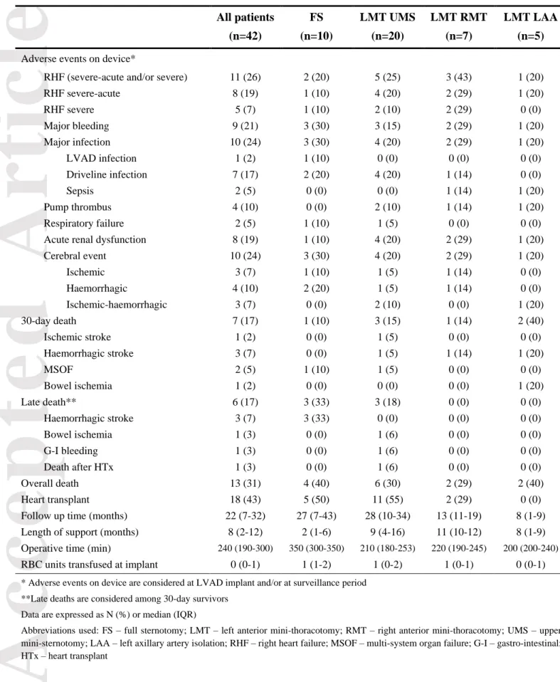
Table 2 – Post implant outcomes by surgical approach adopted All patients (n=42) FS (n=10) LMT UMS (n=20) LMT RMT (n=7) LMT LAA (n=5)
Adverse events on device*
RHF (severe-acute and/or severe) 11 (26) 2 (20) 5 (25) 3 (43) 1 (20)
RHF severe-acute 8 (19) 1 (10) 4 (20) 2 (29) 1 (20) RHF severe 5 (7) 1 (10) 2 (10) 2 (29) 0 (0) Major bleeding 9 (21) 3 (30) 3 (15) 2 (29) 1 (20) Major infection 10 (24) 3 (30) 4 (20) 2 (29) 1 (20) LVAD infection 1 (2) 1 (10) 0 (0) 0 (0) 0 (0) Driveline infection 7 (17) 2 (20) 4 (20) 1 (14) 0 (0) Sepsis 2 (5) 0 (0) 0 (0) 1 (14) 1 (20) Pump thrombus 4 (10) 0 (0) 2 (10) 1 (14) 1 (20) Respiratory failure 2 (5) 1 (10) 1 (5) 0 (0) 0 (0)
Acute renal dysfunction 8 (19) 1 (10) 4 (20) 2 (29) 1 (20)
Cerebral event 10 (24) 3 (30) 4 (20) 2 (29) 1 (20) Ischemic 3 (7) 1 (10) 1 (5) 1 (14) 0 (0) Haemorrhagic 4 (10) 2 (20) 1 (5) 1 (14) 0 (0) Ischemic-haemorrhagic 3 (7) 0 (0) 2 (10) 0 (0) 1 (20) 30-day death 7 (17) 1 (10) 3 (15) 1 (14) 2 (40) Ischemic stroke 1 (2) 0 (0) 1 (5) 0 (0) 0 (0) Haemorrhagic stroke 3 (7) 0 (0) 1 (5) 1 (14) 1 (20) MSOF 2 (5) 1 (10) 1 (5) 0 (0) 0 (0) Bowel ischemia 1 (2) 0 (0) 0 (0) 0 (0) 1 (20) Late death** 6 (17) 3 (33) 3 (18) 0 (0) 0 (0) Haemorrhagic stroke 3 (7) 3 (33) 0 (0) 0 (0) 0 (0) Bowel ischemia 1 (3) 0 (0) 1 (6) 0 (0) 0 (0) G-I bleeding 1 (3) 0 (0) 1 (6) 0 (0) 0 (0) Death after HTx 1 (3) 0 (0) 1 (6) 0 (0) 0 (0) Overall death 13 (31) 4 (40) 6 (30) 2 (29) 2 (40) Heart transplant 18 (43) 5 (50) 11 (55) 2 (29) 0 (0)
Follow up time (months) 22 (7-32) 27 (7-43) 28 (10-34) 13 (11-19) 8 (1-9)
Length of support (months) 8 (2-12) 2 (1-6) 9 (4-16) 11 (10-12) 8 (1-9)
Operative time (min) 240 (190-300) 350 (300-350) 210 (180-253) 220 (190-245) 200 (200-240)
RBC units transfused at implant 0 (0-1) 1 (1-2) 1 (0-2) 1 (0-1) 0 (0-1)
* Adverse events on device are considered at LVAD implant and/or at surveillance period **Late deaths are considered among 30-day survivors
Data are expressed as N (%) or median (IQR)
Abbreviations used: FS – full sternotomy; LMT – left anterior mini-thoracotomy; RMT – right anterior mini-thoracotomy; UMS – upper mini-sternotomy; LAA – left axillary artery isolation; RHF – right heart failure; MSOF – multi-system organ failure; G-I – gastro-intestinal; HTx – heart transplant
Accepted
Article
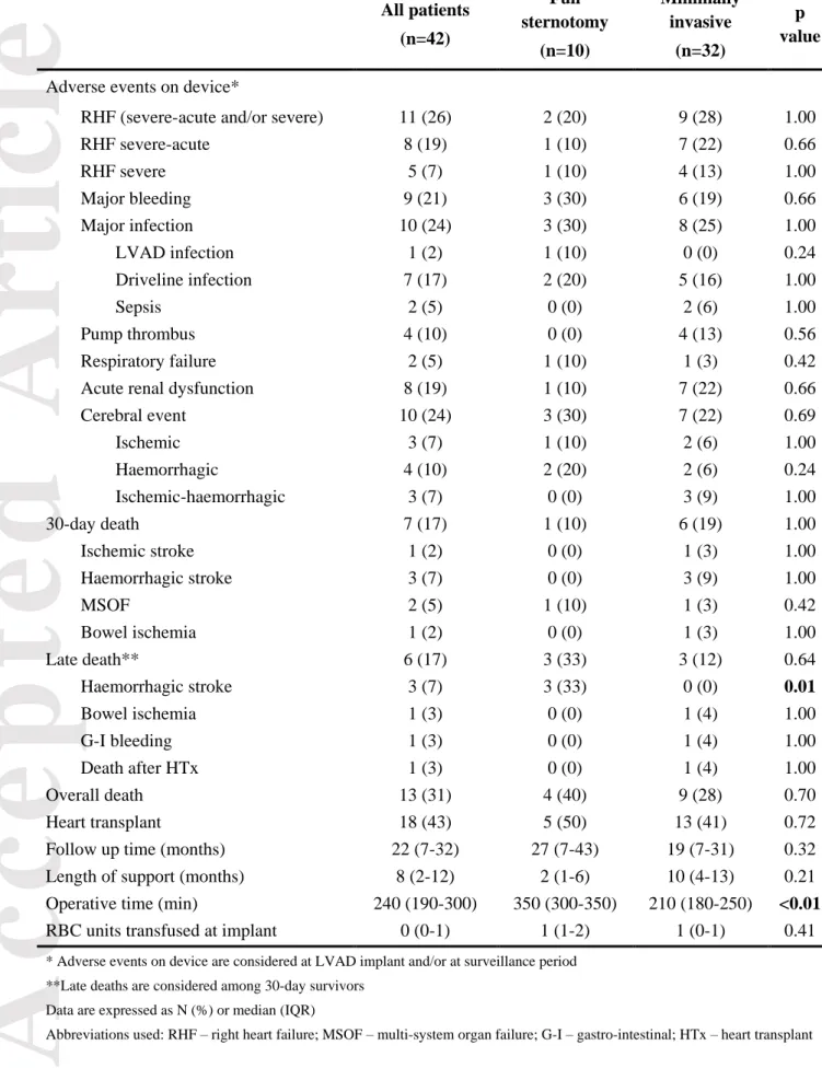
Table 3 – Univariate analysis of post implant outcomes All patients (n=42) Full sternotomy (n=10) Minimally invasive (n=32) p value Adverse events on device*
RHF (severe-acute and/or severe) 11 (26) 2 (20) 9 (28) 1.00
RHF severe-acute 8 (19) 1 (10) 7 (22) 0.66 RHF severe 5 (7) 1 (10) 4 (13) 1.00 Major bleeding 9 (21) 3 (30) 6 (19) 0.66 Major infection 10 (24) 3 (30) 8 (25) 1.00 LVAD infection 1 (2) 1 (10) 0 (0) 0.24 Driveline infection 7 (17) 2 (20) 5 (16) 1.00 Sepsis 2 (5) 0 (0) 2 (6) 1.00 Pump thrombus 4 (10) 0 (0) 4 (13) 0.56 Respiratory failure 2 (5) 1 (10) 1 (3) 0.42
Acute renal dysfunction 8 (19) 1 (10) 7 (22) 0.66
Cerebral event 10 (24) 3 (30) 7 (22) 0.69 Ischemic 3 (7) 1 (10) 2 (6) 1.00 Haemorrhagic 4 (10) 2 (20) 2 (6) 0.24 Ischemic-haemorrhagic 3 (7) 0 (0) 3 (9) 1.00 30-day death 7 (17) 1 (10) 6 (19) 1.00 Ischemic stroke 1 (2) 0 (0) 1 (3) 1.00 Haemorrhagic stroke 3 (7) 0 (0) 3 (9) 1.00 MSOF 2 (5) 1 (10) 1 (3) 0.42 Bowel ischemia 1 (2) 0 (0) 1 (3) 1.00 Late death** 6 (17) 3 (33) 3 (12) 0.64 Haemorrhagic stroke 3 (7) 3 (33) 0 (0) 0.01 Bowel ischemia 1 (3) 0 (0) 1 (4) 1.00 G-I bleeding 1 (3) 0 (0) 1 (4) 1.00 Death after HTx 1 (3) 0 (0) 1 (4) 1.00 Overall death 13 (31) 4 (40) 9 (28) 0.70 Heart transplant 18 (43) 5 (50) 13 (41) 0.72
Follow up time (months) 22 (7-32) 27 (7-43) 19 (7-31) 0.32
Length of support (months) 8 (2-12) 2 (1-6) 10 (4-13) 0.21
Operative time (min) 240 (190-300) 350 (300-350) 210 (180-250) <0.01
RBC units transfused at implant 0 (0-1) 1 (1-2) 1 (0-1) 0.41
* Adverse events on device are considered at LVAD implant and/or at surveillance period **Late deaths are considered among 30-day survivors
Data are expressed as N (%) or median (IQR)
Accepted
Article
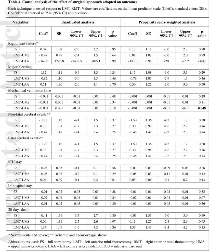
Table 4. Causal analysis of the effect of surgical approach adopted on outcomes
Each technique is tested respect to LMT-RMT. Values are coefficients on the linear predictor scale (Coeff), standard errors (SE), Confidential Interval at 95% (95% CI) and p-values.
Variables Unadjusted analysis Propensity score weighted analysis
Coeff SE Lower 95% CI Upper 95% CI p value Coeff SE Lower 95% CI Upper 95% CI p value
Right heart failure*
FS 0.07 1.07 -2.0 2.2 0.95 0.13 1.11 -2.0 2.3 0.90 LMT-UMS - 0.47 0.99 -2.4 1.5 0.64 0.01 1.02 -2.0 2.0 0.99 LMT-LAA - 16.70 1745.8 -3438.5 3405.1 0.99 - 18.10 0.98 -20 -16.2 <0.01 Major bleeding FS 1.32 1.11 -0.9 3.5 0.24 1.15 1.08 -1.0 3.3 0.29 LMT-UMS - 0.82 1.10 -3.0 1.3 0.46 - 0.79 1.07 -2.9 1.3 0.46 LMT-LAA 0.51 1.30 -2.0 3.1 0.70 0.50 1.25 -2.0 3.0 0.69
Mechanical ventilation time
FS - 0.001 0.001 -0.01 0.01 0.48 - 0.001 0.001 -0.01 0.01 0.28
LMT-UMS - 0.001 0.001 -0.01 0.01 0.34 - 0.001 0.001 -0.01 0.01 0.11
LMT-LAA - 0.001 0.001 -0.01 0.01 0.18 - 0.001 0.001 -0.01 -0.01 0.049
Non-fatal cerebral events**
FS - 1.28 1.42 -4.1 1.5 0.37 - 1.50 1.36 -4.2 1.2 0.28
LMT-UMS 0.30 1.01 -1.7 2.3 0.77 0.30 0.99 -1.6 2.2 0.76
LMT-LAA - 0.47 1.47 -3.4 2.4 0.75 - 0.48 1.41 -3.2 2.3 0.74
Fatal cerebral events**
FS - 1.28 1.42 -4.1 1.5 0.37 - 1.50 1.36 -4.2 1.2 0.28 LMT-UMS 0.30 1.01 -1.7 2.3 0.77 0.30 0.98 -1.6 2.2 0.76 LMT-LAA - 0.47 1.47 -3.4 2.4 0.75 - 0.48 1.41 -3.2 2.3 0.74 ICU stay FS - 0.03 0.05 -0.1 0.1 0.56 - 0.03 0.03 -0.09 0.03 0.26 LMT-UMS - 0.05 0.07 -0.2 0.1 0.29 - 0.05 0.03 -0.11 0.01 0.12 LMT-LAA 0.04 0.09 -0.1 0.2 0.61 0.05 0.06 -0.1 0.2 0.43 In-hospital stay FS - 0.01 0.02 -0.05 0.03 0.58 - 0.01 0.01 -0.03 0.01 0.35 LMT-UMS - 0.02 0.01 -0.04 0.01 0.24 - 0.02 0.01 -0.04 0.01 0.07 LMT-LAA - 0.01 0.02 -0.05 0.03 0.80 - 0.01 0.01 -0.03 0.01 0.56 30-days death FS - 0.41 1.59 -3.5 2.7 0.80 - 0.03 1.53 -3.0 3.0 0.99 LMT-UMS 0.06 1.31 -2.5 2.6 0.97 0.11 1.27 -2.4 2.6 0.93 LMT-LAA 1.37 1.49 -1.6 4.3 0.36 1.36 1.43 -1.4 4.2 0.35
* Severe-acute and severe; ** ischemic and haemorrhagic stroke
Abbreviations used: FS – full sternotomy; LMT – left anterior mini-thoracotomy; RMT – right anterior mini-thoracotomy; UMS – upper mini-sternotomy; LAA – left axillary artery isolation; ICU – intensive care unit
Accepted
Article
Figure legends Figure 1
Evolution of the surgical approach for the HeartWare HVAD implantation. A) Techniques entailing outflow graft anastomosis to the ascending aorta: A1 – full sternotomy; A2 – left anterior mini-thoracotomy and upper mini-sternotomy; A3 – left and right anterior mini-mini-thoracotomy. B) Left anterior mini-thoracotomy with outflow graft anastomosis to the left axillary artery.
Figure 2
Surgical techniques for the outflow graft anastomosis of HeartWare HVAD to the left axillary artery (LAA). A) Procedure that applies when the LAA calibre is >8 mm: end-to-end outflow graft anastomosis and end-to-side distal LAA connection. B) Procedure used when the LAA calibre is <8mm: 8 mm-graft interposition to LAA and end-to-side outflow graft anastomosis.
Figure 3