저작자표시-비영리-변경금지 2.0 대한민국 이용자는 아래의 조건을 따르는 경우에 한하여 자유롭게 l 이 저작물을 복제, 배포, 전송, 전시, 공연 및 방송할 수 있습니다. 다음과 같은 조건을 따라야 합니다: l 귀하는, 이 저작물의 재이용이나 배포의 경우, 이 저작물에 적용된 이용허락조건 을 명확하게 나타내어야 합니다. l 저작권자로부터 별도의 허가를 받으면 이러한 조건들은 적용되지 않습니다. 저작권법에 따른 이용자의 권리는 위의 내용에 의하여 영향을 받지 않습니다. 이것은 이용허락규약(Legal Code)을 이해하기 쉽게 요약한 것입니다. Disclaimer 저작자표시. 귀하는 원저작자를 표시하여야 합니다. 비영리. 귀하는 이 저작물을 영리 목적으로 이용할 수 없습니다. 변경금지. 귀하는 이 저작물을 개작, 변형 또는 가공할 수 없습니다.
의학석사 학위논문
Biomechanical Properties of Glenohumeral
Joint Capsule in Diabetic and non-Diabetic
Patients with Adhesive Capsulitis
당뇨병이 있는 유착성 견관절낭염 환자에서 견관절낭의
생역학적 특성
2020년 8월
서울대학교 대학원
임상의과학과
장 원 기
Thesis for the Degree of Master of Science
in Clinical Medical Sciences
Biomechanical Properties of Glenohumeral
Joint Capsule in Diabetic and non-Diabetic
Patients with Adhesive Capsulitis
당뇨병이 있는 유착성 견관절낭염 환자에서 견관절낭의
생역학적 특성
August 2020
Seoul National University College of Medicine
Clinical Medical Sciences
Wonkee Chang, M.D.
당뇨병이 있는 유착성 견관절낭염
환자에서 견관절낭의 생역학적 특성
지도교수 정 선 근
이 논문을 의학석사 학위논문으로 제출함
2020년 5월
서울대학교 대학원
임상의과학과
장 원 기
장 원 기 석사 학위논문을 인준함
2020년 7월
위
원
장
(인)
부 위 원 장
(인)
위 원
(인)
Biomechanical Properties of Glenohumeral
Joint Capsule in Diabetic and non-Diabetic
Patients with Adhesive Capsulitis
By
Wonkee Chang, M.D.
A Thesis Submitted in Partial Fulfillment of the
Requirements for the Degree of Master of Science in
Clinical Medical Sciences at the Seoul National University
College of Medicine
July 2020
Approved by thesis committee:
Professor Chairman
Professor Vice Chairman
Professor
Abstract
Introduction: Diabetes mellitus (DM) is a well-known risk factor for the development of Adhesive capsulitis (AC) of the shoulder. However, the relationships between DM and the severity of the AC has not been fully established yet.
Objectives: To investigate whether DM incurs more severe AC, the differences in capsular stiffness were compared between DM and non-DM AC patients and the associations of DM parameters with capsular stiffness among DM patients were determined.
Design: Case series.
Setting: A tertiary university hospital outpatient clinic dedicated to intra-articular hydraulic distension (IHD).
Participants: A total of 114 patients (DM 24, non-DM 90) with AC who underwent IHD.
Methods: Pressure-volume profiles were obtained using a real-time pressure monitoring technique during IHD and the capsular stiffness (Kcap) was defined as the slope of elastic phase from the pressure-volume curve. Demographic, clinical and
DM parameters were collected by retrospective chart review.
Main Outcome Measurements: Comparison of averages was used to investigate
whether there was difference of Kcap between DM and non-DM patients with AC.
Multiple linear regression model was established to evaluate the association
between Kcap and DM after adjusting for other covariates. Spearman rho test and
Mann-Whitney test were used to analyze the relationship between Kcap and DM
duration, HbA1c, time integrated HbA1c and insulin usage.
Results: The mean Kcap had no significant difference between DM and non-DM AC
patients. (33.03 ± 20.63 versus 25.99 ± 14.08 , p=0.141). Presence of DM was
associated with higher Kcap after adjusting covariates(β=9.37, p=0.014). Duration of
DM had positive correlation with Kcap (r=0.436, p=0.048) while HbA1c, time
integrated HbA1c and insulin usage had no significant relationship with the capsular stiffness in the AC patients with DM.
Conclusions: Duration of DM was positively correlated with the capsular stiffness, suggesting longer duration of DM is associated with more severe AC in the patients with DM. There was no significant difference of capsular stiffness between AC patients with and without DM, however after adjusting covariates, presence of DM
was associated with higher capsular stiffness. Further prospective researches with larger numbers of subjects are required to delineate relationships between DM and the severity of AC.
Keywords: adhesive capsulitis, diabetes mellitus, IHD, biomechanical properties, capsular stiffness, DM duration
Table of Contents
List of Tables …..……… 5
List of Figures ………... 6
I. Introduction …...……… 7
II. Methods ……….……….……...… 10
III. Results ……….……… 16
IV. Discussion ……….……... 22
V. Conclusion ………….……….……….. 28
VI. References ……….……….. 29
국문초록
……….……….. 35
List of Tables
Table 1. Demographic, clinical and biomechanical properties of
the
patients
(N=114)
………. 17
Table 2. Demographic, clinical and biomechanical properties of
the
patients
after
age,
gender
matching
(N=72)
………. 18
Table 3. Factors associated with
K
capin the patients with adhesive
capsulitis
………….………..……….
19
List of Figures
Figure 1. An example of P-V waveform acquired during IHD ….…
15
Figure 2. Scatter plot of capsular stiffness in relationship with
diabetes associated parameters ……….……….
21
Figure 3. Examples of “Serrated” Pressure-Time curve ………
26
I. Introduction
Adhesive capsulitis (AC) of shoulder is a condition characterized by a painful,
gradual loss of both active and passive glenohumeral joint range of motion (ROM)1,
2. It is a self-limiting condition which lasts between 1 to 3 years untreated3, and is
known to affect patients’ activities of daily living and the qualities of life.
Diabetes mellitus (DM) is a well-established risk factor for the development of AC along with age, female gender, thyroid disease, congestive heart disease and
dyslipidemia4-6. A recent meta-analysis has revealed the patients with DM were five
times more likely to have AC than those without DM7. However, the impact of DM
on the severity of AC has not been studied thoroughly and remains unclear. Several literatures have reported the shoulder pain and the quality of life were poorer
among AC patients with DM than the patients without DM8, 9, while others
demonstrated no significant difference in the disability or pain between the two
groups10, 11. It has also been widely accepted that the duration and the severity of
DM are related to the prevalence of AC among DM patients12, 13, but the underlying
mechanisms how DM affects AC have not been properly elucidated yet. Researches have speculated advanced glycosylation end product (AGE) accumulation, a
long-term complication of DM, as an important factor in developing AC in DM patients14. In-vitro studies have revealed incubation time and glucose concentration are
responsible for the formation of AGE15 and the accumulation of the AGE in the soft
tissues results in decreased elasticity16 . From these reports, we developed a
hypothesis that higher blood glucose level might exacerbate capsular fibrosis to cause higher capsular stiffness.
Intra-articular hydraulic distension (IHD) of the glenohumeral joint is a widely used
non-operative treatment option for treating AC17, 18, and its therapeutic effect has
been well-proven in the literatures19, 20. To improve the therapeutic effect of IHD, we
have developed a real-time intra-articular pressure monitoring technique21, which
has also allowed us to obtain the real-time intra-articular pressure-volume (P-V) data, from which biomechanical properties including capsular stiffness of the
glenohumeral joint were quantitatively calculated22-26. Since IHD was adopted in our
frozen shoulder clinic, approximately 500 IHD procedures have been undertaken. Before prospective researches are planned and executed, we delved into accumulated IHD data to explore the hypothesis.
In this study, we aimed to test the hypothesis that DM may affect the severity of AC by increasing capsular stiffness of the glenohumeral joint. For the purpose, we
compared capsular stiffness of AC patients with and without DM. In addition, the relationship of capsular stiffness with exposure to higher blood glucose level determined by duration of DM, blood levels of HbA1c, time integrated HbA1c levels, and insulin usage status.
II. Methods
Study Participants
A total of 194 patients’ medical data and P-V profiles who underwent IHD procedure at the outpatient clinic for adhesive capsulitis from October 2009 to January 2011 were retrospectively reviewed. From the patients who underwent multiple IHD procedures, only the P-V profiles from the first IHD were included in the study. AC was diagnosed when the passive shoulder ROM of the affected
shoulder was limited in the external rotation (ER) with 1 or more directions27.
Limitation of shoulder ROM was defined as passive shoulder ROMs measured by goniometer as following; abduction < 80°, flexion < 130°, internal rotation level L1
and below and external rotation < 30 ° or scratch test level C7 and above22, 25.
Patients were excluded when they had previous history of trauma or major surgery (n=33), breast cancer (n=12) or full-thickness tear (n=2) on the affected shoulder. Patients who did not reach phase 2 during the IHD procedure (n=9) and patients with irregular P-V profiles which were not analyzable (n=24) were also excluded from the study. In final, 114 patients were included in the study analyzation and of them, 24 patients had DM. Initial analysis was done with 114 patients and there was
significant difference of age between DM and non-DM group. To minimize the effect of age and gender on the capsular stiffness, we selected non-DM group using propensity-score matching technique. A logistic regression model was created to derive a propensity score with age and gender as independent variables. A DM group patient was matched with 2 non-DM patient (1:2 matching). In final, 72 (DM n=24, non-DM n=48) patients’ data were analyzed. Among DM patients, HbA1c within 3 months of IHD procedure, DM duration, time integrated HbA1c which was defined as the value of HbA1c integrated by time period throughout the entire medical record and finally, type of medication (Insulin usage status) were obtained from the medical record review. The study was approved by the institutional review board of Seoul National University Hospital.
Clinical Evaluation
All patients went through passive ROM measurement and sonographic evaluation prior to the IHD procedure by the senior author (S.G.C). Passive ROMs were measured with goniometer while the patient was sitting upright on a stool. Abduction, flexion and external rotation ROMs were measured with the goniometer and the sum of the ROMs (ROM_sum) was defined as the summation of the
degrees of ROMs in 3 directions. Scratch tests were used to evaluate the external and internal rotation ROMs which were measured as the lowest and the highest anatomy reached with the thumb and the middle finger, respectively.
The sonographic evaluation on the affected shoulder was performed after ROM measurement. Specific findings including long biceps tendon sheath swelling, rotator cuff tear, calcification, subacromial and subdeltoid bursa swelling were assessed to exclude other pathologic diseases of the shoulder.
IHD procedure
IHD procedures were performed by the senior author (S.G.C.) at the outpatient clinic in the following setting. The subject was sitting on a stool with the arm resting on the thigh to have fully relaxed during the procedure. A 22-gauge spinal needle was connected to a pressure sensor and a 50-mL syringe via a 3-way stopcock. The needle was inserted 1 cm lateral to the ultrasound transducer, penetrating the infraspinatus tendon and the posterior capsule. After placing the needle tip in the joint space, a fluid mixture (total 50 mL) of 1mL of 40mg triamcinolone; 10 mL of 1% lidocaine and 39 mL of normal saline was infused with a constant-speed pump at the rate of 7 mL / min. The infusion was terminated in
the following conditions: when the P-V curve reached the third phase; when the intra-articular pressure increased abruptly indicating impending rupture; when the patient asked for the termination of the procedure.
P-V data analysis
The P-V profiles were analyzed and reviewed in the following process by the 2 authors (W.K.C and S.G.C.). The P-V curve were derived from the Pressure-Time(P-T) curve obtained during the IHD. Then the P-V curve was divided into 3 phases: phase I the initial filling phase; phase II the elastic deformation phase; phase III the plastic deformation phase. The slope of the phase II was considered as the stiffness of the joint capsule (Kcap). The volume and pressure at the infusion termination
point were defined as the maximal volume (Vmax) and the maximal pressure (Pmax),
respectively. Figure 1 demonstrates a typical P-V curve with definitions of
biomechanical properties (Kcap, Vmax and Pmax). A detailed processing methods of P-V
profile analysis are described in the previous studies23-26.
Statistical Analysis
We compared demographic variables (age, gender, symptom duration and dominant hand), clinical parameters (ROM of abduction, flexion, external rotation
and the sum of three ROMs) and biomechanical properties (Kcap, Vmax and Pmax)
between the DM group and non-DM group using independent t-test for the
numeric variables and chi-square test for categorical variables. A multiple linear
regression model was used to evaluate the association between Kcap and DM after
adjusting for other covariates. The correlation between Kcap and DM parameters
(HbA1c, DM duration, time integrated HbA1c and insulin usage status) were analyzed with Spearman rho test and Mann-Whitney test because the sample size
was not large enough to guarantee normal distribution. p-values of <0.05 were
considered statistically significant. All analyses were performed using open-source R software.
Figure 1. An example of P-V waveform acquired during IHD. The slope (black solid line) of the elastic deformation phase (between the gray dotted lines) was defined as Kcap, while the volume and pressure at the infusion termination point were
III. Results
Comparison of Demographic, Clinical and Biomechanical
properties between the DM and non-DM group
There was significant difference of age between DM and non-DM group on
analysis with 114 patients. (63.46 vs 57.16, p=0.005). Otherwise, there were no
significant difference in the demographic, clinical and biomechanical variables (Pmax,
Vmaxand Kcap) (Table 1).
There were no significant differences in the demographic and clinical characteristics between the AC patients with and without DM after propensity score matching was done (Table 2). All the biomechanical variables were also not
significantly different between the two patient groups (434.15 vs 436.10 p=0.947,
22.57 vs 21.04 p=0.478 and 33.03 vs 25.99 p=0.141 respectively).
Factors associated with Capsular Stiffness
Table 3 shows the multiple linear regression model between Kcap and age,
gender(female), symptom duration and presence of DM. Presence of DM as well as
Table 1 Demographic, clinical and biomechanical properties of the patients (N = 114) DM (N = 24) Non-DM (N = 90) P Demographic / Clinical properties Sex ratio, M:F 11:13 28:62 0.268 Age (yr) 63.46(9.49) 57.16(9.62) 0.005* Involved side, R:L 9:15 51:39 0.778
Symptom duration (mo) 11.83(9.33) 7.85(6.74) 0.060
Abduction ROM (‘) 61.17(16.68) 53.73(13.79) 0.211
Forward flexion ROM (‘) 98.13(13.69) 100.10(19.80) 0.574
External rotation ROM (‘) 32.21(13.55) 34.21(11.37) 0.463
Sum of 3 ROMs (‘) 191.50(33.51) 188.04(31.64) 0.640
Biomechanical properties
Pmax(mmHg) 434.15(125.93) 439.08(105.77) 0.862
Vmax(ml) 22.57(10.40) 20.03(7.66) 0.273
Kcap(mmHg/ml) 33.03(20.63) 27.65(14.87) 0.241
*P<0.05, ROM = range of motion; Pmax = pressure at the maximal volume; Vmax =
Table 2 Demographic, clinical and biomechanical properties of the patients after age, gender matching (N = 72)
DM (N = 24) Non-DM (N = 48) P Demographic / Clinical properties Sex ratio, M:F 11:13 19:29 .800 Age (yr) 63.46(9.49) 62.42(8.49) .639 Involved side, R:L 9:15 22:26 .674
Symptom duration (mo) 11.83(9.33) 8.42(7.32) .093
Abduction ROM (‘) 61.17(16.68) 54.94(12.22) .076
Forward flexion ROM (‘) 98.13(13.69) 100.54(19.67) .592
External rotation ROM (‘) 32.21(13.55) 33.56(9.80) .629
Sum of 3 ROMs (‘) 191.50(33.51) 189.04(29.27) .750
Biomechanical properties
Pmax(mmHg) 434.15(125.93) 436.10(112.56) .947
Vmax(ml) 22.57(10.40) 21.04(7.52) .478
Kcap(mmHg/ml) 33.03(20.63) 25.99(14.08) .141
*P<0.05, ROM = range of motion; Pmax = pressure at the maximal volume; Vmax =
Table 3 Factors associated with Kcap in the patients with adhesive capsulitis. β±SE Age -0.43±0.15† Gender (Female) 8.55±3.10† Symptom duration -0.01±0.20 Presence of DM 9.37±3.76*
*P<0.05, †P<0.01 by multiple linear regression analysis after adjusting for other covariates.
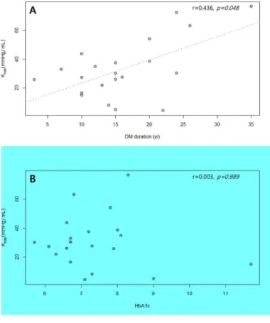
Correlation between the Capsular Stiffness and DM parameters
DM duration had positive correlation with Kcap (r = 0.436, p=0.048) while HbA1c
and time integrated HbA1c showed no significant correlation with Kcap (p=0.989,
p=0.238 respectively). There was no significant difference of Kcapbetween the insulin usage group and oral hypoglycemic agent(OHA) only group (38.60 vs 21.45, p=0.216). (Figure 2A-2D)
Figure 2. Scatterplots showing relationship between capsular stiffness (Kcap) and DM duration (A), HbA1c (B) time integrated HbA1c (C) and the Boxplot of capsular stiffness according to Insulin usage status (D).
IV. Discussion
This study aimed to unveil the relationship between DM and the severity of AC by comparing the capsular stiffness of patients with and without DM, and examining
the DM parameters that were related to the capsular stiffness. Kcap, which is
considered as a biomechanical and quantitative indicator of the severity of AC, was calculated from the P-V curve obtained during IHD. We have hypothesized that the glenohumeral joint capsule would be stiffer in the patients with DM and both duration of DM and glycemic status would be correlated to the capsular stiffness.
There was no significant difference of Kcap between the AC patients with and
without DM, although linear multiple regression analysis has revealed presence of
DM is associated with higher Kcap after adjusting covariates. Among DM parameters,
only duration of DM had positive correlation with Kcap.
Duration of DM had positive correlation with Kcap while HbA1c and insulin usage
status were not related to the Kcap. Considering AC is a condition with insidious
onset and gradual progression2, 3, HbA1c and insulin status which represents the
glycemic status at the time of examination, might not fully appreciate the long-term glycemic burden related to the severity of AC in the patient with DM. These findings are concurrent with the previous studies on the relationship between DM
parameters and prevalence of AC12, 13, 28, which implies DM duration is an important factor in both development and aggravation of AC in the patients with DM.
Incubation time and glucose concentration are two key factors in the accumulation
of AGE in the soft tissues15. Time integrated HbA1c, which incorporates both factors,
seemed to be a good proxy for reflecting the degree of AGE accumulation in the glenohumeral joint capsule. Time integrated HbA1c showed good correlation with prevalence of adhesive capsulitis in the type 1 DM patients with over 45 years of
follow-up29, thus we supposed it would also present a good correlation with the Kcap,
which did not in result. Since our study was performed at tertiary hospital OPD clinic for AC, we were not able to trace the glycemic status for the whole period of DM duration in the most of our patients, resulting in less accurate approximations of glycemic burden. A more controlled study including patients’ medical data on whole duration of DM might give us more information on the relationship between DM and capsular stiffness.
Although DM duration was associated with capsular stiffness, there was no
difference of Kcap between DM and non-DM patient with AC which might seem to
be somewhat conflicting. We believe the patient pool selection of our study is
during IHD procedures which were performed to the AC patients with moderate to severe degree of pain on limitation of ROMs. For mild AC patients, physiotherapy including shoulder stretching on the directions of LOM and NSAIDs were treatment
of choice in our clinic, thus Kcap of these mild AC patients were unobtainable. The
influence of confounding factors such as age and gender on Kcap might also
account for this result. As reported in the previous study25, female gender was
associated with higher Kcap and after adjusting covariates (age, female gender,
duration of symptom), presence of DM was associated with higher Kcap. From these
results, we can speculate AC patients with DM would have stiffer glenohumeral joint capsule than those without DM.
The real-time pressure monitoring and P-V analysis technique we implemented in this study has an advantage to be acknowledged. It allowed us to measure the tightness by the degree of stretching of the soft tissues in the glenohumeral joint capsule during IHD. The tightness of the soft tissues including coracohumeral
ligament and rotator interval are considered as the key features of AC30, 31, thus this
technique has advantage of appreciating the severity of the AC in a biomechanical and physiological manner. Since previous studies on the relationship between DM
Disability Index; SPADI)8, 10, 11 as main outcomes to evaluate the severity of AC. Our study’s strength lies in the evaluation method of the severity of AC and to our knowledge, this is the first study on the impact of DM on the severity of AC using a biomechanical, quantitative outcome measure.
On analyzing the pressure-volume profiles acquired during IHD, we found a few
notable waveforms which were observed among patients with very high Kcap (Figure
3A,3B). Patients with these “Serrated” P-T curves had very high Kcap (mean: 66.727),
which could implicate a certain subgroup of patients might have stiffer capsule than the others. Although this study has failed to identify the characteristics of such
subgroup with high Kcap, further research is required to uncover such subgroups of
Figure 3. Examples of “Serrated” Pressure-Time curve shown in the patients with high Kcap
Limitations
There are several limitations to be mentioned in this study. First, there was a fair difference of baseline age between DM and non-DM patients (63.46 vs 57.16, p=0.005) and similar age differences were shown in the previous studies carried out in the tertiary hospital settings[10][32]. To minimize the effect of the age and gender, which are well-known factors affecting the capsular stiffness25, this study adopted propensity-score matching technique.
Secondly, since the medical history was obtained retrospectively, several known risk
factors of AC such as hyperlipidemia33, familial history of AC and Dupuytren’s
disease6 were not assessed.
Finally, due to the small number of DM patients, the statistical analysis between
DM parameters and Kcap were nonparametric, which resulted in reduced statistical
power. Further prospective research on larger sample size will provide a clearer relationship between DM parameters and capsular stiffness.
V. Conclusion
In this study, duration of DM was positively correlated with the Kcap, suggesting longer duration of DM was associated with more severe AC in the patients with DM.
There was no significant difference of Kcap between AC patients with and without
DM, although presence of DM was associated with higher Kcap after adjustment of
confounding factors. Further prospective research with larger subjects is require to conclude the relationship between DM and the severity of AC.
VI. Reference
1. Neviaser AS, Hannafin JA. Adhesive capsulitis: a review of current treatment.
Am J Sports Med 2010;38:2346–56.
2. Hannafin J a, Chiaia T a. Adhesive capsulitis. A treatment approach. Clin
Orthop Relat Res 2000:95–109.
3. Dias R, Cutts S, Massoud S. Frozen shoulder. BMJ 2005;331:1453–6..
4. Huang Y-P, Fann C-Y, Chiu Y-H, Yen M-F, Chen L-S, Chen H-H, et al.
Association of diabetes mellitus with the risk of developing adhesive capsulitis of the shoulder: a longitudinal population-based followup study. Arthritis Care Res (Hoboken) 2013;65:1197–202.
5. Tighe CB, Oakley WS. The prevalence of a diabetic condition and adhesive
capsulitis of the shoulder. South Med J 2008;101:591–5.
6. Wang K, Ho V, Hunter-Smith DJ, Beh PS, Smith KM, Weber AB. Risk factors in
idiopathic adhesive capsulitis: a case control study. J Shoulder Elb Surg 2013;22:e24-9.
7. Zreik NH, Malik RA, Charalambous CP. Adhesive capsulitis of the shoulder
2016;6:26–34.
8. Cole A, Gill TK, Shanahan EM, Phillips P, Taylor AW, Hill CL. Is diabetes
associated with shoulder pain or stiffness? Results from a population based study. J Rheumatol 2009;36:371–7.
9. Griggs SM, Ahn A, Green A. Idiopathic adhesive capsulitis. A prospective
functional outcome study of nonoperative treatment. J Bone Joint Surg Am 2000;82-A:1398–407.
10. Laslett LL, Burnet SP, Jones JA, Redmond CL, McNeil JD. Musculoskeletal
morbidity: The growing burden of shoulder pain and disability and poor quality of life in diabetic outpatients. Clin Exp Rheumatol 2007;25:422–9.
11. Uddin MM, Khan AA, Haig AJ, Uddin MK. Presentation of frozen shoulder
among diabetic and non-diabetic patients. J Clin Orthop Trauma 2014;5:193– 8.
12. Balci N, Balci MK, Tüzüner S. Shoulder Adhesive Capsulitis and Shoulder
Range of Motion in Type II Diabetes Mellitus. J Diabetes Complications 1999;13:135–40.
shoulder capsulitis in insulin- and non-insulin-dependent diabetes mellitus. Br J Rheumatol 1986;25:147–51.
14. Hwang KR, Murrell GAC, Millar NL, Bonar F, Lam P, Walton JR. Advanced
glycation end products in idiopathic frozen shoulders. J Shoulder Elb Surg 2016;25:981–8.
15. Vlassara H, Brownlee M, Cerami A. Nonenzymatic glycosylation: role in the
pathogenesis of diabetic complications. Clin Chem 1986;32:527–38.
16. Lee JM, Veres SP. Advanced glycation end-product cross-linking inhibits
biomechanical plasticity and characteristic failure morphology of native tendon. J Appl Physiol 2019;126:832–41.
17. Rizk TE, Gavant ML, Pinals RS. Treatment of adhesive capsulitis (frozen
shoulder) with arthrographic capsular distension and rupture. Arch Phys Med Rehabil 1994;75:803–7.
18. Mulcahy KA, Baxter AD, Oni OO, Finlay D. The value of shoulder distension
arthrography with intraarticular injection of steroid and local anaesthetic: a follow-up study. Br J Radiol 1994;67:263–6.
the treatment of adhesive capsulitis of the shoulder]. Can Assoc Radiol J 1989;40:84–6.
20. Quraishi NA, Johnston P, Bayer J, Crowe M, Chakrabarti AJ. Thawing the
frozen shoulder: A randomised trial comparing manipulation under anaesthesia with hydrodilatation. J Bone Jt Surg - Ser B 2007;89:1197–200.
21. Lee KJ, Lee HD, Chung SG. Real-time pressure monitoring of intraarticular
hydraulic distension for painful stiff shoulders. J Orthop Res 2008;26:965–70.
22. Chung SG, Lee KJ, Kim HC, Seo KS, Lee YT. Intra-articular Pressure Profiles of
Painful Stiff Shoulders Compared With Those of Other Conditions. PM R 2009;1:297–307.
23. Kim K, Lee KJ, Kim HC, Lee K-J, Kim D-K, Chung SG. Capsule preservation
improves short-term outcome of hydraulic distension in painful stiff shoulder. J Orthop Res 2011;29:1688–94.
24. Koh ES, Chung SG, Kim TU, Kim HC. Changes in biomechanical properties of
glenohumeral joint capsules with adhesive capsulitis by repeated capsule-preserving hydraulic distensions with saline solution and corticosteroid. PM R 2012;4:976–84.
25. Lee SY, Lee KJ, Kim W, Chung SG. Relationships Between Capsular Stiffness and Clinical Features in Adhesive Capsulitis of the Shoulder. PM R
2015;7:1226–34.
26. Yi Y, Lee KJ, Kim W, Oh B-M, Chung SG. Biomechanical properties of the
glenohumeral joint capsule in hemiplegic shoulder pain. Clin Biomech (Bristol, Avon) 2013;28:873–8.
27. Buchbinder R, Green S, Forbes A, Hall S, Lawler G. Arthrographic joint
distension with saline and steroid improves function and reduces pain in patients with painful stiff shoulder: Results of a randomised, double blind, placebo controlled trial. Ann Rheum Dis 2004;63:302–9.
28. Thomas SJ, McDougall C, Brown IDM, Jaberoo M-C, Stearns A, Ashraf R, et al.
Prevalence of symptoms and signs of shoulder problems in people with diabetes mellitus. J Shoulder Elb Surg 2007;16:748–51.
29. Juel NG, Brox JI, Brunborg C, Holte KB, Berg TJ. Very High Prevalence of
Frozen Shoulder in Patients With Type 1 Diabetes of ≥45 Years’ Duration: The Dialong Shoulder Study. Arch Phys Med Rehabil 2017;98:1551–9.
changes in the rotator cuff interval with arthroscopic correlation. Skeletal Radiol 2005;34:522–7.
31. Ozaki J, Nakagawa Y, Sakurai G, Tamai S. Recalcitrant chronic adhesive
capsulitis of the shoulder. Role of contracture of the coracohumeral ligament and rotator interval in pathogenesis and treatment. J Bone Joint Surg Am 1989;71:1511–5.
32. Rauoof MA, Lone NA, Bhat BA, Habib S. Etiological factors and clinical profile
of adhesive capsulitis in patients seen at the Rheumatology clinic of a tertiary care hospital in India. Saudi Med J 2004;25:359–62.
33. Lo SF, Chu SW, Muo CH, Meng NH, Chou LW, Huang WC, et al. Diabetes
mellitus and accompanying hyperlipidemia are independent risk factors for adhesive capsulitis: A nationwide population-based cohort study (version 2). Rheumatol Int 2014;34:67–74.
국문 초록
당뇨병이 있는 유착성 견관절낭염 환자에
서 견관절낭의 생역학적 특성
서론: 유착성 견관절낭염(adhesive capsulitis)은 통증을 동반하며 서서히 진행하는 견 관절의 가동범위의 감소를 특징으로 하는 질환으로 흔히 ‘ 오십견 ’ 이라 불린다. 이전 연구들을 통해 당뇨병이 유착성 견관절낭염의 위험인자임이 잘 알려졌으나 당 뇨병이 유착성 견관절낭염의 심각도에 미치는 영향에 대해서는 보고된 바 많지 않 다. 이에 본 연구에서는 당뇨병이 있는 유착성 견관절낭염 환자와 그렇지 않은 환 자의 관절낭의 강직도(stiffness)의 차이가 있는지를 비교하고 당뇨병이 있는 유착성 견관절낭염 환자에서 관절낭의 강직도에 영향을 미치는 당뇨병 관련 지표가 무엇이 있는지를 알아보았다. 방법: 유착성 견관절낭염 환자 총 114명 (당뇨 환자 24명, 비당뇨 환자 90명)에 대하 여 수압팽창술을 시행하는 도중 압력 센서를 이용하여 실시간으로 관절낭 내부의 압력-용량 데이터를 얻었고 이를 분석하여 관절낭의 최대 부피, 최대 부피에서의 압력, 관절낭의 강직도를 확인하였다. 후향적 리뷰를 통해 환자들의 역학적, 임상적 변수들을 확인하였다. 결과: 당뇨병이 있는 유착성 견관절낭염 환자와 당뇨병이 없는 환자의 관절낭의 강 직도는 유의미한 차이가 없었다(33.03 ± 20.63 대 25.99 ± 14.08, p=0.141). 그러나 다 중선형회귀모형을 통하여 공변인들을 보정하자 당뇨병은 견관절낭의 강직도와 연관 성을 보였다. (β=9.37, p=0.014)당뇨병이 있는 환자에서 당뇨병 유병 기간, 혈당화색 소(HbA1c), 시간으로 적분한 혈당화색소, 인슐린 사용 여부와 관절낭의 강직도와의 관계에서는 당뇨병 유병 기간만이 의미 있는 상관관계를 나타내었다(r=0.436, p=0.048).결론: 당뇨병의 유병기간과 관절낭의 강직도는 양의 상관관계를 지니며 이는 당뇨 병의 유병기간이 길수록 유착성 견관절낭염의 심각도가 커짐을 시사한다. 당뇨병이 있는 유착성 견관절낭염 환자와 그렇지 않은 환자간의 관절낭의 강직도의 유의미한 차이는 없었으나 공변인들을 보정한 후 당뇨병은 견관절의 강직도와 연관성을 보였 다. 당뇨병과 유착성 견관절낭염의 심각도간의 관계에 대한 결론을 내기 위해서는 향후 더 많은 환자를 대상으로 전향적인 연구가 필요하다. 주요어: 유착성 견관절낭염, 당뇨병, 수압팽창술, 관절낭의 강직도, 당뇨병 유병기간 학번: 2014-22019