Asian Pac. J. Health Sci., 2018; 5(4):65-84 e-ISSN: 2349-0659, p-ISSN: 2350-0964 ____________________________________________________________________________________________________________________________________________
____________________________________________________________________________________________________________________________________________ Singh et al ASIAN PACIFIC JOURNAL OF HEALTH SCIENCES, 2018; 5(4):65-84
www.apjhs.com
65
`
Document heading doi: 10.21276/apjhs.2018.5.4.12 Review Article
Downregulation of
Bcr-Abl
oncogene in Chronic Myeloid Leukemia by microRNAs
Priyanka Singh
1, Sonu Kumar Gupta
1, Villayat Ali
1And Malkhey Verma
1*1
Department of Biochemistry and Microbial Sciences, Central University of Punjab, Bhatinda, India Received: 10-09-2018 / Revised: 25-10-2018 / Accepted: 8-11-2018
Abstract
The well-known myeloproliferative malignancy, chronic myeloid leukemia (CML), causes due to the formation of short and modified Philadelphia chromosome having the Bcr-Abl oncogene. Many therapeutic approaches have been made for the treatment of CML, the best one was the development of Tyrosine Kinase Inhibitors (TKIs), mainly Imatinib. But after the development of mutation against Imatinib, researchers moved towards RNA interference (RNAi) of BCR-ABL mRNA via microRNAs. In this review, we identified 105 miRNAs by Target Scan, miRbase and miRNAMap, which target the proteins of CML signaling pathway. These are selected on the basis of their constitutive activation in the Bcr-Abl positive cell lines. Targeting these proteins by miRNAs might effectively enhance chemotherapy-induced cytotoxicity in CML cells. Out of these 105 miRNAs, 21 were found to commonly effective against those proteins. These 21 microRNAs may or may not have been studied in CML cases, but have been studied in other solid or myeloid tumors. This review might be helpful in extending the studies regarding regulation of CML signaling proteins by miRNAs.
Keywords: Chronic Myeloid Leukemia (CML), MicroRNAs (miRNAs), Target Scan, miRbase, miRNAMap Introduction
Long-lasting myeloid blood cancer, chronic myeloid leukemia (CML), happens due to the translocation between the long arms of chromosomes 9 (ch9) and 22 (ch22). This abnormal chromosome which formed after translocation was first observed by Peter C. Nowel and David A. Hungerford at Fox Chase Cancer Centre, Philadelphia, USA, because of this it is also termed as the Philadelphia (Ph) chromosome [1]. The chromosome translocation is found in 95-96% of CML cases [2]. This chromosome is unusually short and has a fused gene called Bcr-Abloncogene. This gene code for a protein BCR-ABL, a tyrosine kinase signaling protein that is ―always on,‖ causing the cell to divide uncontrollably [3].
____________________________ *Correspondence
Dr. Malkhey Verma Associate Professor
Department of Biochemistry and Microbial Sciences, Central University of Punjab, Bhatinda, India-151001 E-Mail: malkhey@yahoo.com ; malkhey@cup.edu.in
The protein also activates several intracellular signaling pathways after enhancing the expression of growth proteins such as interleukin-3 or granulocyte colony-stimulating factor by binding to their receptors [4], and affects the DNA double-strand break repair process which leads drug resistance after therapy [5]. There are three clinical phases of CML: an initial chronic phase (during which the disease process is easily controlled), a transitional accelerated phase, and blast crisis phase. In Western countries, 90% of CML patients are diagnosed in the chronic phase. The therapies for treatment and management of CML includes Tyrosine Kinase Inhibitors (TKIs), Protein Translation Inhibitors, Myelosuppressive agents, Leukapheresis, Interferon-alfa (INF-), Transplantation and Splenectomy. Among all the therapies the highly recommended one is TKIs therapy. Imatinib, a kind of TKI, blocks ATP binding on the Abl part of the fused oncogene Bcr-Abl, as a result, the constitutive tyrosine kinase activity responsible for the development of CML get exaggerated [6].But, afterward, the Imatinib resistance was observed in the CML patients due to the inability of the Imatinib to tackle the Bcr-Abl over expression. The Imatinib
Asian Pac. J. Health Sci., 2018; 5(4):65-84 e-ISSN: 2349-0659, p-ISSN: 2350-0964 ____________________________________________________________________________________________________________________________________________
____________________________________________________________________________________________________________________________________________ Singh et al ASIAN PACIFIC JOURNAL OF HEALTH SCIENCES, 2018; 5(4):65-84
www.apjhs.com
66
resistance may be ABL independent and BCR-ABL dependent. The BCR-ABL independent mechanism is due to high Bcr-Abl kinase levels [7,8], while the BCR-ABL dependent mechanisms include Abl kinase mutations like T315I (threonine to isoleucine missense at position 315) [6]. The independent resistance mechanism depends on the intercellular concentration of this drug. In this mechanism, the cells either activate various signaling pathways or overexpress multidrug resistance proteins, such as P‐ glycoprotein (MDR‐ 1), which controls the drug's entry and exit in the cell [9,10].
In order to eradicate Imatinib resistance, multiple strategies have been proposed at the protein level as well as the transcript level [6]. In October 2012, to cope up with the TKIs resistance, the US Food and Drug Administration (FDA) approved Omacetaxine as a protein translation inhibitor that is specified for chronic or accelerated phases of CML patients. Another myelosuppressive agents/therapy used for CML treatment comprises Hydroxyurea (Hydrea), a deoxynucleotide synthesis inhibitor; Busulfan, an alkylating agent; Leukapheresis, a method to decrease WBC count in the leukemic patients by using a cell separator; INF- along with Imatinib; bone marrow transplantation (BMT) and splenectomy. But, due to the failure of the above-mentioned therapies somewhere, scientists moved towards RNA interference (RNAi) via small naturally occurring RNA molecules called microRNAs (miRNAs/ miRs). MicroRNAs (miRNAs) are endogenous non-coding RNAs of 19–25 nucleotides in length which regulate gene expression. Lin-4 and let-7 were the first two microRNAs that isolated from Caenorhabditis elegans [11,12,13]. Since then, a list of microRNAs has been discovered and the investigations of their functions are going on. The miRNAs get transcribed in the nucleus by polymerase II into pri-miRNAs and later get transported into the cytoplasm in the form of pre-miRNA. In the cytoplasm, pre-miRNA is cleaved by Dicer (RNase III endonuclease) to produce RNA duplex of 19-23 nucleotides. The RNA duplex consists of two strands, one is guide strand, which gets integrated into miRISC, while the other strand, the anti-guide or passenger strand, get degraded by the RISC. The guide strand binds to the complementary nucleotide sequence on the strand and causes mRNA degradation.
In this review, we have tried to discuss the monitoring role of miRNAs in the signaling pathways of CML. For this, we have highlighted the miRNA mediated regulation of different proteins involved in signaling, such as STAT5A/B, Shc, Grb2, Ras, Gab2, CRKL, CRK, CBL, as well as PI3K. The knowledge about the expression pattern and regulative role of miRNAs in monitoring the components of CML signaling pathways might help to design the approaches needed to combat CML.
MicroRNA and CML Signaling Pathways
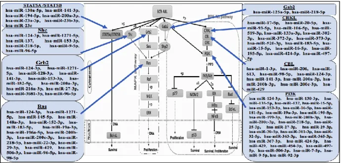
CML comprises an incredibly complex network of signaling cascade mechanism. The hybrid protein BCR-ABL having tyrosine kinase characteristics and transforming ability, which activates the downstream signal transduction pathways involved in CML (Fig. 1) [14].
In Bcr-Abl oncogene, c-Abl acts as the catalytic domain while the Bcr acts as the interacting site for various substrates, and so after enhancement of the ‗biological portfolio‘, BCR-ABL activates various signaling pathways.
JAK2-STAT5 was one of the primary pathways that are being constitutively activated by BCR-ABL [15,16]. The C-terminus of BCR-ABL binds to JAK2 physically and cause its phosphorylation; after activation, JAK2 itself enhances the downstream molecules including STAT3 and STAT5. BCR-ABL also activates STAT5 directly and thus independent of JAK2 [17].STAT5 activation has been shown to be correlated with the activation of anti-apoptotic protein Bcl-xL, and finally develop drug resistance by activation of Rad51 [5]. BCR-ABL directly stimulates the tyrosine-phosphorylation and STAT5 dimerization, the STAT5 dimer then translocated into the nucleus and binds to DNA and endorses activation of downstream target genes [18]. The study proved that the STAT5 overexpression leads to the TKI-resistant phenotype while STAT3 and STAT1 have no effect [19]. The mRNA expression of STAT5A and STAT5B (two homologous STAT5 gene products) found to be increased at advanced CML stage.
Asian Pac. J. Health Sci., 2018; 5(4):65-84 e-ISSN: 2349-0659, p-ISSN: 2350-0964 ____________________________________________________________________________________________________________________________________________
____________________________________________________________________________________________________________________________________________ Singh et al ASIAN PACIFIC JOURNAL OF HEALTH SCIENCES, 2018; 5(4):65-84
www.apjhs.com
67
Fig. 1 Signaling cascade in Chronic Myeloid Leukemia Hematopoietic Stem Cell The mammalian Shc gene codes for adaptor proteins,
which stimulates the cytoplasmic transduction of mitogenic stimuli from RTKs Ras[20]. Shc proteins to get phosphorylated by Receptor Tyrosine Kinases (RTKs) after ligand stimulation [20]; and then interacts with the SH2 domain of Grb2 and acts as an alternative docking site for the Grb2/Sos complex [21-23]. The reports suggest that the Shc/Grb2/Sos complex is involved in Ras stimulation. Shc proteins are found to be constitutively tyrosine-phosphorylated in cancer that causes continuous activation of oncogenes with TK activity [24,25]. Shc polypeptides are found to be good substrates of the BCR-ABL kinase activity, and the constitutive phosphorylation of Shc found to be express in the Bcr-Abl expressing cells [26,27]. MAPK pathway is the main and well known downstream signaling cascade in various cancers and other cellular processes [28]. This signaling cascade also plays a pivotal role in Ph+ cells and is necessary for the transcription of proliferating and anti-apoptotic genes [29]. In Ph+ cells, the autophosphorylation of tyrosine 177 on BCR-ABL fusion protein provides the binding site for growth factor receptor-bound protein 2 (Grb2). Subsequently, Grb2 binds to the SOS protein, which stabilizes RAS (GTPase) in its active form. Activated RAS provokes the kinase activity of RAF, which later on initiates a signaling pathway via the kinases MEK1/MEK2 and ERK (MAPK), finally begins the activation of gene transcription [30].
One critical signal transducer of BCR-ABL is Gab2. Through many interaction motifs, Gab2 proteins link growth factor and cytokine receptors to downstream effectors such as the Shp2/Ras/ERK, PI3K/AKT/mTOR and JAK/STAT pathways [31].The
Grb2 acts as the connecting link between BCR-ABL and Gab2, by binding to the SH2 domain of the Bcr and SH3 domain of Gab2 [32,33]. Gab2 is also involved in oncogenic signaling as it gets express in multiple solid tumors and also in acute myeloid leukemia (AML) [31]. Gab2 get silenced by the shRNAs, that inhibit proliferation and colony formation of CD34+ cells in CML patients [34], and this supports that human CML might be dependent on the BCR-ABL-driven Gab2 signaling and may be Gab2 signaling directs the TKI sensitivity of CML cells. Similar to previously discussed networks, PI3K/Akt also play an important role in cell cycle progression, differentiation, transcription, translation and finally apoptosis [35,36]. BCR-ABL along with PI3K, CBL, CRK, and CRKL form multimeric complexes and that lead to the activation of PI3K pathway [37]. Later, AKT a serine-threonine kinase activates the anti-apoptotic signaling by binding and phosphorylating the pro-apoptotic protein, Bad. After that, Bad becomes inactive because it is trapped by cytoplasmic 14-3-3 proteins and no longer free to bind anti-apoptotic proteins [38].
The study on the regulation by microRNAs in cancer has increased unexpectedly over the last 10 years. Similar to diabetes, cancer is also found to be the common disease for today's world; at least one-third of the population is suffering from cancer. Despite progress in the treatment of cancer, most people with advanced cancer get eventually die. So, the need of new treatments for cancer lead to the discovery of miRNAs and siRNAs, that approved as the new tool for the diagnosis and treatment of cancer. A total of
Asian Pac. J. Health Sci., 2018; 5(4):65-84 e-ISSN: 2349-0659, p-ISSN: 2350-0964 ____________________________________________________________________________________________________________________________________________
____________________________________________________________________________________________________________________________________________ Singh et al ASIAN PACIFIC JOURNAL OF HEALTH SCIENCES, 2018; 5(4):65-84
www.apjhs.com
68
38,589 mature miRNAs have been registered in the miRBase (as of March 2018), which were recognized from humans, primates, rodents, birds, fish, worms, flies, and viruses. The miRNAs sequences are found to be highly conserved across species. The first link between miRNA and cancer was recognized when the gene locations of miR-15 and miR-16 found to be deleted on chromosome 13 in the Chronic
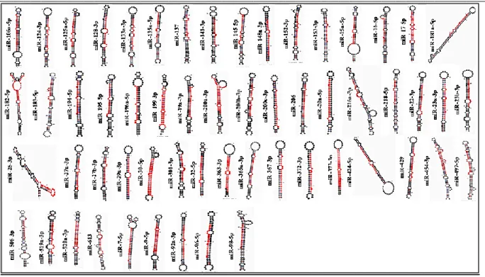
Lymphocytic Leukemia (CLL), so both these miRNAs recognized as the tumor suppressors [39]. Several miRNAs (having the sites of 8-mer, 7mer-m8 and 7mer-A1), which play a significant role in the signaling pathway of CML identified with the help of tools such as Target Scan (target predicting tool), miRbase (alignment tool) and miRNAMap (secondary structures determining tool) (Fig. 2) (Table 1) (Fig. 3).
Fig. 2 miRNAs that target the components of CML signaling pathways
Table1: Different Human miRNAs (hsa-miRs), which target the genes of CML pathways, alongwith their accession no., mature sequence, chromosomal location and gene location
miRNAs Accession no. Mature sequence
(5’ 3’) Chromoso mal Location Gene Location (Exon/Intron/ UTR) Target gene miR-130a-5p MIMAT0004593 gcucuuuucacauugugcuacu chr 11 Intergenic
STAT5A / STAT5B miR-141-3p MIMAT0000432 uaacacugucugguaaagaugg chr 12 Intergenic
miR-194-5p MIMAT0000460 uguaacagcaacuccaugugga chr 1 Intron-12/-1 miR-200a-3p MIMAT0000682 uaacacugucugguaacgaugu chr 1 Intergenic
miR-23a-3p MIMAT0000078 aucacauugccagggauuucc chr 19 Intergenic miR-23b-3p MIMAT0000418 aucacauugccagggauuaccac chr 9 Intron+14/+6/+
15/+5/+4 miR-23c MIMAT0018000 aucacauugccagugauuaccc chr X ---- miR-124-3p MIMAT0000422 uaaggcacgcggugaaugccaa chr 8 Intergenic
Shc miR-1271-5p MIMAT0005796 cuuggcaccuagcaagcacuca chr 5
----miR-137 MIMAT0000429 uuauugcuuaagaauacgcguag chr 1 Intergenic miR-153-3p MIMAT0000439 uugcauagucacaaaagugauc chr 2 Intergenic miR-218-5p MIMAT0000275 uugugcuugaucuaaccaugu chr 4 Intron+14/+15
miR-9-5p MIMAT0000441 ucuuugguuaucuagcuguauga chr 1 Intron +1/+2 miR-96-5p MIMAT0000095 uuuggcacuagcacauuuuugcu chr 7 Intergenic
Asian Pac. J. Health Sci., 2018; 5(4):65-84 e-ISSN: 2349-0659, p-ISSN: 2350-0964 ____________________________________________________________________________________________________________________________________________
____________________________________________________________________________________________________________________________________________ Singh et al ASIAN PACIFIC JOURNAL OF HEALTH SCIENCES, 2018; 5(4):65-84
www.apjhs.com
69
miR-124-3p MIMAT0000422 uaaggcacgcggugaaugccaa chr 8 Intergenic
Grb2 miR-1271-5p MIMAT0005796 cuuggcaccuagcaagcacuca chr 5
----miR-128-3p MIMAT0000424 ucacagugaaccggucucuuu chr 2 Intron+15/+18 miR-141-3p MIMAT0000432 uaacacugucugguaaagaugg chr 12 Intergenic miR-153-3p MIMAT0000439 uugcauagucacaaaagugauc chr 2 Intergenic miR-182-5p MIMAT0000259 uuuggcaaugguagaacucacacu chr 7 Intergenic miR-200a-3p MIMAT0000682 uaacacugucugguaacgaugu chr 1 Intergenic miR-216a-3p MIMAT0022844 ucacaguggucucugggauuau chr 2 Intergenic miR-27a-3p MIMAT0000084 uucacaguggcuaaguuccgc chr 19 Intergenic miR-27b-3p MIMAT0000419 uucacaguggcuaaguucugc chr 9 Intron+14/+6/+
15/+5/+4 miR-3681-3p MIMAT0018109 acacagugcuucauccacuacu chr 2
----miR-96-5p MIMAT0000095 uuuggcacuagcacauuuuugcu chr 7 Intergenic miR-124-3p MIMAT0000422 uaaggcacgcggugaaugccaa chr 8 Intergenic
Ras miR-1271-5p MIMAT0005796 cuuggcaccuagcaagcacuca chr 5
----miR-145-5p MIMAT0000437 guccaguuuucccaggaaucccu chr 5 Intergenic miR-148a-3p MIMAT0000243 ucagugcacuacagaacuuugu chr 7 Intergenic miR-152-3p MIMAT0000438 ucagugcaugacagaacuugg chr 17 Intron +2/+1 miR-183-5p MIMAT0000261 uauggcacugguagaauucacu chr 7 Intergenic miR-19a-3p MIMAT0000073 ugugcaaaucuaugcaaaacuga chr 13 3UTR+2
Intron +2/+3 miR-196a-5p MIMAT0000226 uagguaguuucauguuguuggg chr 17 Intergenic miR-200b-3p MIMAT0000318 uaauacugccugguaaugauga chr 1 Intergenic
hsa-miR-200c-3p
MIMAT0000617 uaauacugccggguaaugaugga chr 12 Intergenic miR-218-5p MIMAT0000275 uugugcuugaucuaaccaugu chr 4 Intron +14/+15
miR-22-3p MIMAT0000077 aagcugccaguugaagaacugu chr 17 3UTR+3/+2 Exon+3 miR-29a-3p MIMAT0000086 uagcaccaucugaaaucgguua chr 7 Intergenic
miR-429 MIMAT0001536 uaauacugucugguaaaaccgu chr 1 Intergenic miR-506-3p MIMAT0002878 uaaggcacccuucugaguaga chr X Intergenic miR-96-5p MIMAT0000095 uuuggcacuagcacauuuuugcu chr 7 Intergenic miR-98-5p MIMAT0000096 ugagguaguaaguuguauuguu chr X Intron
+59/+60/+61/+ 34 miR-125a-5p MIMAT0000443 ucccugagacccuuuaaccuguga chr 19 Intergenic
Gab2 miR-218-5p MIMAT0000275 uugugcuugaucuaaccaugu chr 4 Intron +14/+15
miR-17-5p MIMAT0000070 caaagugcuuacagugcagguag chr 13 3UTR+2 Intron +2/+3
CRKL miR-20a-5p MIMAT0000075 uaaagugcuuauagugcagguag chr 13 3UTR+2
Intron +2/+3 miR-93-5p MIMAT0000093 caaagugcuguucgugcagguag chr 7 Intron +13/+8 miR-106a-5p MIMAT0000103 aaaagugcuuacagugcagguag chr X Intergenic miR-519a-3p MIMAT0002869 aaagugcauccuuuuagagugu chr 19 Intergenic miR-133a-3p MIMAT0000427 uuugguccccuucaaccagcug chr 18 Intron-12/+3 miR-302a-3p MIMAT0000684 uaagugcuuccauguuuugguga chr 4 Intron-10/-11 miR-372-3p MIMAT0000724 aaagugcugcgacauuugagcgu chr 19 Intergenic miR-373-3p MIMAT0000726 gaagugcuucgauuuuggggugu chr 19 Intergenic
Asian Pac. J. Health Sci., 2018; 5(4):65-84 e-ISSN: 2349-0659, p-ISSN: 2350-0964 ____________________________________________________________________________________________________________________________________________
____________________________________________________________________________________________________________________________________________ Singh et al ASIAN PACIFIC JOURNAL OF HEALTH SCIENCES, 2018; 5(4):65-84
www.apjhs.com
70
miR-520a-3p MIMAT0002834 aaagugcuucccuuuggacugu chr 19 Intergenic miR-183-5p MIMAT0000261 uauggcacugguagaauucacu chr 7 Intergenic miR-15a-5p MIMAT0000068 uagcagcacauaaugguuugug chr 13 Intron+3/+4/+5
Exon+3/+4 miR-16-5p MIMAT0000069 uagcagcacguaaauauuggcg chr 13 Intron+3/+4/+5 miR-195-5p MIMAT0000461 uagcagcacagaaauauuggc chr 17 Intron+1 miR-424-5p MIMAT0001341 cagcagcaauucauguuuugaa chr X Exon+1 miR-497-5p MIMAT0002820 cagcagcacacugugguuugu chr 17 Intron+1
miR-1-3p MIMAT0000416 uggaauguaaagaaguauguau chr 18
----CBL miR-206 MIMAT0000462 uggaauguaaggaagugugugg chr 6 Intergenic
miR-613 MIMAT0003281 aggaauguuccuucuuugcc chr 12 Intron+2/+1 miR-98-5p MIMAT0000096 ugagguaguaaguuguauuguu chr X Intron
+59/+60/+61/+ 34 miR-124-3p MIMAT0000422 uaaggcacgcggugaaugccaa chr 8 Intergenic miR-141-3p MIMAT0000432 uaacacugucugguaaagaugg chr 12 Intergenic miR-200a-3p MIMAT0000682 uaacacugucugguaacgaugu chr 1 Intergenic miR-200b-3p MIMAT0000318 uaauacugccugguaaugauga chr 1 Intergenic miR-200c-3p MIMAT0000617 uaauacugccggguaaugaugga chr 12 Intergenic miR-429 MIMAT0001536 uaauacugucugguaaaaccgu chr 1 Intergenic miR-124-3p MIMAT0000422 uaaggcacgcggugaaugccaa chr 8 Intergenic
PI3K miR-130a-3p MIMAT0000425 cagugcaauguuaaaagggcau chr 11 Intergenic
miR-135a-5p MIMAT0000428 uauggcuuuuuauuccuauguga chr 3 Intergenic miR-124-3p MIMAT0000422 uaaggcacgcggugaaugccaa chr 8 Intergenic miR-130-3p MIMAT0000425 cagugcaauguuaaaagggcau chr 11 Intergenic miR-135a-5p MIMAT0000428 uauggcuuuuuauuccuauguga chr 3 Intergenic miR-137 MIMAT0000429 uuauugcuuaagaauacgcguag chr 1 Intergenic miR-15a-5p MIMAT0000068 uagcagcacauaaugguuugug chr 13 Intron+3/+4/+5
Exon+3/+4 miR-153-3p MIMAT0000439 uugcauagucacaaaagugauc chr 2 Intergenic
Asian Pac. J. Health Sci., 2018; 5(4):65-84 e-ISSN: 2349-0659, p-ISSN: 2350-0964 ____________________________________________________________________________________________________________________________________________
____________________________________________________________________________________________________________________________________________ Singh et al ASIAN PACIFIC JOURNAL OF HEALTH SCIENCES, 2018; 5(4):65-84
www.apjhs.com
71
miR-181a-5p MIMAT0000256 aacauucaacgcugucggugagu chr 9 Intron-2/+1
PI3K miR-19a-3p MIMAT0000073 ugugcaaaucuaugcaaaacuga chr 13 3UTR+2
Intron+3/+2 miR-195-5p MIMAT0000461 uagcagcacagaaauauuggc chr 17 Intron+1
miR-199-3p MIMAT0000232 acaguagucugcacauugguua chr 19 Intron-14/-15/-16 miR-200b-3p MIMAT0000318 uaauacugccugguaaugauga chr 1 Intergenic miR-200c-3p MIMAT0000617 uaauacugccggguaaugaugga chr 12 Intergenic
miR-218-5p MIMAT0000275 uugugcuugaucuaaccaugu chr 4 Intron+14/+15 miR-25-3p MIMAT0000081 cauugcacuugucucggucuga chr 7 Intron+13/+8 miR-27a-3p MIMAT0000084 uucacaguggcuaaguuccgc chr 19 Intergenic miR-29a-3p MIMAT0000086 uagcaccaucugaaaucgguua chr 7 Intergenic miR-30-5p MIMAT0000087 uguaaacauccucgacuggaag chr 6 Intron+3 miR-301-3p MIMAT0000688 cagugcaauaguauugucaaagc chr 17 Intron+1/-1
miR-32-5p MIMAT0000090 uauugcacauuacuaaguugca chr 9 Intron+12/+13/ +14 miR-363-3p MIMAT0000707 aauugcacgguauccaucugua chr X Intergenic miR-365a-3p MIMAT0000710 uaaugccccuaaaaauccuuau chr 16 Intergenic miR-367-3p MIMAT0000719 aauugcacuuuagcaaugguga chr 4 Intron-10/-11 miR-424-5p MIMAT0001341 cagcagcaauucauguuuugaa chr X Exon+1
miR-429 MIMAT0001536 uaauacugucugguaaaaccgu chr 1 Intergenic miR-454-3p MIMAT0003885 uagugcaauauugcuuauagggu chr 17 Intron+1/-1 miR-497-5p MIMAT0002820 cagcagcacacugugguuugu chr 17 Intron+1
miR-506-3p MIMAT0002878 uaaggcacccuucugaguaga chr X Intergenic miR-7-5p MIMAT0000252 uggaagacuagugauuuuguugu
u
chr 9 Intergenic miR-9-5p MIMAT0000441 ucuuugguuaucuagcuguauga chr 1
----miR-92a-3p MIMAT0000092 uauugcacuugucccggccugu chr 13 3UTR+2 Intron+3/+2
Asian Pac. J. Health Sci., 2018; 5(4):65-84 e-ISSN: 2349-0659, p-ISSN: 2350-0964 ____________________________________________________________________________________________________________________________________________
____________________________________________________________________________________________________________________________________________ Singh et al ASIAN PACIFIC JOURNAL OF HEALTH SCIENCES, 2018; 5(4):65-84
www.apjhs.com
72
Fig. 3 Secondary structures of miRNAs that target the components of CML signaling pathways (Note: The red colour segment in the structure represents the mature sequence)
Few miRNAs (21 out of 105) which are found to be common, includes hsa-miR-124-3p, hsa-miR-1271-5p, miR-137, miR-141-3p, miR-15a-5p, miR-153-3p, miR-16-5p, miR-183-5p, 195-5p, 19a-3p, 200, hsa-miR-218-5p, hsa-miR-27-3p, hsa-miR-29-3p, hsa-miR-424-5p, hsa-miR-429, hsa-miR-497-hsa-miR-424-5p, hsa-miR-506-3p, hsa-miR-9-5p, hsa-miR-96-5p and hsa-miR-98-5p (Table 1). The reports on the function of these miRNAs in CML have not been found generally, but the role of them in other cancers has been reported. These miRNAs may possess oncogenic or tumor suppressive characteristics.
MiR-124 is found to act as the tumor suppressor in glioma, medulloblastoma, oral squamous cell carcinomas and hepatocellular carcinoma (HCC) [40-43]. The miR-124 was also reported to affect the invasive and metastatic potential of breast cancer [44]. MiR-124 also downregulates the pancreatic cancer progression after binding to 3‘UTR of Rac1, which proved as the tumor promoter in pancreatic cancer [45]. Using bioinformatics, miR-1271 was predicted to target the FOXQ1, a transcription factor that plays a significant role in growth, aging, metabolism, and cancer [46,47]. Several findings proved different expression patterns of miR-1271. It gets upregulated in
head and neck tumor cells [48] downregulated in hepatocellular cancer [49], and gastric cancer cell lines [50]. This miRNA also affects the PI3K-Akt pathway in the Non-small cell lung cancer (NSCLC) cell lines, by acting against mTOR. Akt gets phosphorylated by mTOR and carry out the PI3K-Akt pathway of the cell signaling. miR-1271 was found to affect the translation of mTOR only without causing the degradation of mTOR mRNA [51].
A negative correlation was found between the paxillin (Pxn) gene and miR-137 in the Colorectal cancer (CRC) [52]. The Pxn codes for a focal adhesion molecule, that forms link between the extracellular matrix (ECM) and actin cytoskeleton [53]. Similar to miR-1271, the expression of miR-137 also vary according to cancer. Downregulation of miR-137 has also been observed in oral cancer cells [54], glioblastoma cell lines [52], while it gets upregulated in squamous cell carcinoma of the tongue [55]. In the cell cycle, this miRNA affects the cyclin D1–CDK4/CDK6 complex, by targeting CDK6, as a result, in cell cycle the G1/S transition gets halted [56].
MiR-141 found to act as an antagonist in the Ovarian Cancer. OC is a lethal disease in women, which occur mainly due to Epithelial-mesenchymal transition (EMT). During this transition, make the cells to gain
Asian Pac. J. Health Sci., 2018; 5(4):65-84 e-ISSN: 2349-0659, p-ISSN: 2350-0964 ____________________________________________________________________________________________________________________________________________
____________________________________________________________________________________________________________________________________________ Singh et al ASIAN PACIFIC JOURNAL OF HEALTH SCIENCES, 2018; 5(4):65-84
www.apjhs.com
73
the capacity of migration and invasion. Overexpression of miR-141 in the OC cell line SKOV3 significantly inhibit the cell proliferation by affecting the EMT [57]. miR-141 is found to inhibit the proliferation of melanoma cells [58], and gastric cancer cells [59]. miR-141 also get downregulated in the renal cell carcinoma (RCC) in comparison with the normal kidney. miR-141 inhibit ZFHX1B, a transcriptional repressor of E‐ cadherin, and thus suppress the EMT transition in RCC [60].
MicroRNAs-15 and -16 belong to a common predecessor family. Both act as an antagonist on the downstream target of the p53 signaling pathway. Upregulation of miR-15/16 block cell cycle and also accomplish caspase-3 dependent apoptosis. Involuntary expression of miR-15/16 led the low expression of RPS6KB1 mRNA and thus affect the mTOR pathway [61]. miR-15/miR-16 is found to be removed in more than 65% of Chronic Lymphocytic Leukemia (CLL) cell lines which suggest the suppressive role of miR-15 in this cancer [39]. These miRNAs target the antiapoptotic protein Bcl2, which get overexpressed in most of the nondividing B cells of CLL [62]. miR-15a/16-1 target the genes involving in the ‗‗cell cycle‘‘, in both a leukemic cell model and in primary CLLs, and categorizes a signature of common genes whose suppressing characterizes the miR-15a/16-1-stimulated phenotype in CLL [63]. The miR-16 upregulation decreases Cyclin D1 and BCL2 at mRNA and protein levels in MCF-7 cell line (breast cancer cell line). It also affects cell proliferation and provokes apoptosis in MCF-7 cells [64].
High‐ throughput method recognized miR‐ 153 as an oncogene in the prostate cancers. miR-153 inhibits the expression of PTEN (a tumor suppressor gene); and as a result, enhances the G1/S transition [65]. miR-153 also found to act as the tumor suppressor. It was reported that miR-153 reduce EMT transition and hence destroy tumor cells in gastric cancer and breast cancer; and suppress metastasis in human non-small-cell lung cancer [66-68]. miR-153 also promote cisplatin-mediated apoptosis in breast cancer cell lines by suppressing Homologous to the E6-associated protein carboxyl terminus domain containing 3 (HECTD3) expression [69]. miR-153-3p was found to inhibit metastasis in the Oral cavity squamous cell carcinoma (OSCC) by targeting Nrf2. Nrf2 protein gets constitutively upregulated in the tumor under oxidative stress. The miR-153-3p/Nrf2 interaction might be considered as a potential therapeutic target in OSCC [70].
The expression of miR-183 gets over-express or under-express depending on the types of cancer. MiR-183 have been found to be upregulated in leukemia, hepatic and colorectal cancer [71-74], and downregulated in lung cancer [75]. miR-183 binds to 3‘UTR of VIL2/Ezrin a member of the ezrin/radixin/moesin (ERM) family of proteins that controls cytoskeletal-membrane interactions and cell signaling [76]. Ezrin is a membrane cytoskeleton crosslinker and controls the actin during adhesion and motility in breast cancer cells [77]. Hence it is reported that miR-183 diminishes migration in breast cancer cells.
The miR-195-5p have been studied in several cancer cell lines as the tumor suppressor. miR‐ 195‐ 5p was identified to inhibit the glucose uptake in the bladder cancer T24 cells by directly targeting the glucose transporter member 3 (GLUT3) [78]. miR-195-5p is also involved in the suppression of human prostate cancer (PCa). The miR-195 target the ribosomal protein S6 kinase, 70 kDa, polypeptide 1 (RPS6KB1) gene in PCa and reduce the cell invasion and migration and also enhance the apoptosis [79]. miR-195-5p regulate the PHD finger protein 19 (PHF19) in the Hepatocellular carcinoma (HCC) [80]. PHF19, a component of polycomb repressive complex 2 (PRC2), promote the hepatoma cell migration, invasion, and proliferation; also regulate the growth of xenograft tumors [81].
MiR-19a-3p regulate the 5-Lipoxygenase (5-LO), the key enzyme in leukotriene biosynthesis [82]. Leukotrienes are mediators of the innate immune system and inflammatory processes, and they might also be involved in cancer development. miRNA-19a/b acts as an oncogene in oral cancer and it negatively regulates SOCS3 by JAK-STAT pathway [83].
MiR-200a induce apoptosis in the renal cell carcinoma by directly targeting SIRT1 [84,85]. SIRT1 has been shown to deacetylate and thereby deactivate the p53 protein. The regulatory relationship between miR-200c and Heme oxygenase-1 (HO-1) determined in Renal cell carcinoma (RCC). miR-200c might sensitize RCC cells to Sorafenib and Imatinib to hinder cell proliferation by targeting HO-1 [85]. HO-1 is a heme degrading enzyme; and acts as the anti-inflammatory and anti-apoptotic protein, due to which it may lead to cancer. HO-1 was found to express in lung cancer [86], and it was recently reported that HO- 1 nuclear localization was involved in imatinib resistance in chronic myeloid leukemia cells [87], indicating the increased significance of HO-1 in cancer biology. Chen et al., found the low expression of miR-200a in
Asian Pac. J. Health Sci., 2018; 5(4):65-84 e-ISSN: 2349-0659, p-ISSN: 2350-0964 ____________________________________________________________________________________________________________________________________________
____________________________________________________________________________________________________________________________________________ Singh et al ASIAN PACIFIC JOURNAL OF HEALTH SCIENCES, 2018; 5(4):65-84
www.apjhs.com
74
hepatocellular carcinoma cells, and it might inhibit the metastasis in the hepatocellular carcinoma cells by targeting FOX2 [88]. Ovarian cancer is found to be the most lethal among gynecological tumors, and it is due to its metastatic behavior. The metastasis occurs in cancer cells due to the EMT. The miR-200 family induce the mesenchymal to epithelial transition (MET), therefore, miR-200 family members found to be effective against metastasis [89].
MiR-218 is found to be Lamin targeting miRNA. In the mammalian cells, Lamin, the protein of nuclear lamina is of two types: Lamin A (Lamin A and C, which are all products of alternative splicing from the LMNA gene) and Lamin B (Lamin B1 and B2, which are encoded by two separate genes, LMNB1 and LMNB2, respectively) [90]. These proteins provide shape and stability to the nucleus; and also control protein complexes involved in replication, transcription and nuclear positioning. Lamin A is often found to be low expressed in breast, ovarian, colon and gastric cancers [91-93], while Lamin B1 is overexpressed in liver, pancreas and prostate cancers [94-96].Breast cancer emerged as the most commonly diagnosed life-threatening cancer in women, especially the Triple-negative breast cancer (TNBC). But the treatment of TNBC remains challenging, so the miRNAs have emerged as a potential tool for the diagnosis and treatment of breast cancer. miR-218 is downregulated in TNBC samples and it directly targets the Lamin B1, so the report revealed that miR-218 has a tumor suppressive activity in breast cancer [97].
MiRNA-27a-3p found to act against Yes-associated protein-1(YAP1) in oral squamous cell carcinoma (OSCC) patients and OSCC cell lines [98]. YAP1 act as the co-activator for TEAD transcription factor, which activate cell growth genes and anti-apoptotic genes. Increased expression of miR-27a-3p also inhibit the EMT related molecules in OSCC cell lines, including Snail and Twist; and hence manipulate the metastasis through the EMT inhibition. miR-27a also get downregulated in cervical cancer by obstructing TGF-βRI expression and TGF-β signaling. miR-27a is also found to act as antitumor in colorectal cancer [99,100], oral squamous carcinoma [54], and esophageal carcinoma [101]. miR-27a was also found to act as an oncogene in many tumors by regulating cell proliferation, metastasis and drug resistance [102-104]. The role of miR-27a in human colon cancer was found to be as the key regulator of lymph angiogenesis and it functions via the TGF-β-SMAD4 signaling pathway. TGF-β/SMAD4 signaling pathway regulates the signal transduction from cell membrane to nucleus
and controls a wide series of cellular activities, as well as cancer progression and metastasis [105]. These findings indicated the key roles of miR-27a in tumorigenesis and metastasis in human colon cancer and implicated miR-27a as a potential target for the development of new anticancer therapies [106]. MiR-29a/b/c is considered as tumor suppressive in head and neck squamous cell carcinoma (HNSCC), where it targets the Lysyl oxidase-like 2 (LOXL2), and thus affect the metastasis in this carcinoma [107]. LOXL2 gene encodes for an extracellular copper-dependent amine oxidase that catalyzes the initial stage in the formation of crosslinks between collagen and elastin, and this cause remodeling in ECM and enhances the cellular metastasis [108]. A study reported the role of miR-29a/b/c in EMT also after interacting with the transcription factor Snail1, which further repressed E-cadherin [109]. miR-29 act against anti-apoptotic protein Mcl-1in bile duct epithelial cancer [110]; low expression of miR-29 found in breast cancer patients [111]; the downregulation of miR-29 is associated with metastasis and so this miRNA can be used as the biomarker for tumor advancement [112]. MiR-424-5p act as both tumor suppressor and oncogene depending upon the tissues. The downregulation of miR-424-5p was found in hepatocellular carcinoma [113], cervical cancer [114],esophageal squamous cell carcinoma [115], compared to the upregulation in pancreatic cancer [116]. In Epithelial Ovarian Cancer (EOC), miR-424-5p arrest G1/S transition in cell cycle by targeting CCNE1, which code for cyclin that bind to CDK2 and cause cell cycle progression [117]. miR-424 acts as the oncogene in non-small cell lung cancer (NSCLC) by enhancing cell migration and invasion after targeting Tumor Necrosis Factor Alpha-Induced Protein 1(TNFAIP1) [118].
Like other miRNAs, mir-429 perform as both oncogenic and tumor suppressive. It gets over expressed in endometrial carcinoma [119], and colorectal cancer [120]; while low expressed in osteosarcoma [121], renal cell carcinoma [122] and cervical cancer [123]. In the case of bladder cancer, the behavior of miR-429 is controversial. This considered having a tumor suppressive role based on the earlier report in which miR-429 was found to inhibit the EMT by restoring the expression of E-cadherin in bladder cancer [124], while in another report miR-429 enhance bladder cancer by targeting CDKN2B [125]. Mir-429 is also showing the oncogenic effect in prostate cancer by targeting p27kip1 [126]. p27kip1 regulate cell cycle
Asian Pac. J. Health Sci., 2018; 5(4):65-84 e-ISSN: 2349-0659, p-ISSN: 2350-0964 ____________________________________________________________________________________________________________________________________________
____________________________________________________________________________________________________________________________________________ Singh et al ASIAN PACIFIC JOURNAL OF HEALTH SCIENCES, 2018; 5(4):65-84
www.apjhs.com
75
after interacting with cyclins or cyclin/CDK complexes to inhibit their kinase activities that normally promote G1/S phase progression [127,128].
The tumor suppressive role of miR-497-5p along with miR-195-5p and miR-455-3p found in melanoma A375 cells by targeting hTERT, which encode for the catalytic subunit of telomerase [129]. Its expression is involved in the process of cell immortalization and cancer tumorigenesis, growth, migration, invasion, and prognostic evaluation, although the underlying mechanism remains unclear [130]. MiR-497-5p acts as the negative regulator for SMAD3 and affect the TGFβ signaling pathway and finally lead to the G0/G1 arrest [131]. MiR-497-5p has been reported to suppress the tumor cell proliferation and metastasis in prostate cancer, hepatocellular carcinoma, and non-small cell lung cancer (NSCLC) tissues [132-135].
MiR-506-3p repress the activity of long non-coding RNA (lncRNA) NEAT1, which is found to play a crucial role in Pancreatic cancer (PC) [136]. Long non-coding RNAs consist of 200 nucleotides and play an important role in tumorigenesis. miR-506 is considerably downregulated in many cancers and act as a tumor suppressor by targeting oncogenes like N-Ras, PIM3, SPHK1, ROCK1, and ETS-1, hence regulate cell proliferation, apoptosis, metastasis and drug resistance [137]. In cervical cancer, miR-506 acts as an anti-proliferative agent by directly targeting the Gli3 transcription factor of the hedgehog pathway [138]. MiR-9 (miR-9-5p and miR-9-3p) get overexpressed in tumors in comparison to the normal breast tissues (NBTs), but both miR-9-5p and miR-9-3p found to differentially expressed in the breast cancer subgroups as identified by the expression of estrogen receptor (ER) and progesterone receptor (PgR) [139]. In Human Papilloma Virus (HPV)-positive cancer, miR-9 along with HPV E6 oncoprotein increase cell migration by downregulating follistatin-like 1 (FSTL1) and Activate Leukocyte Cell Adhesion Molecule (ALCAM) mRNAs [140].
The role of miR-96-5p is found suspicious in the case of hepatocellular carcinoma (HCC) because according to a data it suppresses apoptosis in hepatocellular carcinoma (HCC) by inhibiting caspase 9, an important protein for mitochondria regulated apoptosis [141], while another study reveals its metastatic role along with miR-182-5p in HCC by targeting 5′-UTR and 3′-UTR of three key Insulin Growth Factor (IGF) axis genes, namely IGF-1R, IGF Binding Protein-3 (IGFBP-3) and IGF-II transcripts [142]. In TNBC,
highly aggressive form of breast cancer miR-96 including miR-557, and miR-3182 inhibit the mTOR and S6K1 genes of PI3K/Akt/mTOR signaling pathway and so affect the cellular functions like transcription, translation, division, metabolism, metastasis, proliferation, and development of tumor [143]. miR-96 inhibit EMT by targeting astrocyte elevated gene 1 (AEG 1) in Glioblastoma cancer cells [144]. AEG-1 triggers anchorage-independent growth and invasiveness of tumor cells.
Few reports revealed miR-98-5p as an oncogene in non-small lung cancer (NSCLC). Cisplatin emerged as an effective drug for lung cancer, but later drug-resistance and side effects of cisplatin led to the discovery of new drugs. And then the discovery of Epigallocatechin-3- gallate (EGCG) in green tea proved as the chemo preventive agent. EGCG has displayed tumor suppressive effect in the prostate, head, and neck; and lung cancer [145-147]. EGCG is found to enhance the effect of cisplatin and also induce apoptosis in tumor cells. EGCG could improve the therapeutic effect of cisplatin through inhibiting miR-98–5p, signifying that miR-98–5p could be a target in clinical cisplatin treatment of NSCLC [148]. EGCG also increase the capacity of cisplatin by inducing the cisplatin transporter copper transporter 1 (CTR1). NEAT1, a long non-coding RNA (lncRNA) upregulates CTR1 to boost cisplatin sensitivity. Mir-98-5p bind to NEAT1 at the complementary site and lowers the expression of CTR1 [149]. In comparison to NSCLC, the tumor suppressive role of miR-98-5p was observed in pancreatic ductal adenocarcinoma (PDAC). MiR-98-5p was downregulated in the cancerous tissues and it is interpreted that miR-98-5p inhibited PDAC cell proliferation and metastasis by negatively regulating MAPK4 in PDAC cells [150]. Conclusion
Imatinib was found to be superior to conventional medications for CML treatment, but Imatinib resistance led to the development of other TKIs, that appear to overwhelmed Imatinib resistance for many but not all mutations [151-153]. So, the research in this field bent towards gene interference by miRNAs [154,155]. Anti-BCR-ABL RNAi assists Imatinib in inducing cell death in Imatinib-resistant CML cells [153,155].
To search for an alternative treatment, we reviewed miRNAs which regulates the expression of STAT5, Shc, Grb2, Ras, CRKL, CBL and PI3K. These (STAT5, Shc, Grb2, Ras, CRKL, CBL and PI3K) belong to different functional classes of proteins
Asian Pac. J. Health Sci., 2018; 5(4):65-84 e-ISSN: 2349-0659, p-ISSN: 2350-0964 ____________________________________________________________________________________________________________________________________________
____________________________________________________________________________________________________________________________________________ Singh et al ASIAN PACIFIC JOURNAL OF HEALTH SCIENCES, 2018; 5(4):65-84
www.apjhs.com
76
(tyrosine phosphatase, transcription factor, adaptor protein); and these are selected on the basis of their constitutive activation in the Bcr-Abl positive cell lines. Targeting these molecules by miRNAs might effectively enhance chemotherapy-induced cytotoxicity in CML cells, and a time may come when we will be able to control the CML population.
Abbreviations
CCNE: cyclin E; CDK: Cyclin Dependent Kinase; CDKN2B: Cyclin-dependent kinase 4 inhibitor B; FOXQ1: Forkhead Box Q1 protein; Gab2: GRB2-associated-binding protein 2; Grb2: Growth factor receptor-bound protein 2; hsa: Homo sapiens; hTERT: human telomerase reverse transcriptase; IB: inhibitor of kappa B; IKK: Inhibitor of nuclear factor kappa-B kinase subunit alpha ; JAK: Janus Kinase; MAPK: Mitogen Activated Protein Kinase; MDM2: Mouse double minute 2 homolog protein; MDR: Multidrug Resistance protein; MEK: MAPK/ERK kinase; mTOR: mammalian Target Of Rapamycin; N-Ras: Neuroblastoma Ras protein; NEAT1: Nuclear Enriched Abundant Transcript 1; NFB: nuclear factor kappa-light-chain-enhancer of activated B cells; PI3K: Phosphatidylinositol-4,5-bisphosphate 3-kinase; PIM3: Pim-3 proto-oncogene, serine/threonine kinase; PKB: Protein Kinase B; PTEN: Phosphatase and Tensin homolog protein; PTPN11: Protein Tyrosine Phosphatase Non-Receptor Type 11; Rac1: Ras-Related C3 Botulinum Toxin Substrate 1 protein; RISC: RNA-Induced Silencing Complex; ROCK1: Rho Associated Protein Kinase 1; RPS6KB1: Ribosomal Protein S6 Kinase Beta-1 protein; RTK: Receptor Tyrosine Kinase; SH2: Src Homology 2; Shp2: Src Homology Region 2-Containing Protein Tyrosine Phosphatase 2; SOCS3: Suppressor Of Cytokine Signaling 3 protein; SOS: Son Of Sevenless protein; SPHK1:Sphingosine Kinase 1 protein; STAT: Signal Transducer and Activator of Transcription protein; TEAD: TEA domain family member 1 protein; TGF-: Transforming Growth Factor Beta; ZFHX1B: Zinc Finger E-Box-Binding Homeobox 1B protein
Acknowledgment: No source of funding used in the preparation of this review. The authors acknowledge all researchers who have contributed to CML therapy and apologize to them whose effort in this field was not directly cited in this review due to space confinement. The authors also appreciate the support received from the Central University of Punjab, Bhatinda, India, in writing this manuscript.
References
1. Rowley, J. D. A new consistent chromosomal abnormality in chronic myelogenous leukaemia identified by quinacrine fluorescence and Giemsa staining. Nature, 1973;243(5405):290-293.
2. Groffen, J., Stephenson, J. R., Heisterkamp, N., de Klein, A., Bartram, C. R., &Grosveld, G. (1984). Philadelphia chromosomal breakpoints are clustered within a limited region, bcr, on chromosome 22. Cell, 1984;36(1):93-99. 3. Nowell, C. The minute chromosome (Ph 1) in
chronic granulocytic leukemia. Annals of Hematology,1962; 8(2):65-66.
4. Donato, N. J., Wu, J. Y., Zhang, L., Kantarjian, H., &Talpaz, M. Down-regulation of interleukin-3/granulocyte-macrophage colony-stimulating factor receptor β-chain in BCR-ABL+ human leukemic cells: association with loss of cytokine-mediated Stat-5 activation and protection from apoptosis after BCR-ABL inhibition. Blood,2001; 97(9):2846-2853. 5. Slupianek, A., Schmutte, C., Tombline, G.,
Nieborowska-Skorska, M., Hoser, G., Nowicki, M. O., Pierce, A. J., Fishel, R.,&Skorski, T. BCR/ABL regulates mammalian RecA homologs, resulting in drug resistance. Molecular cell, 2001; 8(4):795-806. 6. Vaidya, S., Ghosh, K., &Vundinti, B. R. Recent
developments in drug resistance mechanism in chronic myeloid leukemia: a review. European journal of haematology,2011; 87(5):381-393. 7. Jorgensen, H. G., & Holyoake, T. L. A
comparison of normal and leukemic stem cell biology in chronic myeloid leukemia. Hematological oncology, 2001;19(3): 89-106. 8. Copland, M., Hamilton, A., Elrick, L. J., Baird,
J. W., Allan, E. K., Jordanides, N., Barow, M., Mountford, J. C., & Holyoake, T. L.. Dasatinib (BMS-354825) targets an earlier progenitor population than imatinib in primary CML but does not eliminate the quiescent fraction. Blood, 2006;107(11):4532-4539. 9. Faderl, S., Talpaz, M., Estrov, Z., O'brien, S.,
Kurzrock, R., &Kantarjian, H. M. The biology of chronic myeloid leukemia. New England Journal of Medicine, 1999;341(3):164-172. 10. Melo, J. V. (1996). The diversity of BCR-ABL
fusion proteins and their relationship to leukemia phenotype [editorial; comment]. 11. Reinhart, B. J., Slack, F. J., Basson, M.,
Pasquinelli, A. E., Bettinger, J. C., Rougvie, A. E., Horvitz, H. R., &Ruvkun, G. The
21-Asian Pac. J. Health Sci., 2018; 5(4):65-84 e-ISSN: 2349-0659, p-ISSN: 2350-0964 ____________________________________________________________________________________________________________________________________________
____________________________________________________________________________________________________________________________________________ Singh et al ASIAN PACIFIC JOURNAL OF HEALTH SCIENCES, 2018; 5(4):65-84
www.apjhs.com
77
nucleotide let-7 RNA regulates developmental timing in Caenorhabditis elegans. nature, 2000;403(6772):901-906. 12. Blahna, M. T., &Hata, A. Regulation of miRNA
biogenesis as an integrated component of growth factor signaling. Current opinion in cell biology, 2013;25(2): 233-240.
13. O'carroll, D., & Schaefer, A. (2013). General principals of miRNA biogenesis and regulation in the brain. Neuropsychopharmacology, 2013;38(1):39-54.
14. Melo, J. V., Hughes, T. P., &Apperley, J. F. Chronic myeloid leukemia. ASH Education Program Book, 2003:(1); 132-152.
15. Ilaria, R. L., & Van Etten, R. A. P210 and P190BCR/ABL induce the tyrosine phosphorylation and DNA binding activity of multiple specific STAT family members. Journal of Biological Chemistry, 1996; 271(49): 31704-31710.
16. Shuai, K., Halpern, J., Rao, X., & Sawyers, C. L.Constitutive activation of STAT5 by the BCR-ABL oncogene in chronic myelogenous leukemia. Oncogene, 1996;13(2):247-254. 17. Hantschel, O., Warsch, W., Eckelhart, E.,
Kaupe, I., Grebien, F., Wagner, K. U., Superti-Furga, G.,& Sexl, V. BCR-ABL uncouples canonical JAK2-STAT5 signaling in chronic myeloid leukemia. Nature chemical biology, 2012;8(3):285.
18. Valent, P. Targeting the JAK2-STAT5 pathway in CML. Blood, 2014;124(9), 1386-1388. 19. Warsch, W., Kollmann, K., Eckelhart, E.,
Fajmann, S., Cerny-Reiterer, S., Holbl, A., Gleixner, K. V., Dworzak, M., Mayerhofer, M., Hoermann, G.,& Herrmann, H. High STAT5 levels mediate imatinib resistance and indicate disease progression in chronic myeloid leukemia. Blood, 2011;117(12): 3409–3420. 20. Bonfini, L., Migliaccio, E., Pelicci, G.,
Lanfrancone, L., &Pelicci, P. Not all Shc's roads lead to Ras. Trends in biochemical sciences, 1996;21(7): 257-261.
21. Rozakis-Adcock, M., McGlade, J., Mbamalu, G., Pelicci, G., Daly, R., Li, W., Batzer, A., Thomas, S., Brugge, J., Pelicci, P. G., & Schlessinger, J. Association of the Shc and Grb2/Sem5 SH2-containing proteins is implicated in activation of the Ras pathway by tyrosine kinases. Nature, 1992;360(6405), 689-692.
22. Egan, S. E., Giddings, B. W., Brooks, M. W., Buday, L., Sizeland, A. M., & Weinberg, R. A. Association of SosRas exchange protein with
Grb2 is implicated in tyrosine kinase signal transduction and transformation. Nature, 1993;363(6424), 45-51.
23. Salcini, A. E., McGlade, J., Pelicci, G., Nicoletti, I., Pawson, T., & Pelicci, P. G. Formation of Shc-Grb2 complexes is necessary to induce neoplastic transformation by overexpression of Shc proteins. Oncogene, 1994;9(10): 2827-2836.
24. Pelicci, G., Lanfrancone, L., Salcini, A. E., Romano, A., Mele, S., Grazia, M. B., Segatto, O., Di, P. F.,& Pelicci, P. G. Constitutive phosphorylation of Shc proteins in human tumors. Oncogene, 1995;11(5), 899-907. 25. Jucker, M., Schiffer, C. A., & Feldman, R. A. A
tyrosine-phosphorylated protein of 140 kD is constitutively associated with the phosphotyrosine binding domain of Shc and the SH3 domains of Grb2 in acute myeloid leukemia cells. Blood, 1997;89(6):2024-2035. 26. Matsuguchi, T., Salgia, R., Hallek, M., Eder, M.,
Druker, B., Ernst, T. J., & Griffin, J. D. Shc phosphorylation in myeloid cells is regulated by granulocyte macrophage colony-stimulating factor, interleukin-3, and steel factor and is constitutively increased by p210BCR/ABL. Journal of Biological Chemistry, 1994;269(7): 5016-5021.
27. Tauchi, T., Boswell, H. S., Leibowitz, D., & Broxmeyer, H. E. Coupling between p210bcr-abl and Shc and Grb2 adaptor proteins in hematopoietic cells permits growth factor receptor-independent link to ras activation pathway. Journal of Experimental Medicine, 1994;179(1):167-175.
28. Koul, H. K., Pal, M., & Koul, S. Role of p38 MAP kinase signal transduction in solid tumors. Genes & cancer, 2013;4(9-10): 342-359. 29. Kim, E. K., & Choi, E. J. Pathological roles of
MAPK signaling pathways in human diseases. BiochimicaetBiophysicaActa (BBA)-Molecular Basis of Disease, 2010;1802(4): 396-405.
30. Cahill, M. A., Janknecht, R., &Nordheim, A. Signalling pathways: jack of all cascades. Current Biology, 1996;6(1):16-19. 31. Wohrle, F. U., Daly, R. J., &Brummer, T.
Function, regulation and pathological roles of the Gab/DOS docking proteins. Cell Communication and Signaling, 2009;7(1): 22-40.
32. Pendergast, A. M., Quilliam, L. A., Cripe, L. D., Bassing, C. H., Dai, Z., Li, N., Batzer, A., Rabun, K. M., Der, C. J., Schlessinger,
Asian Pac. J. Health Sci., 2018; 5(4):65-84 e-ISSN: 2349-0659, p-ISSN: 2350-0964 ____________________________________________________________________________________________________________________________________________
____________________________________________________________________________________________________________________________________________ Singh et al ASIAN PACIFIC JOURNAL OF HEALTH SCIENCES, 2018; 5(4):65-84
www.apjhs.com
78
J.,&Gishizky, M. L. BCR-ABL-induced oncogenesis is mediated by direct interaction with the SH2 domain of the GRB-2 adaptor protein. Cell, 1993;75(1): 175-185.
33. Harkiolaki, M., Tsirka, T., Lewitzky, M., Simister, P. C., Joshi, D., Bird, L. E., Jones, E. Y., O'Reilly, N.,& Feller, S. M. Distinct binding modes of two epitopes in Gab2 that interact with the SH3C domain of Grb2. Structure, 2009; 17(6):809-822.
34. Scherr, M., Chaturvedi, A., Battmer, K., Dallmann, I., Schultheis, B., Ganser, A., & Eder, M. Enhanced sensitivity to inhibition of SHP2, STAT5, and Gab2 expression in chronic myeloid leukemia (CML). Blood, 2006;107(8): 3279-3287.
35. Brazil, D. P., Yang, Z. Z., & Hemmings, B. A. (2004). Advances in protein kinase B signalling: AKTion on multiple fronts. Trends in biochemical sciences,2004; 29(5):233-242. 36. Hanada, M., Feng, J., & Hemmings, B. A.
Structure, regulation and function of PKB/ AKT—a major therapeutic target. Biochimicaet Biophysica Acta (BBA)-Proteins and Proteomics,2004; 1697(1-2):3-16.
37. Sattler, M., Salgia, R., Durstin, M. A., Prasad, K. V., & Griffin, J. D.Thrombopoietin induces activation of the phosphatidylinositol‐ 3′ kinase pathway and formation of a complex containing p85PI3K and the protooncoprotein p120CBL. Journal of cellular physiology,1997 ; 171(1): 28-33.
38. Franke, T. F., Cantley, L. C., &Toker, A. Direct regulation of the Akt proto-oncogene product by phosphatidylinositol-3,4 bisphosphate. Science, 1997; 275(5300):665-668.
39. Calin, G. A., Dumitru, C. D., Shimizu, M., Bichi, R., Zupo, S., Noch, E., Aldler, H., Rattan, S., Keating, M., Rai, K.,&Rassenti, L. Frequent deletions and down-regulation of micro-RNA genes miR15 and miR16 at 13q14 in chronic lymphocytic leukemia. Proceedings of the National Academy of Sciences,2002;99(24), 15524-15529.
40. Xia, H., Cheung, W. K., Ng, S. S., Jiang, X., Jiang, S., Sze, J., Leung, G. K., Lu, G., Chan, D. T., Bian, X. W., & Kung, H. F. Loss of brain-enriched miR-124 microRNA enhances stem-like traits and invasiveness of glioma cells. Journal of Biological Chemistry, 2012; 287(13):9962-9971.
41. Hunt, S., Jones, A. V., Hinsley, E. E., Whawell, S. A., & Lambert, D. W. MicroRNA‐ 124 suppresses oral squamous cell carcinoma
motility by targeting ITGB1. FEBS letters,2011; 585(1):187-192.
42. Furuta, M., Kozaki, K. I., Tanaka, S., Arii, S., Imoto, I., &Inazawa, J. miR-124 and miR-203 are epigenetically silenced tumor-suppressive microRNAs in hepatocellular carcinoma. Carcinogenesis, 2009;31(5):766-776.
43. Zheng, F., Liao, Y. J., Cai, M. Y., Liu, Y. H., Liu, T. H., Chen, S. P., Bian, X. W., Guan, X. Y., Lin, M. C., Zeng, Y. X.,& Kung, H. F. The putative tumour suppressor microRNA-124 modulates hepatocellular carcinoma cell aggressiveness by repressing ROCK2 and EZH2. Gut,2012; 61(2): 278-289.
44. Liang, Y. J., Wang, Q. Y., Zhou, C. X., Yin, Q. Q., He, M., Yu, X. T., Cao, D. X., Chen, G. Q., He, J. R., & Zhao, Q. MiR-124 targets Slug to regulate epithelial–mesenchymal transition and metastasis of breast cancer. Carcinogenesis, 2012;34(3):713-722.
45. Wang, P., Chen, L., Zhang, J., Chen, H., Fan, J., Wang, K., Luo, J., Chen, Z., Meng, Z., & Liu, L. Methylation-mediated silencing of the miR-124 genes facilitates pancreatic cancer progression and metastasis by targeting Rac1. Oncogene , 2014;33(4):514.
46. Jonsson, H., & Peng, S. L. Forkhead transcription factors in immunology. Cellular and Molecular Life Sciences CMLS,2005; 62(4):397-409.
47. Feuerborn, A., Srivastava, P. K., Kuffer, S., Grandy, W. A., Sijmonsma, T. P., Gretz, N., Brors, B., &Grone, H. J. The Forkhead factor FoxQ1 influences epithelial differentiation. Journal of cellular physiology, 2011; 226(3): 710-719.
48. Nurul-Syakima, Α. Μ., Yoke-Kqueen, C., Sabariah, A. R., Shiran, M. S., Singh, A., & Learn-Han, L. Differential microRNA expression and identification of putative miRNA targets and pathways in head and neck cancers. International journal of molecular medicine, 2011;28(3):327-336.
49. Maurel, M., Jalvy, S., Ladeiro, Y., Combe, C., Vachet, L., Sagliocco, F., Bioulac‐ Sage, P., Pitard, V., Jacquemin‐ Sablon H, Zucman‐ Rossi, J., &Laloo, B. A functional screening identifies five microRNAs controlling glypican‐ 3: role of miR‐ 1271 down‐ regulation in hepatocellular carcinoma. Hepatology, 2013;57(1):195-204. 50. Xiang, X. J., Deng, J., Liu, Y. W., Wan, L. Y.,
Feng, M., Chen, J., &Xiong, J. P. (2015). MiR-1271 inhibits cell proliferation, invasion and
Asian Pac. J. Health Sci., 2018; 5(4):65-84 e-ISSN: 2349-0659, p-ISSN: 2350-0964 ____________________________________________________________________________________________________________________________________________
____________________________________________________________________________________________________________________________________________ Singh et al ASIAN PACIFIC JOURNAL OF HEALTH SCIENCES, 2018; 5(4):65-84
www.apjhs.com
79
EMT in gastric cancer by targeting FOXQ1. Cellular Physiology and Biochemistry, 2015; 36(4):1382-1394.
51. Zhou, Z., Niu, X., Li, C., Sheng, S., & Lu, S. Inhibition of the growth of non-small cell lung cancer by miRNA-1271. American journal of translational research, 2015;7(10):1917-1924 52. Chen, D. L., Wang, D. S., Wu, W. J., Zeng, Z.
L., Luo, H. Y., Qiu, M. Z., Ren, C., Zhang, D. S., Wang, Z. Q., Wang, F. H., & Li, Y. H. Overexpression of paxillin induced by miR-137 suppression promotes tumor progression and metastasis in colorectal cancer. Carcinogenesis, 2012;34(4):803-811.
53. Schaller, M. D. Paxillin: a focal adhesion-associated adaptor protein. Oncogene, 2001; 20(44): 6459-6472.
54. Kozaki, K. I., Imoto, I., Mogi, S., Omura, K., &Inazawa, J. (2008). Exploration of tumor-suppressive microRNAs silenced by DNA hypermethylation in oral cancer. Cancer research, 2008;68(7): 2094-2105.
55. Wong, T. S., Liu, X. B., Wong, B. Y. H., Ng, R. W. M., Yuen, A. P. W., & Wei, W. I. Mature miR-184 as potential oncogenic microRNA of squamous cell carcinoma of tongue. Clinical Cancer Research,2008; 14(9): 2588-2592. 56. Babu, J. M., Prathibha, R., Jijith, V. S.,
Hariharan, R., & Pillai, M. R. A miR-centric view of head and neck cancers. Biochimicaet Biophysica Acta (BBA)-Reviews on Cancer, 2011;1816(1):67-72.
57. Ye, Q., Lei, L., Shao, L., Shi, J., Jia, J., & Tong, X. MicroRNA-141 inhibits epithelial- mesenchymal transition, and ovarian cancer cell migration and invasion. Molecular medicine reports, 2017; 16(5):6743-6749.
58. Poell, J. B., Van Haastert, R. J., De Gunst, T., Schultz, I. J., Gommans, W. M., Verheul, M., Gommans WM, Verheul M, Cerisoli. F., Van Noort, P. I., Prevost, G. P., Schaapveld, R. Q.,& Cuppen, E. A functional screen identifies specific microRNAs capable of inhibiting human melanoma cell viability. PloS one, 2012; 7(8):e43569.
59. Tamura, M., Watanabe, M., Nakajima, A., Kurai, D., Ishii, H., Takata, S.,Nakamoto, K., Sohara, E., Honda, K., Nakamura, M.,& Inui, T. Serial quantification of procalcitonin (PCT) predicts clinical outcome and prognosis in patients with community-acquired pneumonia (CAP). Journal of Infection and Chemotherapy, 2014; 20(2):97-103.
60. Nakada, C., Matsuura, K., Tsukamoto, Y., Tanigawa, M., Yoshimoto, T., Narimatsu, T., Nguyen, L. T., Hijiya, N., Uchida, T., Sato, F.,&Mimata, H.Genome‐ wide microRNA expression profiling in renal cell carcinoma: significant down‐ regulation of miR‐ 141 and miR‐ 200c. The Journal of Pathology: A Journal of the Pathological Society of Great Britain and Ireland, 2008;216(4):418-427.
61. Ramaiah, M. J., Lavanya, A., Honarpisheh, M., Zarea, M., Bhadra, U., &Bhadra, M. P. (2014). miR-15/16 complex targets p70S6 kinase1 and controls cell proliferation in MDA-MB-231 breast cancer cells. Gene, 2014;552(2): 255-264. 62. Kitada, S., Andersen, J., Akar, S., Zapata, J. M., Takayama, S., Krajewski, S., Wang, H. G., Zhang, X., Bullrich, F., Croce, C. M.,& Rai, K. Expression of apoptosis-regulating proteins in chronic lymphocytic leukemia: correlations with in vitro and in vivo chemoresponses. Blood, 1998; 91(9); 3379-3389.
63. Calin, G. A., Cimmino, A., Fabbri, M., Ferracin, M., Wojcik, S. E., Shimizu, M., Taccioli, C., Zanesi, N., Garzon, R., Aqeilan, R. I., &Alder, H.MiR-15a and miR-16-1 cluster functions in human leukemia. Proceedings of the National Academy of Sciences,2008; 105(13):5166-5171. 64. Mobarra, N., Shafiee, A., Rad, S. M. A. H., Tasharrofi, N., Soufi-zomorod, M., Hafizi, M., Movahed, M.,&Soleimani, M. Overexpression of microRNA-16 declines cellular growth, proliferation and induces apoptosis in human breast cancer cells. In Vitro Cellular & Developmental Biology-Animal,2015; 51(6): 604-611.
65. Wu, Z., He, B., He, J., & Mao, X. Upregulation of miR‐ 153 promotes cell proliferation via downregulation of the PTEN tumor suppressor gene in human prostate cancer. The Prostate, 2013;73(6):596-604.
66. Li, W., Zhai, L., Zhao, C., &Lv, S. miR-153 inhibits epithelial–mesenchymal transition by targeting metadherin in human breast cancer. Breast cancer research and treatment, 2015; 150(3): 501-509.
67. Shan, N., Shen, L., Wang, J., He, D., &Duan, C. MiR-153 inhibits migration and invasion of human non-small-cell lung cancer by targeting ADAM19. Biochemical and biophysical research communications,2015; 456(1):385-391. 68. Wang, Z., & Liu, C.MiR-153 regulates metastases of gastric cancer through Snail. Tumor Biology,2016;37(12): 15509-15515.