Staphylococcus aureus
Endophthalmitis:
Antibiotic Susceptibilities, Methicillin Resistance, and
Clinical Outcomes
JAMES C. MAJOR, JR, MICHAEL ENGELBERT, HARRY W. FLYNN, JR, DARLENE MILLER, WILLIAM E. SMIDDY, AND JANET L. DAVIS
● PURPOSE: To investigate the antibiotic susceptibility and clinical outcomes of endophthalmitis caused by methicillin-sensitiveStaphylococcus aureus(MSSA) ver-sus methicillin-resistant (MRSA)S. aureus.
● DESIGN:Retrospective, consecutive case series. ● METHODS:Charts of 32 patients with culture-proven S. aureus endophthalmitis seen at the Bascom Palmer Eye Institute from January 1, 1995, through January 1, 2008, were reviewed. Antibiotic susceptibility profiles, identified using standard microbiologic protocols, and visual acuity at 1 and 3 months were the main outcome measures.
● RESULTS:MSSA was recovered from 19 (59%) of 32 patients and MRSA was recovered from 13 (41%) of 32 patients. Causes included cataract surgery in 18 (56%) of 32 patients, endogenous in 5 (16%) of 32 patients, bleb association in 4 (13%) of 32 patients, pars plana vitrectomy and ganciclovir implantation in 3 (9%) of 32 patients, and trauma in 2 (6%) of 32 patients. All isolates were sensitive to vancomycin. MSSA isolates were sen-sitive to all tested antibiotics, except one that exhibited fluoroquinolone resistance. In the MRSA group, fre-quent resistance occurred with the fourth-generation fluoroquinolones (moxifloxacin, 5 of 13 patients [38%]; gatifloxacin, 5 of 13 patients [38%]). The median pre-senting visual acuity was approximately hand movements for both MSSA and MRSA eyes. All eyes received intravitreal antibiotics. Pars plana vitrectomy was per-formed on 47% of MSSA and 61% of MRSA patients. A final visual acuity of 20/400 or better at 3 months was achieved in 59% of MSSA and 36% of MRSA patients (Pⴝ.5).
● CONCLUSIONS: Although all MSSA and MRSA iso-lates were sensitive to vancomycin, fewer than half of MRSA isolates were sensitive to the fourth-generation fluoroquinolones. Visual acuity outcomes between
MRSA and MSSA eyes were not significantly different. (Am J Ophthalmol 2010;149:278 –283. © 2010 by Elsevier Inc. All rights reserved.)
S
TAPHYLOCOCCUS AUREUS IS AN IMPORTANT ANDfrequent cause of acute-onset endophthalmitis.1This bacterium is most commonly encountered after cat-aract surgery and often is associated with a poor out-come.2,3S. aureushas a variety of potent virulence factors that allow it to adhere to and penetrate into host tissue, evade immune mechanisms to cause host tissue damage, and resist antimicrobial agents.4 –7Resistance may occur in clusters facilitated by the so-called pathogenicity island, or genes encoding 1 or more virulence factors that are distinct genomic islands acquired by horizontal transfer.6 The documented incidence of methicillin-resistant strains ofS. aureus (MRSA) is of particular concern because they are also more likely to exhibit resistance to the fourth-generation fluoroquinolones.8,9 There is also a suggestion of increased virulence of certain strains of MRSA, at least in the setting of pneumonia, bacteremia, and necrotizing fasciitis.5,10 –12 MRSA ocular infections, both in total numbers and percentage of overallS. aureusinfections, are becoming increasingly prevalent.13,14
With regard to endophthalmitis caused by S. aureus, 3 questions are of particular interest to the practicing oph-thalmologist: Are these strains also more resistant to current antibiotics, such as the fourth-generation fluoro-quinolones commonly used for the prevention and treat-ment of endophthalmitis? Are these strains still sensitive to vancomycin? And lastly, is there a difference in the pathogenicity, as measured by clinical outcomes after treatment? With these questions in mind, the antibiotic susceptibilities and clinical outcomes of MRSA versus methicillin-sensitiveS. aureus(MSSA) inducing endoph-thalmitis from different causes in a consecutive case series were examined.
METHODS
A COMPUTER SEARCH OF THE BASCOM PALMER EYE INSTI-tute Microbiology Department data base and correspond-ing medical records identified 32 cases of patients with Accepted for publication Aug 19, 2009.
From the Department of Ophthalmology, Bascom Palmer Eye Institute, University of Miami Miller School of Medicine, Miami, Florida (J.C.M., H.W.F., D.M., W.E.S., J.L.D.); and the Department of Ophthalmology, Edward S. Harkness Eye Institute, College of Physicians and Surgeons, Columbia University, New York, New York (M.E.).
Inquiries to Harry W. Flynn, Jr, Department of Ophthalmology, Bascom Palmer Eye Institute, University of Miami Miller School of Medicine, 900 NW 17th Street, Miami, FL 33136; e-mail: hflynn@ med.miami.edu
S. aureus endophthalmitis between January 1, 1995, and January 1, 2008.
Treatments consisted of either tap and inject, that is, aspiration of a vitreous specimen with subsequent injection of antibiotics (vancomycin 1.0 mg and ceftazidime 2.25 mg) or pars plana vitrectomy with subsequent injection of the same antibiotics. Treatment was at the discretion of the treating physician, which generally followed Endoph-thalmitis Vitrectomy Study (EVS) guidelines for cataract-associated cases, but there was no specific protocol in the current study.2
Intraocular specimens had been obtained from all pa-tients, either through needle aspiration from the vitreous during the tap-and-inject procedure or through pars plana vitrectomy. Vitreous samples were plated on thioglycolate, blood, chocolate, anaerobic blood, and Sabouraud agar and were incubated at 37 C. All isolates were incubated for 18 to 24 hours in a carbon dioxide incubator. Cultures were observed daily for up to 7 days for visible growth. Vitek automated microbial identification and susceptibility test-ing system (bioMérieux, Inc, Durham, North Carolina, USA) and disc diffusion were used to determine and compare susceptibility patterns. Interpretations of culture results were in accordance with guidelines from the Clinical Laboratory Standards Institute (Wayne, Pennsylvania, USA). Coagulase testing was used to identify isolates asS. aureus.
To establishS. aureusas the causative organism, growth of the organism had to be present on 2 or more culture media or semiconfluent growth on 1 or more solid media and had to demonstrate positive smear results. Cases with polymicrobial growth were excluded from the analysis.
Clinical data were compiled using a standard data collection sheet. Data collected included cause, gender, age, and time from inciting event to initial presentation where determinable. Visual acuity (VA) was recorded at presentation, as well as 1 week, 1 month, and 3 months after treatment. For statistical analysis, Snellen VA was converted into logMAR units. VA outcomes were com-pared between MRSA and MSSA from all causes and in the postcataract-only subgroups.
Statistical analysis was performed using Graph Pad Prism software (Graph Pad Software, Inc, La Jolla, Cali-fornia, USA).tTests were used for analyzing variables with Gaussian distribution. The Mann–Whitney U test was used for nonparametric data.
RESULTS
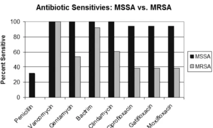
THIRTY-TWO CASES OF ENDOPHTHALMITIS RESULTING fromS. aureuswere identified, including 13 (41%) caused by MRSA and 19 (59%) caused by MSSA. Men and women were distributed equally in both the MRSA and the MSSA group (7 men and 6 women in the MRSA group, 9 men and 10 women in the MSSA group). Also, there was no difference in age between the 2 groups, with FIGURE 1. Bar graph showing endophthalmitis caused by Staphylococcus aureus: antibiotic sensitivity profiles.
MRSAⴝ methicillin-resistant S. aureus; MSSA ⴝ methi-cillin-sensitiveS. aureus.
FIGURE 2. Graph showing endophthalmitis caused by Staph-ylococcus aureus: logarithm of the minimal angle of resolution (logMAR) visual acuity (VA) outcomes.
MRSAⴝ methicillin-resistant S. aureus; MSSA ⴝ methi-cillin-sensitiveS. aureus.
TABLE 1.Endophthalmitis Caused byStaphylococcus aureusin Various Categories of the Current Study
Cause No. MSSA MRSA
Cataract 18 10 8
Endogenous 5 3 2
Bleb associated 4 4 0
Trauma 2 1 1
Pars plana vitrectomy 2 1 1 Ganciclovir 1 0 1
Total 32 19 13
MRSA⫽methicillin-resistantS. aureus; MSSA⫽ methicillin-sensitiveS. aureus.
a mean age of 66 years in the MRSA group and of 67 years in the MSSA group.
Overall, most endophthalmitis cases caused byS. aureus
(18 of 32 cases or 56%) were associated with cataract surgery (Table 1). Of these, 10 (56%) of 18 were caused by MSSA, and 8 (44%) of 18 were caused by MRSA. Five cases were endogenous (5 of 32 cases or 16%), including MRSA in 2 cases and MSSA in 3 cases. Four cases were bleb associated (12%), which were exclusively MSSA. Another 2 cases resulted from trauma (1 MSSA, 1 MRSA), 2 occurred after vitrectomy (1 MSSA, 1 MRSA), and 1 occurred after vitrectomy combined with ganciclovir implant placement (MRSA).
Of the 19 isolates in the MSSA group, 11 (68%) were resistant to penicillin, but all were sensitive to vancomy-cin, gentamivancomy-cin, trimethoprim-sulfa, and clindamycin (Figure 1). One MSSA cataract surgery-related endoph-thalmitis patient exhibited resistance to the fluoroquino-lones (oxifloxacin, gatifloxacin, and moxifloxacin). All other MSSA isolates were sensitive to the fluoroquinolo-nes, accounting for the 95% sensitivity (Figure 2). All MRSA isolates were resistant to penicillin. Among MRSA isolates, only 7 (54%) of 13 were susceptible to gentami-cin, and 8 (61%) of 13 were susceptible to clindamycin. Among all MRSA isolates, sensitivity to the fourth-generation fluoroquinolones was 38% (5/13) for gatifloxa-cin and moxifloxagatifloxa-cin. MSSA isolates were 100% sensitive to trimethoprim-sulfa, whereas MRSA isolates also were sensitive at 92% (12/13). Both MRSA and MSSA isolates were still sensitive to vancomycin.
Treatment in the MSSA group consisted of initial tap and inject in 10 cases (53%) and pars plana vitrectomy followed by the same intravitreal antibiotics in the other 9 cases (47%). In contrast, in the MRSA group, only 5 patients received tap-and-inject treatment (39%), and most (8 of 13 or 61%) were treated with vitrectomy and intraocular antibiotics. Mean time to diagnosis was essen-tially identical in the 2 post– cataract surgery groups, the setting in which the inciting event was easiest to deter-mine (6.1 days in the MSSA group and 5.7 days in the MRSA group).
Overall, visual acuity at presentation ranged from 20/ 200 to no light perception (NLP) in both the MRSA and MSSA groups. These measurements were not statistically different when comparing the MRSA and MSSA groups overall (mean logMAR visual acuity of 1.8 and 1.6, respectively; P ⫽ .87, Mann–WhitneyU test; Figure 2). One week after presentation, VA ranged from 20/25 to NLP in the MSSA group and from 20/100 to NLP in the MRSA group. The mean VA of the MRSA and MSSA groups were identical at this point. One MRSA patient underwent enucleation 1 week after diagnosis secondary to pain after corneal perforation and a limited VA potential because of anterior ischemic optic neuropathy. One month after initial presentation, VA ranged between 20/40 and light perception for the MRSA group (n⫽9 of initial 13)
and 20/20 to NLP in the MSSA group (n⫽ 18 of initial 19). Again, there was no statistically significant difference between the MRSA and MSSA groups from all causes (mean logMAR visual acuity, 1.7 and 1.2, respectively; P ⫽.11, unpairedttest; Figure 2).
After 3 months, VA ranged between 20/70 and light perception in the MRSA group (n⫽6 of initially 13) and 20/60 to NLP in the MSSA group (n⫽ 9 of initially 19; mean logMAR visual acuity, 1.8 and 0.5, respectively;P⫽ .065, unpaired t test). A small difference between the mean logMAR visual acuities in the post– cataract-only MRSA group (n⫽6 of initially 8) and MSSA group (n⫽ 5 of initially 10) was found (Figure 2). Although the mean visual acuity in the MRSA group was between hand movements and 20/400 (logMAR, 1.8), visual acuity in MSSA-infected eyes was 20/60 (logMAR, 0.5). This dif-ference approached statistical significance (P ⫽ .065, unpaired t test). A final VA of 20/400 or better at 3 months was achieved in 59% of MSSA and 36% of MRSA patients (P ⫽ .5). See Table 2 for a summary of visual
TABLE 2.Methicillin-ResistantStaphylococcus aureusand Methicillin-SensitiveStaphylococcus aureusGeneral
Comparison Cause logMAR VAp logMAR VA3 Resistance to 4F (%) Primary Source
MSSA 1.6 0.5 5 Postoperative ECCE MRSA 1.8 1.8 62 Postoperative ECCE 4F⫽fourth-generation fluoroquinolones; ECCE⫽ extracap-sular cataract extraction; logMAR⫽logarithm of the minimal angle of resolution; MRSA ⫽ methicillin-resistant S. aureus; MSSA⫽methicillin-sensitiveS. aureus; VA3⫽visual acuity at 3
months; VAp⫽visual acuity at presentation.
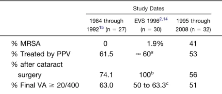
TABLE 3.Endophthalmitis Caused byStaphylococcus aureus: Selected Studies and Clinical Outcomes
Study Dates 1984 through 199215(n⫽27) EVS 19962,14 (n⫽30) 1995 through 2008 (n⫽32) % MRSA 0 1.9% 41 % Treated by PPV 61.5 ⬇60a 53 % after cataract surgery 74.1 100b 56 % Final VAⱖ20/400 63.0 50 to 63.3c 51 EVS⫽Endophthalmitis Vitrectomy Study; MRSA⫽ methicil-lin-resistantS. aureus; PPV⫽pars plana vitrectomy; VA⫽visual acuity.
a
Reported as “other gram-positive growth only.” Includes
Streptococcusspecies. (Approximation fromTable 1, ref.14).
b
After cataract or secondary intraocular lens.
cData taken from EVS Table 5. Fifty percent hadⱖ20/100,
acuity, fluoroquinolone resistance, and source of endo-phthalmitis.
DISCUSSION
THE CURRENT STUDY OF 32 PATIENTS REPRESENTS THE
largest reported clinical case series of S. aureus endoph-thalmitis (Table 3). In the EVS reports of acute-onset endophthalmitis associated with cataract or secondary intraocular lens implantation, S. aureus was the second most common organism after coagulase-negative staphylo-cocci. In the 30 EVS patients with S. aureus, less than 20/100 vision was found in half of the patients.15
Over the current study period from 1995 through 2008, MRSA accounted for more than one third of cases overall and 44% of post-cataract cases. In the period from 1984 through 1992, not a single case of MRSA was identified at the same institution.16 In the EVS, which enrolled pa-tients between 1990 and 1994, MRSA accounted for 1.9% of S. aureus isolates.15 Interestingly, in contrast to the present study from a United States center, a more recent study from Europe did not find any MRSA isolates from 13 S. aureusendophthalmitis isolates recovered between 1995 and 1999.17 This is interesting in light of the frequent preoperative topical antibiotic use in both geographic regions.18,19 However, these observations are in keeping with the striking difference in the overall epidemiologic features and multiresistant gram-positive cocci between the United States and Europe.20The increasing frequency of MRSA endophthalmitis cases mirrors the increasing incidence and changing epidemiologic factors of MRSA infections in general.21
A significant proportion ofS. aureuscases not only were resistant to methicillin, but those resistant isolates also were resistant to the commonly used fourth-generation
fluoroquinolones (gatifloxacin [Zymar; Allergan Inc., Ir-vine, California, USA] and moxifloxacin [Vigamox; Alcon Labs, Inc., Fort Worth, Texas, USA]).22–24 All isolates were still sensitive to vancomycin, the intravitreal antibiotic of choice to treat gram-positive bacterial infections.25 In 2007, the first MSSA endophthalmitis isolate that exhibited resistance to the fourth-generation fluoroquinolones was identified (Figure 3).
Risk factors for the acquisition of MRSA include ad-vanced age, recent hospitalization, and antibiotic use,20 but they were not present in most cases in this retrospec-tive series. In the current study, there was no significant difference in age between the patients in the MRSA and MSSA groups. This may be because the current study population is younger than the age of 75 to 80 years that frequently is cited as an independent predictor of MRSA colonization.26 Second, most endophthalmitis cases were not strictly nosocomial because they followed cataract surgery, which is an outpatient procedure not requiring hospitalization. Last, it was difficult to determine whether patients had used fluoroquinolones or cephalosporins in the previous year, or if they had been hospitalized or institutionalized before treatment. Invasive MRSA, how-ever, still may occur in patients with no established health care risk factors and may be associated with both a community and health care origin.21
Only 2 of 5 endogenous endophthalmitis cases were the result of MRSA.27 It was not surprising to find that the vitreous isolate of the patient who had received a ganci-clovir implant grew MRSA, because immunosuppression or AIDS is a recognized risk factor for MRSA acquisi-tion.28 All bleb-associated endophthalmitis cases were caused by MSSA.
In this retrospective case series, there was no statistically significant difference between MRSA and MSSA patients in presenting and final visual acuity 3 months after FIGURE 3. Photographs showing endophthalmitis caused byStaphylococcus aureus in a 79-year-old female after clear corneal cataract surgery. (Left) Initial clinical appearance 4 days after cataract surgery with a visual acuity of hand movements. The patient was treated with an aqueous tap and vitreous tap and injection of vancomycin 1 mg, ceftazidime 2.25 mg, and dexamethasone 0.4 mg. The isolate was methicillin-sensitive S. aureusand also was resistant to the fourth-generation fluoroquinolones. (Right) Follow-up appearance at 6 months. Final visual acuity was 20/40 after treatment.
treatment, although a trend for worse VA was present in the MRSA cases. Although increased virulence of MRSA strains has been demonstrated in bacteremia, pneumonia, and necrotizing fasciitis,10 –12 the current study failed to demonstrate statistically significant differences in time from cataract surgery to diagnosis, presenting visual acuity, or visual acuity on follow-up. The only difference in this regard was the higher rate of vitrectomy in the MRSA group, possibly reflecting the overall more severe clinical presentation influencing the treating physician to choose initial pars plana vitrectomy in the MRSA group. Al-though not statistically significant, MSSA cases tended to show visual acuity improvement regardless of treatment used, whereas MRSA showed minimal to no improvement trends over follow-up. This approached significance at 3 months. The statistical power of this study is limited by the small sample size and is typical of a rare disease entity that requires long-term follow-up.
Several limitations have to be considered when inter-preting the visual acuity results in the current study. Over the 3-month up, some patients were lost to follow-up, as can be seen in Figure 2. The individual physician
selected the initial treatment without use of a clearly defined protocol for treatment selection or VA testing. MRSA cases tended to present with a significant hy-popyon, pain, and a visible fibrinous exudates in the anterior chamber. The visual acuity tended to be worse on presenta-tion as well. This may well lead the clinician to earlier aggressive management such as a pars plana vitrectomy. The higher rate of vitrectomy in the MRSA group reflects the clinical decision made by individual physicians. One may speculate that if not treated that aggressively, MRSA infec-tions might have fared more poorly. The incidence of patients receiving a pars plana vitrectomy as initial management was remarkably consistent over the study period.
In conclusion, MRSA was identified with increasing frequency and approached one half of all isolates in the current study. There were no MRSA isolates identified in the prior study concluding in 1992 at the same institu-tion.16 Although all MSSA and MRSA isolates were sensitive to vancomycin, fewer than half of MRSA isolates were sensitive to the fourth-generation fluoroquinolones. Visual acuity outcomes between MRSA and MSSA eyes were similar at 3 months of follow-up.
SUPPORTED IN PART BY RESEARCH TO PREVENT BLINDNESS, INC, NEW YORK, NEW YORK. DR FLYNN HAS RECEIVED consulting fees from Alcon Labs, Inc., Fort Worth, Texas, Allergan Inc., Irvine, California, Genentech Inc., South San Francisco, California, and Pfizer Inc., New York, New York, and lecture fees from Alcon and Allergan. The other authors have no proprietary interests to disclose. Involved in Design and conduct of the study (J.C.M., H.W.F., D.M.); Collection (J.C.M., H.W.F., D.M., W.E.S., J.L.D.), management (J.C.M., H.W.F., M.E.), analysis (J.C.M., H.W.F., M.E., D.M.) and interpretation (J.C.M., H.W.F., M.E., D.M., W.E.S., D.M., J.L.D.) of the data; and Preparation, review, or approval of the manuscript (J.C.M., H.W.F., M.E., D.M.). This study was approved by the University of Miami Miller School of Medicine Institutional Review Board. Proper informed consent for both the treatment and participation in the research was obtained. Health Insurance Portability and Accountability Act compliance and adherence to the Declaration of Helsinki and all United States federal or state laws were observed.
REFERENCES
1. Flynn HW Jr, Scott IU, Brod RD, Han DP. Current management of endophthalmitis. Int Ophthalmol Clin 2004; 44:115–137.
2. The Endophthalmitis Vitrectomy Study Group. Microbio-logic factors and visual outcome in the endophthalmitis vitrectomy study. Am J Ophthalmol 1996;122:830 – 846. 3. Lalwani GA, Flynn HWJ, Scott IU, et al. Acute-onset
endophthalmitis after clear corneal cataract surgery (1996 – 2005). Clinical features, causative organisms, and visual acuity outcomes. Ophthalmology 2008;115:473– 476. 4. Callegan MC, Engelbert M, Parke DW 2nd, Jett BD,
Gilmore MS. Bacterial endophthalmitis: epidemiology, ther-apeutics, and bacterium-host interactions. Clin Microbiol Rev 2002;15:111–124.
5. Lindsay JA, Holden MT. Staphylococcus aureus: superbug, super genome? Trends Microbiol 2004;12:378 –385. 6. Chambers HF. Community-associated MRSA—resistance
and virulence converge. N Engl J Med 2005;352:1485–1487. 7. Diep BA, Gill SR, Chang RF, et al. Complete genome sequence of USA300, an epidemic clone of community-acquired methicillin-resistant Staphylococcus aureus. Lancet 2006;367:731–739.
8. Marangon FB, Miller D, Muallem MS, Romano AC, Alfonso EC. Ciprofloxacin and levofloxacin resistance among
methi-cillin-sensitive Staphylococcus aureus isolates from keratitis and conjunctivitis. Am J Ophthalmol 2004;137:453– 458. 9. Ta CN, He L, Nguyen E, De Kaspar HM. Prospective
randomized study determining whether a 3-day application of ofloxacin results in the selection of fluoroquinolone-resistant coagulase-negativeStaphylococcus. Eur J Ophthalmol 2006; 16:359 –364.
10. Francis JS, Doherty MC, Lopatin U, et al. Severe com-munity-onset pneumonia in healthy adults caused by me-thicillin-resistant Staphylococcus aureus carrying the Panton-Valentine leukocidin genes. Clin Infect Dis 2005; 40:100 –107.
11. Gonzalez BE, Martinez-Aguilar G, Hulten KG, et al. Severe Staphylococcal sepsis in adolescents in the era of community-acquired methicillin-resistant Staphylococcus aureus. Pediat-rics 2005;115:642– 648.
12. Miller LG, Perdreau-Remington F, Rieg G, et al. Necrotizing fasciitis caused by community-associated methicillin-resis-tant Staphylococcus aureus in Los Angeles. N Engl J Med 2005;352:1445–1453.
13. Blomquist PH. Methicillin-resistant Staphylococcus aureus infections of the eye and orbit (an American Ophthalmo-logical Society thesis). Trans Am Ophthalmol Soc 2006;104: 322–345.
14. Freidlin J, Acharya N, Lietman TM, Cevallos V, Whitcher JP, Margolis TP. Spectrum of eye disease caused by
methi-cillin-resistant Staphylococcus aureus. Am J Ophthalmol 2007;144:313–315.
15. Han DP, Wisniewski SR, Wilson LA, et al. Spectrum and susceptibilities of microbiologic isolates in the Endoph-thalmitis Vitrectomy Study. Am J Ophthalmol 1996; 122:1–17.
16. Mao LK, Flynn HW Jr, Miller D, Pflugfelder SC. Endoph-thalmitis caused byStaphylococcus aureus. Am J Ophthalmol 1993;116:584 –589.
17. Labit CM, Claeys GW, Verbraeken HE, Verschraegen GL. Methicillin resistance of bacteria isolated from vitreous fluid from patients undergoing vitrectomy. Eur J Ophthalmol 2001;11:160 –165.
18. Masket S. Preventing, diagnosing, and treating endoph-thalmitis. J Cataract Refract Surg 1998;24:725–726. 19. Schmitz S, Dick HB, Krummenauer F, Pfeiffer N.
Endoph-thalmitis in cataract surgery: results of a German survey. Ophthalmology 1999;106:1869 –1877.
20. Harbarth S, Albrich W, Goldmann DA, Huebner J. Control of multiply resistant cocci: do international comparisons help? Lancet Infect Dis 2001;1:251–261.
21. Klevens RM, Morrison MA, Nadle J, et al. Invasive methi-cillin-resistantStaphylococcus aureusinfections in the United States. JAMA 2007;298:1763–1771.
22. Miller D, Flynn PM, Scott IU, Alfonso EC, Flynn HW Jr. In vitro fluoroquinolone resistance in staphylococcal endophthalmitis isolates. Arch Ophthalmol 2006;124: 479 – 483.
23. Deramo VA, Lai JC, Winokur J, Luchs J, Udell IJ. Visual outcome and bacterial sensitivity after methicillin-resistant Staphylococcus aureus-associated acute endophthalmitis. Am J Ophthalmol 2008;145:413– 417.
24. Hori Y, Nakazawa T, Maeda N, et al. Susceptibility compar-isons of normal preoperative conjunctival bacteria to fluoro-quinolones. J Cataract Refract Surg 2009;35:475– 479. 25. Gordon YJ. Vancomycin prophylaxis and emerging
resis-tance: are ophthalmologists the villains? The heroes? Am J Ophthalmol 2001;131:371–376.
26. Harbarth S. Control of endemic methicillin-resistant Staph-ylococcus aureus: recent advances and future challenges. Clin Microbiol Infect 2006;12:1154 –1162.
27. Ness T, Schneider C. Endogenous endophthalmitis caused by methicillin-resistantStaphylococcus aureus (MRSA). Retina 2009;29:831– 834.
28. Pan ES, Diep BA, Charlebois ED, et al. Population dynamics of nasal strains of methicillin-resistant Staphylococcus au-reus—and their relation to community-associated disease activity. J Infect Dis 2005;192:811– 818.
Biosketch
James C. Major, Jr. received his undergraduate degree in Biology from Emory University and Masters and Doctor of Philosophy in Biology at The University of Texas at Austin. Thereafter, he received his medical degree from Baylor College of Medicine. Dr. Major completed his ophthalmology residency, vitreoretinal fellowship, and Chief Residency at the Bascom Palmer Eye Institute in Miami. He served on the faculty at three institutions. He currently is practicing with Retina Consultants of Houston.