Nijmegen
The following full text is a publisher's version.
For additional information about this publication click this link.
http://hdl.handle.net/2066/125214
Please be advised that this information was generated on 2017-12-06 and may be subject to
change.
Aiming to immune elimination of ovarian cancer stem cells
Jiabo Di, Tjitske Duiveman-de Boer, Carl G Figdor, Ruurd Torensma
Jiabo Di, Tjitske Duiveman-de Boer, Carl G Figdor, Ruurd Torensma,Department of Tumor Immunology, Nijmegen Cen-tre for Molecular Life Sciences, Radboud University Nijmegen Medical Centre, GA 6525 Nijmegen, The Netherlands
Author contributions:Di J and Duiveman-de Boer T performed experiments; Di J, Figdor CG and Torensma R designed research and wrote the paper.
Supported by The Dutch government to the Netherlands Insti-tute for Regenerative Medicine, No. FES0908
Correspondence to:Ruurd Torensma, PhD,Department of Tumor Immunology, Nijmegen Centre for Molecular Life Sci-ences, Radboud University Nijmegen Medical Centre, Geert Grooteplein 28, GA 6525 Nijmegen,
The Netherlands. r.torensma@ncmls.ru.nl
Telephone: +31-24-3617600 Fax:+31-24-3540339
Received:February 18, 2013 Revised:June 19, 2013
Accepted:July 18, 2013
Published online: October 26, 2013
Abstract
Ovarian cancer accounts for only 3% of all cancers in
women, but it causes more deaths than any other
gy-necologic cancer. Treatment with chemotherapy and
cytoreductive surgery shows a good response to the
therapy. However, in a large proportion of the patients
the tumor grows back within a few years. Cancer stem
cells, that are less responsive to these treatments, are
blamed for this recurrence of disease. Immune therapy
either cellular or humoral is a novel concept to treat
cancer. It is based on the notice that immune cells
in-vade the tumor. However, the tumor invest heavily to
escape from immune elimination by recruiting several
immune suppressive mechanisms. These processes are
normally in place to limit excessive immune activation
and prevent autoimmune phenomena. Here, we discuss
current knowledge about the immune (suppressive)
status in ovarian cancer. Moreover, we discuss the
im-munological targets of ovarian cancer stem cells.
© 2013 Baishideng. All rights reserved.Key words:
Ovarian cancer; Cancer stem cell; Immune
therapy; Immune suppression; Tumor microenvironment
Core tip:
Ovarian cancer harbors, at a low frequency,
cancer stem cells. Those cancer stem cells express
stem cell specific antigens. Natural immunity against
those antigens exists but is hampered by the
suppres-sive microenvironment that the tumor creates. Erasing
this suppressive microenvironment will make
immuno-logical elimination of those cancer stem cells is an
at-tractive treatment option.
Di J, Duiveman-de Boer T, Figdor CG, Torensma R. Aiming to immune elimination of ovarian cancer stem cells. World J Stem Cells2013; 5(4): 149-162 Available from: URL: http://www. wjgnet.com/1948-0210/full/v5/i4/149.htm DOI: http://dx.doi. org/10.4252/wjsc.v5.i4.149
EPITHELIAL OVARIAN CANCER
Ovarian cancer is the fourth leading cause of death from
cancer in women and the leading cause of death from
gynecological cancer. The lifetime risk to get this disease
is 1 in 60 women in industrial countries but is less
com-mon in Asian and African women. Due to vague
symp-toms and adequate screening methods at the early stages,
more than 60% of the patients are diagnosed at advanced
stage. Most patients respond well to primary treatment,
either cytoreductive surgery followed by chemotherapy or
chemotherapy followed by surgical removal of
remain-ing tumor foci. However, 80% of the patients diagnosed
at late stage will eventually develop recurrent diseases,
the survival is generally poor. The 5-year survival rates
at stage
Ⅲ
and
Ⅳ
are 29% and 13%, respectively. The
relapse of tumor arises the question about the identity of
the cells that give rise to the tumor and somehow escape
from the first line treatment, reside in the body
unde-tected, and finally initiate malignant tumor growth in a
suitable microenvironment.
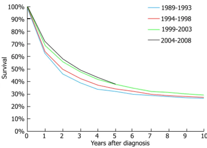
Despite intense efforts to improve chemotherapy,
e.g.
,
the introduction of paclitaxel, and to improve surgical
techniques, over the past 20 years no significant progress
has been made (Figure 1).
REVIEW
Novel therapeutic approaches are urgently needed.
Since ovarian cancer is immunogenic, immunotherapy
should be further pursued and optimized. Stimulating the
immune system to attack ovarian tumor is not a new
con-cept, during the last 20 years numerous
immunological
modalities were involved in clinical trials in ovarian
can-cer treatment
[1]. Targeting a specific tumor antigen plays a
decisive role in the success of immunotherapy.
CANCER STEM CELLS
Tumors are composed of phenotypically and functionally
heterogeneous cells. There are two theories explaining
how this heterogeneity arises
[2,3]. According to the
sto-chastic model, tumor cells are biologically equivalent;
vir-tually every tumor cell is able to generate new tumor cells.
In contrast, the hierarchy model postulates the existence
of tumorigenic as well as non-tumorigenic cells. Only a
subset of cells can initiate tumor growth, and these cells
are considered as tumor-initiating cells (TICs) or cancer
stem cells (CSCs). CSC is a relatively rare cancer cell that
has the ability of self-renewal giving rise to another
ma-lignant stem cell as well as a cell that undergoes massive
proliferation and differentiation to give rise to the
phe-notypically and functionally more mature cancer cells
[4,5].
The similarities of CSCs and normal stem cells (NSCs)
point to the origin of CSCs. There are two hypotheses
[6].
One states that CSCs can be derived from NSCs, so that
they can make use of the already active self-renewal
ma-chinery. Another assumes that the CSCs can be derived
from progenitor cells by regaining the self-renewal
capa-bility. NSCs possess several unique properties. Their
self-renewal enables livelong maintenance of all organs of the
body. In most cases NSC divide slowly. For
hematopoi-etic cells a doubling time of 30 d was reported
[7].
How-ever, for intestinal cells a doubling time of less than 24 h
was reported
[8]. Those fast regenerating organs have stem
cells that are continuously dividing. One of properties
of NSC is the expression of pumps of the ATP binding
cassette (ABC) superfamily
[9-11]. Those pumps can remove
toxic components from the cell. Likewise CSC also
ex-presses members of the ABC family
[10-19]. For melanoma
ABC-B1 and ABC-B5 were reported while other tumors
express other members
[12,13]. This endows CSC with a
nasty property. The pump is able to remove cytotoxic
drugs that are given to patients to kill the tumor. Indeed,
a common property of CSC is their resistance against
cytotoxic drugs, explaining the relapse that is seen in
sev-eral patients. Traditional therapies that kill primarily
non-tumorigenic cancer cells can shrink tumors, but will not
cure the patient because the CSCs that survive the
treat-ment will regenerate the tumor. By prospectively
identify-ing and characterizidentify-ing CSCs, it might be possible to
iden-tify more effective therapies
[20-24]. CSCs can be eliminated
by direct killing, or force them to differentiated cells or by
destroying their niche
[25]. Accordingly, targeting the CSCs
has been put forward as such a new treatment modality
for cancer immunotherapy
[26,27]. Several studies described
in the literature provide several clues for optimizing the
immunotherapy against ovarian cancer.
IDENTIFICATION AND
CHARACTERIZATION
The first experimental evidence suggests the existence
of CSC came from leukemia. Bonnet and co-workers
demonstrated that human leukemias are driven by a small
population of leukemic stem cells capable of transferring
the disease to NOD/SCID mice
[28]. This concept was
ex-tended to solid epithelial tumors by Al-Hajj and
co-work-ers, who demonstrated that a small population of cells
within breast cancer with stem cell properties, bearing the
surface marker CD24
lowCD44
high[4]. Subsequently, CSCs
are identified and prospectively isolated from a variety of
epithelial cancers, including pancreas, colon and prostate
cancers
[29-40].
Ovarian CSC is responsible for ovarian tumor formation
The CSC hypothesis has recently also been explored in
ovarian cancer. In 2008, Zhang
et al
[39]claimed that
epi-thelial ovarian cancers derive from a subpopulation of
CD44
+CD117
+cells. Ferrandina and Curley
independent-ly found that CD133 expression defines a tumor initiating
subpopulation of cells in human ovarian cancer
[41,42]. Gao
and co-workers reported that CD24 could be utilized as
a surface marker to enrich for ovarian CSCs
[32]. Ovarian
CSCs were also detected in the so-called side population,
which are tumorigenic and chemoresistant
[38,43,44].
More-over, Stewart
et al
[45]established a quantitative assay that
enables characterization of TICs from serous ovarian
cancer, and they also found that the tumor initiating cell
phenotype is heterogeneous across patients. And recently,
a gene involved in maintaining stem cell pluripotency,
Nanog, was proved to be expressed by ovarian tumor
cells, and positive Nanog expression indicates poor
pro-gression of patients with ovarian serous carcinoma
[46].
As described above, increasing experimental evidence
suggests that TICs may play a decisive role in the
initia-1989-1993 1994-1998 1999-2003 2004-2008 100% 90% 80% 70% 60% 50% 40% 30% 20% 10% 0% Surviv al
Years after diagnosis
0 1 2 3 4 5 6 7 8 9 10
Figure 1 Survival of patients diagnosed with ovarian carcinoma. The
per-centage of survival after diagnosis is not significantly increased in the past 20 yr
tion and progression of tumors
[4,29-31,35-39,46]. However,
TICs with distinct tumorigenic abilities were
identi-fied
[31,47,48], as well as large variation in their frequency
[49,50].
TICs appear not to be a stable entity but show quite
some plasticity
[2,51-54]. Recently, it was described that the
TIC compartment can be subdivided into long-term
TICs, tumor transient amplifying cells as well as delayed
contributing TICs
[48]. Only the long-term TICs are
capa-ble of maintaining tumor formation in serial xenografts,
and these cells are considered as cancer stem cells.
Phenotypic heterogeneity of ovarian CSCs
CSCs are operationally defined as tumor initiating cells
because the CSC assays rely heavily on
xenotransplan-tation
[55]. Although it was proven that frequency and
tumorigenic ability of melanoma CSCs that can be
de-tected after xenotransplantation were highly dependent
on experimental design
[50,56], current studies on CSCs all
use immunodeficient mice models to check whether
pu-tative CSCs can generate secondary tumors
in vivo
. And
using this method, phenotypically diverse ovarian CSC
populations have been characterized and isolated from
both patient material and immortalized tumor cell lines
with variable stem cell markers
[32,36,38,41,42,46,57,58]. However,
due to the fact that a large number of cells was needed to
establish a secondary tumor in immunodeficient mice, it
is assumed that ovarian CSCs were just enriched in those
cell populations
[59]. Also, it was questionable whether
tu-mor cell lines can represent the status of primary tutu-mor
cells. Moreover, due to the heterogeneity among
indi-viduals, it is important to test CSC markers in significant
numbers of patients.
The expression of well-known CSC markers,
includ-ing, CD44, CD117, CD133, CD24, ABCG2 and aldehyde
dehydrogenase (ALDH), on tumor and ascites derived
cells from patients diagnosed with ovarian cancer is very
diverse and is patient-dependent, and no correlation was
found between marker expression and tumor histological
subtype
[60]. In line with these data, another study
investi-gated epithelial and mesenchymal markers expressed by
primary ovarian tumors, and they also showed different
phenotypic features and expression levels of those
mark-ers in different cellular subsets within tumors
[59].
Addi-tionally, it has been reported that the CSC marker ALDH
show distinct expression pattern in human epithelial
can-cers, and it can only be used to isolate CSCs for tumors
whose corresponding normal tissues express low levels
of ALDH
[61]. Also CD133 as a marker to identify ovarian
CSCs has been questioned, since tumor initiating
activi-ties have been detected in both CD133
+and CD133-
fractions from primary ovarian masses, and CD133
+cell
frequency varies between patients
[45]. Similar doubts of
CD133 as a putative CSC marker has been reported in
colon cancer and melanoma
[56]. Moreover, phenotypic
heterogeneity of breast CSCs was also reported
[34,40,62].
Taken together, these data suggest that CSC phenotypes
are heterogeneous, and experimental variables as well as
xenograft recipients can dramatically influence CSC
fre-quency
[45]. So far a clear set of marker proteins remain to
be identified to target ovarian CSCs.
For better recognition of CSCs, better experimental
methods need to be established. One way to identify CSC
is to focus on genes involved in stem cell pluripotency,
because those genes may be involved in establishment of
tumors and may be inherited by their malignant
counter-parts. Four genes are required for induction of
pluripo-tent stem cells from mouse embryonic or adult fibroblasts
in vitro
, including Oct4, c-Myc, Sox2 and Klf4
[63]. A rare
cell population, in ovarian tumor tissue as well as ascites,
expressing Oct4, Nanog and c-Myc was found. Oct4
ex-pression is crucial for the self-renewing and maintenance
of pluripotent properties of embryonic stem (ES) cells
[64,65]
. The expression of Oct4A indicates that the cells
are undifferentiated
[66]. Recently, abnormal Oct4
expres-sion level was correlated to several cancers
[67-69]. The two
isoforms of Oct4, Oct4A and Oct4B, differ in their
abil-ity to confer self-renewal, only Oct4A can sustain stem
cell properties
[70,71]. Several studies have shown that the
different isoforms and Oct4 may lead to false positive
signals during RT-PCR analysis
[72,73]. In order to rule out
this, a primer set was described to distinguish the Oct4A
from Oct4B and Oct4 pseudogenes
[73]. Oct4A mRNA
expression was detected by us in ascites-derived tumor
cells from all patients tested, regardless of histological
subtypes. The c-Myc protein is normally expressed in the
nucleus and is virtually undetectable in quiescent cells. It
contributes to the long-term maintenance of the ES cell
phenotype and is upregulated in many types of malignant
human cancers
[74]. Moreover, Nanog also sustains ES cell
pluripotency
[75]. Oct4 and Nanog were described to be
higher expressed in side population cells obtained from
ovarian cancer cell lines than the bulk of the cells
[76],
con-firming the expression of stem cell markers as described
here. To sum up, expression of these genes suggests
that those cells are the primitive CSC for ovarian cancer,
because all genes needed for reprogramming to induce
pluripotent stem are present in the same cell.
According to the hierarchy tumor model, the most
“primitive’’ CSCs are able to self-renew, and develop into
more differentiated cells like so-called progenitor cells or
CSC-derived transit-amplifying cells, which are not able
to self-renew but can generate new tumor cells to
sup-port tumor growth
[34,48]. In order to adapt to different host
microenvironments, CSC-derived progenitors may differ
in their phenotypes and functions and in turn
differenti-ate into phenotypically and functionally heterogeneous
tumor cells
[77]. And a different differentiation status might
be generated also to adapt the complicated tumor growth
environment
[78]. These indicate that CSCs and their
prog-enies may differ between different patient tumors and may
be able to change during tumor progression
[55].
Collective-ly, these data may explain why the expression of putative
CSC phenotypes are heterogeneous among patients with
ovarian cancer and why accumulating evidence shows that
solid tumors are initiated by heterogeneous populations
of CSCs, and each CSC subset responsible for distinct
functions in tumor progression
[33,34,40,45,47,48,50,79-83][Engh,
2011 #756].
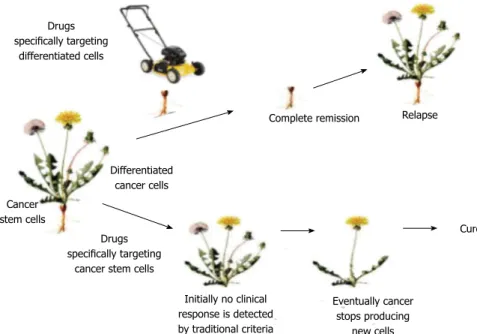
Although CSC phenotypes are heterogeneous,
cur-rent studies suggest ovarian tumor conforms to the CSC
hypothesis
[45,59], and in this scenario, if the most primitive
Oct4-expressing CSC population is eliminated
specifi-cally, the tumor will lose its feeding and eventually fade
away (Figure 2).
Phenotypic plasticity of ovarian tumor cells
CSC may not be a stable entity. Plasticity describes the
dedifferentiation potential of more differentiated cancer
cells to acquire stem cell phenotype and characteristics,
which further contribute to CSC heterogeneity, and
which is an important determinant of the prognosis of
tumors
[55,84,85]. Thus plasticity in CSCs and their
prog-enies make the situation more complex
[51,59]. Two c-Myc
expressing populations were found; one is only highly
positive for c-Myc, the other also express Oct4. The
ire-lationship between these two subpopulations remains to
be investigated. We argue that those intermediate c-Myc
+cells are more differentiated cells than c-Myc
+Oct4
+cells, since in some cases they were not able to survive in
serum-free medium. Also, it is possible that the c-Myc
+cells somehow regain Oct4A expression and become a
primitive CSC. In fact, phenotypic plasticity of ovarian
tumor cells was detected under certain circumstances,
e.g.,
stress created by starvation or co-culture with either
epi-thelial or mesenchymal cells
in vitro
[59].
In line with this, plasticity has been described in other
tumor stem cell studies, showing that non-tumorigenic
cells can convert to a tumorigenic cell
[50,86,87]. For instance,
knocking down of JARID1B in slow cycling melanoma
cells exhausted the tumor, however, expression of
RID1B is dynamic since negative cells can become
JA-RID1B positive
[47]. This indicates that the cancer cells
might reversibly transit between tumorigenic and
non-tumorigenic status, generate reversible heterogeneity
[85,88].
In addition to tumor cells, plasticity was also described in
normal development procedures. Endothelial cells could
simply be converted into multipotent stem-like cells by
Transforming growth factor
β
2 or Bone morphogenetic
protein 4
[89]. Also in spermatogonial development more
differentiated cells can go back to the stem cell state
when the stem cell niche is emptied and the number of
stem cells is decreased. In this way the normal number of
stem cells is recovered by differentiated cells that regain
stem cell properties
[90]. Plasticity would have major
im-plications for the CSC model and for future therapeutic
approaches, as discussed in
[52].
INTERPLAY BETWEEN TUMOR AND THE
IMMUNE SYSTEM
The immune system affects cancer development and
pro-gression. Before the tumor cells cause clinically detectable
disease, they have already resided in the body for a while.
The immune system can recognize and interact with
the transformed cells before and after the formation of
tumormass; this process is termed “cancer
immunoedit-ing’’. Cancer immunoediting consists of three distinct
phases: elimination, equilibrium and escape
[91,92]. During
the elimination phase, tumor specific immune cells and
molecules are recruited to the tumor site and destroy the
developing tumor cells. The equilibrium phase is a
dy-namic state; the interaction between tumor growth and
immune prevention represents a type of tumor
dorman-cy, in which tumor outgrowth is also limited by the
im-mune system
[93]. Meanwhile, due to the immune selection,
some malignant cell can acquire the ability to circumvent
immune recognition, or no longer sensitive to immune
effector mechanisms, and escape. And then their growth
is no longer blocked by the host immunity anymore. In
addition, the malignant tumor cells can even manipulate
the immune system to promote their own growth
[91,92].
Drugsspecifically targeting differentiated cells
Complete remission Relapse
Initially no clinical response is detected by traditional criteria Eventually cancer stops producing new cells Cure Differentiated cancer cells Drugs specifically targeting
cancer stem cells Cancer
stem cells
Figure 2 Killing the mature cancer cells leaves the root intact leading to regrowth of the tumor. Killing the root will exhaust the stem cell pool leading to eradication of the tumor. Reprinted from Jones et al[154].
Immune elimination of tumors
The effectors mechanisms of both cell-mediated
immu-nity and humoral immuimmu-nity have been shown to kill
tu-mors
in vitro
. In several cases also
in vivo
killing of tumor
cells was observed. During the elimination phase of
can-cer immunoediting, different types of immune cells are
recruited to the tumor site, including T cells,
antibody-secreting B cells, different subsets of dendritic cells (DCs),
tumor-associated macrophages (TAMs), myeloid-derived
suppression cells (MDSCs), Th17 cells, natural killer (NK)
cells, NK T cells and
γδ
T cells
[94,95]. And those
intratu-moral T cells were functionally active since interleukin-2
(IL-2) and interferon-
γ
(IFN-
γ
) was produced, which may
enhance T cell proliferation and anti-tumor immunity
[96,97].
An effective antitumor immune response is direct
killing of tumor cells by CD8
+cytotoxic T lymphocytes
(CTLs), which recognize tumor antigens presented by
MHC
Ⅰ
molecules. CD8
+T cell responses specific for
tumor antigens may require cross-presentation of the
tumor antigens by professional antigen presenting cells
(APCs), such as DCs. Most tumor cells do not express
the co-stimulatory molecules needed to initiate T cell
responses or the class
Ⅱ
MHC molecules needed to
stimulate helper T cells that promote the differentiation
of CD8
+T cells. It is possible that tumor cells or their
antigens are ingested by host DCs, the tumor antigens
are then processed inside the DCs, and peptides derived
from these antigens are displayed bound to class
Ⅰ
MHC
molecules for recognition by CD8
+T cells. The APCs
expressing co-stimulatory molecules that provide the
sig-nals needed for differentiation of naïve CD8
+T cells into
anti-tumor effector CTLs, and the APCs express class
Ⅱ
MHC molecules that may present internalized tumor
antigens and activate CD4
+helper T cells as well. Once
effector CTLs are generated, they are able to recognize
and kill the tumor cells without a requirement for
co-stimulation. CTLs mediate lysis of target cells by two
ma-jor mechanisms, the predominant mechanism appears to
be perforin-granzyme-dependent, and the other is FasL
dependent
[98,99]. The ability of CTLs to provide effective
anti-tumor immunity
in vivo
is most clearly seen in animal
experiments. However, tumor-specific CTLs can be
iso-lated from animals and humans with established tumors,
such as melanomas
[100].
The importance of CD4
+helper T cells in tumor
im-munity is less clear. CD4
+cells may play a role in
anti-tumor immune responses by providing cytokines for
effective CTL development. In addition, CD4
+T cells
specific for tumor antigens may secrete cytokines, such as
tumor necrosis factor (TNF) and IFN-
γ
, that can increase
tumor cell class I MHC expression and sensitivity to lysis
by CTLs. IFN-
γ
may also activate macrophages to kill
tu-mor cells. In addition to T cells, tutu-mor-bearing hosts may
produce antibodies against various tumor antigens
[101-104].
Whereas it has also been documented that CD4 T cells
can be more effective than CD8 T cells in tumor killing
in tumor bearing mice
[105]. Moreover, NK cells may kill
many types of tumors, especially “missing’’ cells that have
reduced class I MHC expression and can escape killing
by CTLs
[106,107]. CD4
+T cells cooperate with NK cells to
accomplish the maximum tumor killing
[105]. Macrophages
can kill many tumor cells more efficiently than they can
kill normal cells
[108]. Several studies showed the existence
of tumor infiltrating T cells in ovarian cancer associated
with favorable clinical outcome
[109,110]. Distribution of
tumor infiltrating lymphocytes (TILs) were studied in
patients with late stage ovarian cancer, CD3
+T cells were
detected in more than 50% of the patients and CD4
+and CD8
+T cells were either both present or absent. The
presence of TILs correlates with a better 5 year survival
as well as progression-free survival
[39]. It has also been
documented that patients with higher TIL counts showed
improved overall survival than patients with lower TIL
counts
[111]. Moreover, Sato and co-workers demonstrated
intraepithelial CD8
+TILs and the high CD8
+TIL/
Treg ratio indicates better survival of ovarian cancer
pa-tients
[112].
Immune reactivity towards CSCs
When the immune system is directed to eliminate the
CSC, it will also destroy CSC reverting from more
dif-ferentiated progeny. We consider Oct4 as a suitable
anti-gen for immunological targeting ovarian CSCs, since it is
neither expressed in normal adult stem cells nor somatic
cells. Once the progenitors re-express Oct4 and become
CSCs, they can be recognized and eliminated by
Oct4-reactive T cells. Removing of the CSCs from the pool
will diminish the feeding of more mature tumor cells.
Further understanding of the relationship between CSCs
and their differentiated progenies can help us to develop
better immunotherapeutic strategies that can prevent the
emergence of tumor cell variants that are capable of
gen-erate a new tumor and metastases
[55,113].
OCT4-REACTIVE T CELLS ARE
DETECTABLE
Naturally occurring T cells directed against
tumor-associ-ated antigens (TAAs) can be frequently detected in cancer
patients (reviewed in
[114]). Amazingly, Oct4 reactive CD4
+as well as CD8
+T cells were detected in both healthy
people and patients with ovarian cancer
[115]. This
find-ing suggests that the host immune system has the ability
to target the primitive ovarian CSCs. The frequency of
Oct4 specific T cell was low in peripheral blood, while it
was higher in the ascites of patients. This means those
cells are either recruited to the tumor or proliferate upon
exposure to Oct4. Moreover, lymphocytes isolated from
ascites from patients with ovarian tumor contained Oct4
specific T-cells. It was shown that Oct4-reactive CD8
+T cells produce IFN-
γ
-inducible protein 10 (IP-10) and
IFN-
γ
, and were capable of proliferation upon Oct4
pep-tide loaded or Oct4 mRNA pulsed dendritic cell
stimula-tion. The CD8
+cytotoxic T cells were able to release
ly-sosomal components as indicated by CD107a expression.
Moreover, Oct4-reactive CD4
+T cells were also detected,
and also capable of proliferating upon stimulation. These
results proved the existence of anti-CSC specific T cells
in patients with ovarian cancer.
Natural immunity against genes involved in
pluripo-tency has been shown. Dhodapkar
et al
[115]claim the Oct4
responsive T cells were detected in PBMCs from 83% of
healthy donors, although they showed the Oct4-specific
cells were CD4
+T cells. They also found 38% of patients
with germ-cell tumors had measurable Oct4-specific T
cell immunity at baseline, and after chemotherapy, 83%
of the patients developed Oct4-reactive T cells. Also, it
has been documented that CD8
+Sox2-specific T cells
were frequently detected in patients with monoclonal
gammopathy of undetermined significance (MGUS).
MGUS is a precursor lesion to myeloma, whereas
Sox2-specific T cell immunity was not detectable in patients
with myeloma
[116].
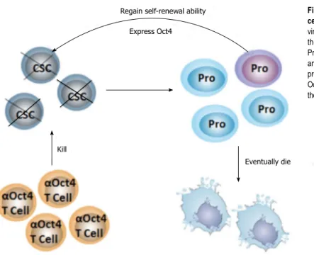
Taken together, these data indicate that the ovarian
CSCs are prone to immunological attack because CSC
specific T cells are present in the T cell repertoire (Figure
3). Meanwhile, this raises the question about why CSCs
and their progenies escape from immune elimination, and
why the already activated Oct4-reactive memory T cells
do not kill those cells.
Immune escape by tumors
Many malignant tumors possess mechanisms that enable
them to disturb the balance in the equilibrium phase and
shift to escape phase, including down-regulation of MHC
Ⅰ
expression on tumor cells, loss or hidden of
tumor-antigen expression, production of immune suppressive
molecules, and inhibition of co-stimulatory or MHC
Ⅱ
molecules expression on APCs, leading to immunologic
tolerance
[92,117,118]. Tumors escape not only from the host
immune system, but also effectively benefit from
infil-trating cells and create a microenvironment that favors
its progression by modifying TIL functions
[119]. Ovarian
tumor can effectively create its suppressive
microenvi-ronment. Curiel
et al
[120]showed the first evidence that
tumor associated CD4
+CD25
+regulatory T cells (Treg)
were correlated with a poor clinical prognosis of ovarian
cancer. They showed the presence of Treg in both tumor
tissue and malignant ascites, and also proved that tumor
cells and microenvironmental macrophages produced the
chemokine CCL22, which attracted Tregs to the tumor
site. Tumor infiltrating Tregs suppress tumor-specific T
cell immunity by blocking T cell proliferation as well as
IFN-
γ
and IL-2 production. Similarly, Woo
et al
[121]found
that CD4
+CD25
+Tregs contribute to CD8
+T cell
dys-function by secreting the immunosuppressive cytokine
transforming growth factor-
β
(TGF-
β
). Later on,
fork-head box protein-3 (FoxP3) expressing Tregs were also
detected and emerged as an independent prognostic
fac-tor for both poor progression-free and overall survival
[122].
Conrad et al. demonstrated that majority of these FoxP3
+Tregs accumulated nearby the tumor and also express
in-ducible co-stimulator (ICOS)
[123]. The expansion and
im-munosuppressive function of these FoxP3
+ICOS
+Treg
cells are dependent on their interaction with plasmacytoid
DCs (pDCs) which provide ICOS-ligand (ICOS-L)
stim-ulation. The presence of immature pDCs was also found
in the vicinity of ovarian tumor and associated with
poor clinical outcome of patients with ovarian tumor
[124].
pDCs are recruited by CXCL12 produced by tumor cells
and produce type
Ⅰ
IFN in response to toll-like receptor
(TLR) ligand triggering
[125,126]. In addition to CD4
+Tregs,
CD8
+Tregs also exist in ascites produced by malignant
ovarian tumor. Wei et al. showed that tumor pDCs
in-duce suppressive CD8
+Tregs in ascites. These CD8
+Tregs inhibit T cell proliferation and IFN-
γ
production,
while they induce IL-10 production
[126]. Moreover,
ovar-ian tumor infiltrating DCs express programmed death 1
(PD-1), which interacts with B7-H1 on tumor-associated
macrophages. This reaction can lead to suppressed NF
κ
B
Regain self-renewal ability Express Oct4
Kill
Eventually die
Figure 3 Hypothesis of specific targeting of primitive can
-cer stem cells. In a non-immunosuppressive tumor
microen-vironment, Oct4-specific T cells (αOct4 T cell) can recognize
the primitive cancer stem cells (CSCs). and destroy them.
Progenitor cells (Pro) differentiate to more mature tumor cells
and will eventually undergo apoptosis or necrosis. Once some progenitors regain the self-renewal machinery and re-express Oct4 to become a CSC, T cells will also eliminate it. In this way, the tumor loses its ability to generate new tumor cells.
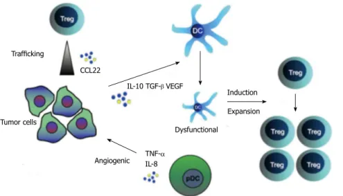
activation and downregulated co-stimulatory molecule
expression on DCs
[127](Figure 4).
Ovarian tumor infiltrating T cells are anergic
A remarkable characteristic of ovarian cancer is the
typi-cal metastasis behavior. Metastases are found but hardly
in other organs. As the tumor spreads in a diffuse
intra-abdominal fashion and even after recurrence, it is in most
cases confined to the peritoneal cavity. There are several
papers that report the presence of metalloproteases in
ascites
[128-130]. Those enzymes are found in metastasizing
tumors by chopping tissues to make room for the
metas-tasis. Moreover, ovarian tumors orchestrate suppressive
mechanisms that enable them to evade or resist host
im-mune responses
[131-135]. The fact that CTLs against human
tumors can be easily generated
in vitro
using peripheral
blood lymphocytes indicates that the tumor
microenvi-ronment has immunosuppressive capacities
[131]. Tumor
infiltrating immune cells together with fibroblasts and
extracellular matrix form a scaffold supporting tumor cell
expansion, contribute to establish an inflammatory milieu
that nourishes the tumor and promotes its growth
[131,136].
And apparently, the weak anti-CSC immunity generated
by Oct4-reactive T cells is counterbalanced (Figure 5).
Collectively, this metastasis behavior suggest that as soon
as tumor cells escape from the immune suppressive
mi-croenvironment in the peritoneal cavity and enter sites
where full immune responses are possible in the
periph-ery, they cannot survive
[132,134,137]. This opens enormous
possibilities to treat patients by boosting the immune
response.
The assumption that without this suppressive
mi-croenvironment the immune system is able to eradicate
tumor cells needs further prove. Furthermore, as argued
for immunotherapy, only boosting the antitumor immune
response is not enough. It is of great importance to
“re-pair’’ the already existing tumor specific T cells
in vivo
. It
was found that ovarian tumor infiltrating lymphocytes
fail to proliferate in response to CD3/CD28 stimulation
and adding IL-2 cannot reverse this unresponsiveness.
The inhibited T cell proliferation was due to reduced
cyclin E expression (unpublished data). So even though
the host immune system can recognize the tumor, they
lack the ability to eliminate it. The observed effects were
reversible after culture of the cells
ex-vivo
for 10 d. This
demonstrates that the impaired functions are reversible
and can be repaired. The results are in line with recent
findings from other groups proved that TIL isolated from
melanoma, oral carcinoma, colorectal carcinomas were
also functionally impaired, as manifested by decreased
proliferative responses and decreased ability to
medi-ate cytotoxicity
[138]. Abnormalities in signal transduction
molecules associated with reduced expression of T-cell
receptor (TCR)
ζ
chain
[139]and/or hampered Fas/FasL
signaling pathway
[140]. Moreover, it has been shown that T
cells isolated from ascites of patients with ovarian tumor
were deficient in expression of
ζ
chain, lower basal
lev-els of protein tyrosine phosphorylation, altered patterns
of protein phosphorylation when stimulated via surface
CD3 or CD16, and declined expression and kinase
activ-ity of p56
lck. These deficiencies in expression and
func-tion of signaling molecules were associated with reduced
proliferation and an altered profile of cytokine secretion
by the NK or T cells isolated from ascites and
stimu-lated with IL-2 or by cross-linking of surface CD3
[141].
In addition, tumor-associated CD8
+T cells might be
dysfunctional due to upregulation of programmed death
1 (PD-1) and T cell immunoglobulin and
mucin-domain-containing molecule 3 (Tim-3)
[142,143]. We could not detect
PD-1 expression in ascites-derived lymphocytes,
how-ever, both ascitic CD4
+and CD8
+cells showed
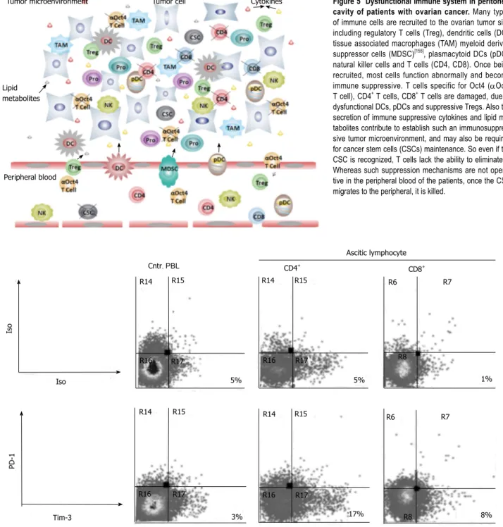
up-regulation of Tim-3 (Figure 6). These findings indicate
that infiltrated immune cells are not only suppressed,
but also impaired in their signaling pathways resulting
from the yet unknown factors present in tumor
associ-ated ascites.
Furthermore, except for harming of immune cells,
ovarian cancer cells also secrete immunosuppressive and
pro-inflammatory cytokines into the tumor
microenvi-ronment to support tumor growth
[144,145]. Previous studies
demonstrated that IL-6 is significantly increased in cyst
fluid, serum as well as ascites of patient with advanced
Trafficking Tumor cells CCL22 Angiogenic IL-10 TGF-β VEGF TNF-α IL-8 Dysfunctional Induction ExpansionFigure 4 Immune-suppressive pathways in ovarian cancer. Tregs are attracted to the tumor
environment by CCL22, secreted by the tumor.
The tumor microenvironment expresses molecules that can convert functional antigen presenting cells
(APCs) into dysfunctional ones. These dysfunction -al APCs in turn stimulate Treg differentiation and expansion. pDCs are also present in the tumor
en-vironment and stimulate tumor growth by releasing
tumor necrosis factor-α and interleukin-8. (modified
from[132]). pDCs also facilitate immunosuppressive
ovarian cancer, and associated with poor prognosis
[144,146].
IL-6 is a pro-inflammatory cytokine. It has multiple
ef-fects on T cell function, and it has already been reported
to be an important factor in promoting the progression
of epithelial of ovarian cancer
[147]. IL-6 also plays a role
in enhancing tumor growth by inducing abnormal c-Myc
expression
in vitro
. It has been shown that IL-6 can
in-duce c-Myc translation in multiple myeloma cells and
meanwhile c-Myc is shuttled to cytoplasm by the
RNA-binding protein, hnRNP A1
[148]. Our research
demon-strated that c-Myc was expressed in both nucleus and
cytoplasm in ovarian tumor tissue as well as ascitic cells,
while c-Myc is only expressed in the nucleus of normal
stem cells. Similarly, except for being expressed in the
nucleus, c-Myc was also detected in the cytoplasm of
leukemia patients
[149]. Regulation of stem cell genes or
even tumor development by cytokine indicates a strong
correlation between the tumor and its microenvironment.
Taken together, these results indicate that in addition to
its suppressive property, the tumor successfully creates a
favorable microenvironment to support tumor growth.
In conclusion, ovarian cancer is an extremely
compli-cated disease, because the tumor growth might be driven
by heterogeneous CSCs and multiple immunosuppressive
Tumor microenvironment
Lipid metabolites
Peripheral blood
Tumor cell Cytokines Figure 5 Dysfunctional immune system in peritoneal cavity of patients with ovarian cancer. Many types
of immune cells are recruited to the ovarian tumor site,
including regulatory T cells (Treg), dendritic cells (DC), tissue associated macrophages (TAM) myeloid derived
suppressor cells (MDSC)[155], plasmacytoid DCs (pDC),
natural killer cells and T cells (CD4, CD8). Once being
recruited, most cells function abnormally and become
immune suppressive. T cells specific for Oct4 (αOct4 T cell), CD4+ T cells, CD8+ T cells are damaged, due to dysfunctional DCs, pDCs and suppressive Tregs. Also the secretion of immune suppressive cytokines and lipid me -tabolites contribute to establish such an
immunosuppres-sive tumor microenvironment, and may also be required
for cancer stem cells (CSCs) maintenance. So even if the
CSC is recognized, T cells lack the ability to eliminate it.
Whereas such suppression mechanisms are not opera-tive in the peripheral blood of the patients, once the CSC migrates to the peripheral, it is killed.
R14 R15 R14 R15 R15 R14 R14 R15 R16 R16 R16 R17 R16 R17 R17 R17 3% 5% 5% 17% R8 8% R6 R7 R8 1% R6 R7 Iso Iso Tim-3 PD-1 CD4+ CD8+ Cntr. PBL Ascitic lymphocyte
Figure 6 PD-1 and Tim-3 expression by ascitic lymphocytes from patients with ovarian cancer. PD-1 expression was undetectable in ascitic lymphocytes. Com
mechanisms are functional in the abdomen. To enable an
immunological attack on CSC either the response has to
strengthened or the immunosuppressive milieu has to be
reversed or both.
FUTURE PERSPECTIVES
For future studies, it is of great importance to investigate
how somatic cells are reprogrammed
in vivo
to become
malignant pluripotent cells, and how the self-renewal
pathways are orchestrated in such transformed cells.
Furthermore, it remains unclear why the pluripotent
genes were upregulated in a small subset of tumor cells.
We sequenced both Oct4 and c-Myc isolated from
ovar-ian patient ascitic cells, however, no mutation was found
(unpublished data). It is important to elucidate what
went wrong in the self-renewal pathways in the patients
and why. Understanding this might help to stop tumor
growth before it happens.
Another challenge is how to boost the favorable host
immune response in the suppressive tumor
microenvi-ronment and train the immune system to fight against
ovarian cancer. To overcome this, it is of great
impor-tance to determine the mechanisms that contribute to
protective immune responses against tumors and to
en-hance these effector mechanisms in a tumor specific way.
And apparently, only boost the immune system is not
enough to eliminate tumors, due to functional crippling
of TILs.
Moreover, the role of ascites in tumor progression
remains to be elucidated. Ascitic fluid is produced by
ovarian tumor. The cellular fraction of ascites consists of
tumor cells, lymphocytes and mesothelial cells; and the
acellular fraction harbors cytokines, growth factors,
bio-active lipids, angiogenic factors, and extracellular matrix
constituents
[150-152]. Although the role of ascites as tumor
cell microenvironment remains poorly understood,
re-cent research suggests that it may affect cell growth,
invasion and induction of resistance of ovarian cancer
cells and thus may play a decisive role in ovarian tumor
progression
[153].
REFERENCES
1 Kandalaft LE, Powell DJ, Singh N, Coukos G.
Immunother-apy for ovarian cancer: what’s next? J Clin Oncol 2011; 29: 925-933 [PMID: 21079136 DOI: 10.1200/JCO.2009.27.2369]
2 Dick JE. Stem cell concepts renew cancer research. Blood
2008; 112: 4793-4807 [PMID: 19064739 DOI: 10.1182/ blood-2008-08-077941]
3 Shackleton M, Quintana E, Fearon ER, Morrison SJ.
Hetero-geneity in cancer: cancer stem cells versus clonal evolution.
Cell 2009; 138: 822-829 [PMID: 19737509]
4 Al-Hajj M, Wicha MS, Benito-Hernandez A, Morrison SJ,
Clarke MF. Prospective identification of tumorigenic breast
cancer cells. Proc Natl Acad Sci USA 2003; 100: 3983-3988 [PMID: 12629218 DOI: 10.1073/pnas.0530291100]
5 Al-Hajj M, Becker MW, Wicha M, Weissman I, Clarke MF.
Therapeutic implications of cancer stem cells. Curr Opin Genet Dev 2004; 14: 43-47 [PMID: 15108804 DOI: 10.1016/ j.gde.2003.11.007]
6 Al-Hajj M, Clarke MF. Self-renewal and solid tumor stem
cells. Oncogene 2004; 23: 7274-7282 [PMID: 15378087 DOI: 10.1038/sj.onc.1207947]
7 Cheshier SH, Morrison SJ, Liao X, Weissman IL. In vivo
pro-liferation and cell cycle kinetics of long-term self-renewing hematopoietic stem cells. Proc Natl Acad Sci USA 1999; 96: 3120-3125 [PMID: 10077647 DOI: 10.1073/pnas.96.6.3120]
8 Hua G, Thin TH, Feldman R, Haimovitz-Friedman A,
Clev-ers H, Fuks Z, Kolesnick R. Crypt base columnar stem cells in small intestines of mice are radioresistant. Gastroenterol-ogy 2012; 143: 1266-1276 [PMID: 22841781 DOI: 10.1053/ j.gastro.2012.07.106]
9 Higgins CF. Multiple molecular mechanisms for multidrug
resistance transporters. Nature 2007; 446: 749-757 [PMID: 17429392 DOI: 10.1038/nature05630]
10 Scharenberg CW, Harkey MA, Torok-Storb B. The ABCG2
transporter is an efficient Hoechst 33342 efflux pump and is
preferentially expressed by immature human hematopoietic progenitors. Blood 2002; 99: 507-512 [PMID: 11781231 DOI: 10.1182/blood.V99.2.507]
11 Kim M, Turnquist H, Jackson J, Sgagias M, Yan Y, Gong M, Dean M, Sharp JG, Cowan K. The multidrug resistance transporter ABCG2 (breast cancer resistance protein 1)
ef-fluxes Hoechst 33342 and is overexpressed in hematopoietic
stem cells. Clin Cancer Res 2002; 8: 22-28 [PMID: 11801536] 12 Chen KG, Valencia JC, Gillet JP, Hearing VJ, Gottesman
MM. Involvement of ABC transporters in melanogenesis and the development of multidrug resistance of melanoma.
Pigment Cell Melanoma Res 2009; 22: 740-749 [PMID: 19725928 DOI: 10.1111/j.1755-148X.2009.00630.x]
13 Dean M, Fojo T, Bates S. Tumour stem cells and drug resis-tance. Nat Rev Cancer 2005; 5: 275-284 [PMID: 15803154 DOI: 10.1038/nrc1590]
14 Stavrovskaya AA, Stromskaya TP. Transport proteins of the ABC family and multidrug resistance of tumor cells.
Biochemistry (Mosc) 2008; 73: 592-604 [PMID: 18605983 DOI: 10.1134/S0006297908050118]
15 Marques DS, Sandrini JZ, Boyle RT, Marins LF, Trindade GS. Relationships between multidrug resistance (MDR) and stem cell markers in human chronic myeloid leukemia cell lines. Leuk Res 2010; 34: 757-762 [PMID: 19969351 DOI: 10.1016/j.leukres.2009.11.004]
16 Borovski T, De Sousa E Melo F, Vermeulen L, Medema JP. Cancer stem cell niche: the place to be. Cancer Res 2011;
71: 634-639 [PMID: 21266356 DOI: 10.1158/0008-5472.
CAN-10-3220]
17 Sottoriva A, Sloot PM, Medema JP, Vermeulen L. Exploring cancer stem cell niche directed tumor growth. Cell Cycle 2010;
9: 1472-1479 [PMID: 20372084 DOI: 10.4161/cc.9.8.11198]
18 Blanpain C, Mohrin M, Sotiropoulou PA, Passegué E. DNA-damage response in tissue-specific and cancer stem cells.
Cell Stem Cell 2011; 8: 16-29 [PMID: 21211780 DOI: 10.1016/ j.stem.2010.12.012]
19 Moore N, Lyle S. Quiescent, slow-cycling stem cell popula-tions in cancer: a review of the evidence and discussion of
significance. J Oncol 2011; 2011: 396076 [PMID: 20936110] 20 Lacerda L, Pusztai L, Woodward W A. The role of tumor
ini-tiating cells in drug resistance of breast cancer: implications for future therapeutic approaches. Drug Resistance Updates
2010; 13: 99-108 [PMID: 20739212]
21 Landen CN, Goodman B, Katre AA, Steg AD, Nick AM, Stone RL, Miller LD, Mejia PV, Jennings NB, Gershenson DM, Bast RC, Coleman RL, Lopez-Berestein G, Sood AK. Targeting aldehyde dehydrogenase cancer stem cells in ovarian cancer. Mol Cancer Ther 2010; 9: 3186-3199 [PMID: 20889728 DOI: 10.1158/1535-7163.MCT-10-0563]
22 Alvero AB, Montagna MK, Holmberg JC, Craveiro V, Brown D, Mor G. Targeting the mitochondria activates two inde-pendent cell death pathways in ovarian cancer stem cells.
10.1158/1535-7163.MCT-11-0023]
23 Hu Y, Fu L. Targeting cancer stem cells: a new therapy to cure cancer patients. Am J Cancer Res 2012; 2: 340-356 [PMID: 22679565]
24 McCubrey JA, Steelman LS, Abrams SL, Misaghian N, Chappell WH, Basecke J, Nicoletti F, Libra M, Ligresti G, Sti-vala F, Maksimovic-Ivanic D, Mijatovic S, Montalto G, Cer-vello M, Laidler P, Bonati A, Evangelisti C, Cocco L, Martelli AM. Targeting the cancer initiating cell: the ultimate target for cancer therapy. Curr Pharm Des 2012; 18: 1784-1795 [PMID: 22394167 DOI: 10.2174/138161212799859701]
25 Pardal R, Clarke MF, Morrison SJ. Applying the principles of stem-cell biology to cancer. Nat Rev Cancer 2003; 3: 895-902 [PMID: 14737120 DOI: 10.1038/nrc1232]
26 Iovino F, Meraviglia S, Spina M, Orlando V, Saladino V, Dieli F, Stassi G, Todaro M. Immunotherapy targeting co-lon cancer stem cells. Immunotherapy 2011; 3: 97-106 [PMID: 21174560 DOI: 10.2217/imt.10.87]
27 Morrison BJ, Schmidt CW, Lakhani SR, Reynolds BA, Lo-pez JA. Breast cancer stem cells: implications for therapy of breast cancer. Breast Cancer Res 2008; 10: 210 [PMID: 18671830 DOI: 10.1186/bcr2111]
28 Bonnet D, Dick JE. Human acute myeloid leukemia is orga-nized as a hierarchy that originates from a primitive hema-topoietic cell. Nat Med 1997; 3: 730-737 [PMID: 9212098 DOI: 10.1038/nm0797-730]
29 Collins AT, Berry PA, Hyde C, Stower MJ, Maitland NJ. Prospective identification of tumorigenic prostate cancer stem cells. Cancer Res 2005; 65: 10946-10951 [PMID: 16322242 DOI: 10.1158/0008-5472.CAN-05-2018]
30 Dalerba P, Dylla SJ, Park IK, Liu R, Wang X, Cho RW, Hoey T, Gurney A, Huang EH, Simeone DM, Shelton AA, Parm-iani G, Castelli C, Clarke MF. Phenotypic characterization of human colorectal cancer stem cells. Proc Natl Acad Sci USA 2007; 104: 10158-10163 [PMID: 17548814 DOI: 10.1073/ pnas.0703478104]
31 Fang D, Nguyen TK, Leishear K, Finko R, Kulp AN, Hotz S, Van Belle PA, Xu X, Elder DE, Herlyn M. A tumorigenic subpopulation with stem cell properties in melanomas.
Cancer Res 2005; 65: 9328-9337 [PMID: 16230395 DOI: 10.1158/0008-5472.CAN-05-1343]
32 Gao MQ, Choi YP, Kang S, Youn JH, Cho NH. CD24+ cells from hierarchically organized ovarian cancer are enriched in cancer stem cells. Oncogene 2010; 29: 2672-2680 [PMID: 20190812 DOI: 10.1038/onc.2010.35]
33 Hermann PC, Huber SL, Herrler T, Aicher A, Ellwart JW, Guba M, Bruns CJ, Heeschen C. Distinct populations of can-cer stem cells determine tumor growth and metastatic activ-ity in human pancreatic cancer. Cell Stem Cell 2007; 1: 313-323 [PMID: 18371365 DOI: 10.1016/j.stem.2007.06.002]
34 Hwang-Verslues WW, Kuo WH, Chang PH, Pan CC, Wang HH, Tsai ST, Jeng YM, Shew JY, Kung JT, Chen CH, Lee EY, Chang KJ, Lee WH. Multiple lineages of human breast
can-cer stem/progenitor cells identified by profiling with stem
cell markers. PLoS One 2009; 4: e8377 [PMID: 20027313 DOI: 10.1371/journal.pone.0008377]
35 Kemper K, Prasetyanti PR, De Lau W, Rodermond H, Clev-ers H, Medema JP. Monoclonal antibodies against Lgr5 iden-tify human colorectal cancer stem cells. Stem Cells 2012; 30: 2378-2386 [PMID: 22969042 DOI: 10.1002/stem.1233] 36 Shi MF, Jiao J, Lu WG, Ye F, Ma D, Dong QG, Xie X.
Iden-tification of cancer stem cell-like cells from human epithe-lial ovarian carcinoma cell line. Cell Mol Life Sci 2010; 67: 3915-3925 [PMID: 20549538 DOI: 10.1007/s00018-010-0420-9] 37 Singh SK, Clarke ID, Terasaki M, Bonn VE, Hawkins C,
Squire J, Dirks PB. Identification of a cancer stem cell in hu -man brain tumors. Cancer Res 2003; 63: 5821-5828 [PMID: 14522905]
38 Szotek PP, Pieretti-Vanmarcke R, Masiakos PT, Dinulescu DM, Connolly D, Foster R, Dombkowski D, Preffer F,
Ma-claughlin DT, Donahoe PK. Ovarian cancer side population
defines cells with stem cell-like characteristics and Mullerian
Inhibiting Substance responsiveness. Proc Natl Acad Sci USA
2006; 103: 11154-11159 [PMID: 16849428 DOI: 10.1073/ pnas.0603672103]
39 Zhang S, Balch C, Chan MW, Lai HC, Matei D, Schilder JM,
Yan PS, Huang TH, Nephew KP. Identification and charac -terization of ovarian cancer-initiating cells from primary hu-man tumors. Cancer Res 2008; 68: 4311-4320 [PMID: 18519691 DOI: 10.1158/0008-5472.CAN-08-0364]
40 Zucchi I, Astigiano S, Bertalot G, Sanzone S, Cocola C, Pelucchi P, Bertoli G, Stehling M, Barbieri O, Albertini A, Schöler HR, Neel BG, Reinbold RA, Dulbecco R. Distinct populations of tumor-initiating cells derived from a tumor generated by rat mammary cancer stem cells. Proc Natl Acad Sci USA 2008; 105: 16940-16945 [PMID: 18957543 DOI: 10.1073/pnas.0808978105]
41 Curley MD, Therrien VA, Cummings CL, Sergent PA, Kou-louris CR, Friel AM, Roberts DJ, Seiden MV, Scadden DT,
Rueda BR, Foster R. CD133 expression defines a tumor initi -ating cell population in primary human ovarian cancer. Stem Cells 2009; 27: 2875-2883 [PMID: 19816957]
42 Ferrandina G, Martinelli E, Petrillo M, Prisco MG, Zannoni G, Sioletic S, Scambia G. CD133 antigen expression in ova-rian cancer. BMC Cancer 2009; 9: 221 [PMID: 19583859 DOI: 10.1186/1471-2407-9-221]
43 Hu L, McArthur C, Jaffe RB. Ovarian cancer stem-like side-population cells are tumourigenic and chemoresistant. Br J Cancer 2010; 102: 1276-1283 [PMID: 20354527 DOI: 10.1038/ sj.bjc.6605626]
44 Patrawala L, Calhoun T, Schneider-Broussard R, Zhou J, Claypool K, Tang DG. Side population is enriched in tumori-genic, stem-like cancer cells, whereas ABCG2+ and ABCG2- cancer cells are similarly tumorigenic. Cancer Res 2005;
65: 6207-6219 [PMID: 16024622 DOI: 10.1158/0008-5472.
CAN-05-0592]
45 Stewart JM, Shaw PA, Gedye C, Bernardini MQ, Neel BG, Ailles LE. Phenotypic heterogeneity and instability of human ovarian tumor-initiating cells. Proc Natl Acad Sci USA 2011; 108: 6468-6473 [PMID: 21451132 DOI: 10.1073/ pnas.1005529108]
46 Lee M, Nam EJ, Kim SW, Kim S, Kim JH, Kim YT. Prog-nostic impact of the cancer stem cell-related marker NANOG in ovarian serous carcinoma. Int J Gynecol Can-cer 2012; 22: 1489-1496 [PMID: 23095773 DOI: 10.1097/ IGJ.0b013e3182738307]
47 Roesch A, Fukunaga-Kalabis M, Schmidt EC, Zabierowski SE, Brafford PA, Vultur A, Basu D, Gimotty P, Vogt T, Her-lyn M. A temporarily distinct subpopulation of slow-cycling melanoma cells is required for continuous tumor growth.
Cell 2010; 141: 583-594 [PMID: 20478252 DOI: 10.1016/ j.cell.2010.04.020]
48 Dieter SM, Ball CR, Hoffmann CM, Nowrouzi A, Herbst F, Zavidij O, Abel U, Arens A, Weichert W, Brand K, Koch M, Weitz J, Schmidt M, von Kalle C, Glimm H. Distinct types of tumor-initiating cells form human colon cancer tumors and metastases. Cell Stem Cell 2011; 9: 357-365 [PMID: 21982235 DOI: 10.1016/j.stem.2011.08.010]
49 Frank NY, Schatton T, Frank MH. The therapeutic promise of the cancer stem cell concept. J Clin Invest 2010; 120: 41-50 [PMID: 20051635 DOI: 10.1172/JCI41004]
50 Quintana E, Shackleton M, Foster HR, Fullen DR, Sabel MS, Johnson TM, Morrison SJ. Phenotypic heterogeneity among tumorigenic melanoma cells from patients that is revers-ible and not hierarchically organized. Cancer Cell 2010; 18: 510-523 [PMID: 21075313 DOI: 10.1016/j.ccr.2010.10.012] 51 Clevers H. The cancer stem cell: premises, promises and
challenges. Nat Med 2011; 17: 313-319 [PMID: 21386835 DOI: 10.1038/nm.2304]
Eradi-cating cancer cells: struggle with a chameleon. Oncotarget
2011; 2: 99-101 [PMID: 21378413]
53 Renkvist N, Castelli C, Robbins PF, Parmiani G. A listing of human tumor antigens recognized by T cells. Cancer Immu-nol Immunother 2001; 50: 3-15 [PMID: 11315507 DOI: 10.1007/ s002620000169]
54 Visvader JE, Lindeman GJ. Cancer stem cells: current status and evolving complexities. Cell Stem Cell 2012; 10: 717-728 [PMID: 22704512 DOI: 10.1016/j.stem.2012.05.007]
55 Tang DG. Understanding cancer stem cell heterogeneity and plasticity. Cell Res 2012; 22: 457-472 [PMID: 22357481 DOI: 10.1038/cr.2012.13]
56 Quintana E, Shackleton M, Sabel MS, Fullen DR, Johnson
TM, Morrison SJ. Efficient tumour formation by single hu -man melanoma cells. Nature 2008; 456: 593-598 [PMID: 19052619 DOI: 10.1038/nature07567]
57 Chang B, Liu G, Xue F, Rosen DG, Xiao L, Wang X, Liu J. ALDH1 expression correlates with favorable prognosis in ovarian cancers. Mod Pathol 2009; 22: 817-823 [PMID: 19329942 DOI: 10.1038/modpathol.2009.35]
58 Fong MY, Kakar SS. The role of cancer stem cells and the side population in epithelial ovarian cancer. Histol Histo-pathol 2010; 25: 113-120 [PMID: 19924647]
59 Strauss R, Li ZY, Liu Y, Beyer I, Persson J, Sova P, Möller T, Pesonen S, Hemminki A, Hamerlik P, Drescher C, Urban N, Bartek J, Lieber A. Analysis of epithelial and mesenchymal markers in ovarian cancer reveals phenotypic heterogeneity and plasticity. PLoS One 2011; 6: e16186 [PMID: 21264259 DOI: 10.1371/journal.pone.0016186]
60 Di J, Yigit R, Figdor CG, Duiveman-de Boer T, Massuger LFAG, Torensma R. Expression compilation of several puta-tive cancer stem cell markers by primary ovarian carvinoma.
J Cancer Ther 2010; 1: 165-173 [DOI: 10.4236/jct.2010.14026] 61 Deng S, Yang X, Lassus H, Liang S, Kaur S, Ye Q, Li C,
Wang LP, Roby KF, Orsulic S, Connolly DC, Zhang Y, Mon-tone K, Bützow R, Coukos G, Zhang L. Distinct expression levels and patterns of stem cell marker, aldehyde dehydroge-nase isoform 1 (ALDH1), in human epithelial cancers. PLoS One 2010; 5: e10277 [PMID: 20422001 DOI: 10.1371/journal. pone.0010277]
62 Lorico A, Rappa G. Phenotypic heterogeneity of breast can-cer stem cells. J Oncol 2011; 2011: 135039 [PMID: 21317983] 63 Takahashi K, Yamanaka S. Induction of pluripotent stem
cells from mouse embryonic and adult fibroblast cultures by defined factors. Cell 2006; 126: 663-676 [PMID: 16904174 DOI: 10.1016/j.cell.2006.07.024]
64 Nichols J, Zevnik B, Anastassiadis K, Niwa H, Klewe-Nebenius D, Chambers I, Schöler H, Smith A. Formation of pluripotent stem cells in the mammalian embryo depends on the POU transcription factor Oct4. Cell 1998; 95: 379-391 [PMID: 9814708 DOI: 10.1016/S0092-8674(00)81769-9] 65 Niwa H, Miyazaki J, Smith AG. Quantitative expression of
Oct-3/4 defines differentiation, dedifferentiation or self-renewal of ES cells. Nat Genet 2000; 24: 372-376 [PMID: 10742100 DOI: 10.1038/74199]
66 Looijenga LH, Stoop H, de Leeuw HP, de Gouveia Brazao CA, Gillis AJ, van Roozendaal KE, van Zoelen EJ, Weber RF, Wolffenbuttel KP, van Dekken H, Honecker F, Bokemeyer C, Perlman EJ, Schneider DT, Kononen J, Sauter G,
Ooster-huis JW. POU5F1 (OCT3/4) identifies cells with pluripotent
potential in human germ cell tumors. Cancer Res 2003; 63: 2244-2250 [PMID: 12727846]
67 Tsai LL, Yu CC, Chang YC, Yu CH, Chou MY. Markedly increased Oct4 and Nanog expression correlates with cis-platin resistance in oral squamous cell carcinoma. J Oral Pathol Med 2011; 40: 621-628 [PMID: 21342274 DOI: 10.1111/ j.1600-0714.2011.01015.x]
68 Atlasi Y, Mowla SJ, Ziaee SA, Bahrami AR. OCT-4, an em-bryonic stem cell marker, is highly expressed in bladder can-cer. Int J Cancer 2007; 120: 1598-1602 [PMID: 17205510 DOI:
10.1002/ijc.22508]
69 Tai MH, Chang CC, Kiupel M, Webster JD, Olson LK, Tro-sko JE. Oct4 expression in adult human stem cells: evidence in support of the stem cell theory of carcinogenesis. Carcino-genesis 2005; 26: 495-502 [PMID: 15513931]
70 Lee J, Kim HK, Rho JY, Han YM, Kim J. The human OCT-4 isoforms differ in their ability to confer self-renewal. J Biol Chem 2006; 281: 33554-33565 [PMID: 16951404]
71 Cauffman G, Liebaers I, Van Steirteghem A, Van de Velde H. POU5F1 isoforms show different expression patterns in human embryonic stem cells and preimplantation embryos.
Stem Cells 2006; 24: 2685-2691 [PMID: 16916925]
72 Liedtke S, Stephan M, Kögler G. Oct4 expression revisited: potential pitfalls for data misinterpretation in stem cell re-search. Biol Chem 2008; 389: 845-850 [PMID: 18627312] 73 Zhao S, Yuan Q, Hao H, Guo Y, Liu S, Zhang Y, Wang
J, Liu H, Wang F, Liu K, Ling EA, Hao A. Expression of OCT4 pseudogenes in human tumours: lessons from glioma and breast carcinoma. J Pathol 2011; 223: 672-682 [PMID: 21341266]
74 Cartwright P, McLean C, Sheppard A, Rivett D, Jones K, Dalton S. LIF/STAT3 controls ES cell self-renewal and pluri-potency by a Myc-dependent mechanism. Development 2005;
132: 885-896 [PMID: 15673569 DOI: 10.1242/dev.01670]
75 Chambers I, Colby D, Robertson M, Nichols J, Lee S, Tweed-ie S, Smith A. Functional expression cloning of Nanog, a pluripotency sustaining factor in embryonic stem cells. Cell
2003; 113: 643-655 [PMID: 12787505]
76 Vathipadiekal V, Saxena D, Mok SC, Hauschka PV, Ozbun
L, Birrer MJ. Identification of a potential ovarian cancer stem
cell gene expression profile from advanced stage papillary serous ovarian cancer. PLoS One 2012; 7: e29079 [PMID: 22272227]
77 Pietras K, Ostman A. Hallmarks of cancer: interactions with the tumor stroma. Exp Cell Res 2010; 316: 1324-1331 [PMID: 20211171]
78 Scheel C, Weinberg RA. Phenotypic plasticity and epithelial-mesenchymal transitions in cancer and normal stem cells?
Int J Cancer 2011; 129: 2310-2314 [PMID: 21792896 DOI: 10.1002/ijc.26311]
79 Penchev VR, Rasheed ZA, Maitra A, Matsui W. Het-erogeneity and targeting of pancreatic cancer stem cells.
Clin Cancer Res 2012; 18: 4277-4284 [PMID: 22896694 DOI: 10.1158/1078-0432.CCR-11-3112]
80 Leth-Larsen R, Terp MG, Christensen AG, Elias D, Kühl-wein T, Jensen ON, Petersen OW, Ditzel HJ. Functional het-erogeneity within the CD44 high human breast cancer stem cell-like compartment reveals a gene signature predictive of distant metastasis. Mol Med 2012; 18: 1109-1121 [PMID: 22692575 DOI: 10.2119/molmed.2012.00091]
81 Engh JA. A heterogeneous population of stem cells within glioblastoma tumors in the setting of disease relapse. Neuro-surgery 2011; 68: N15-N16 [PMID: 21792098]
82 Pietras A. Cancer stem cells in tumor heterogeneity. Adv Cancer Res 2011; 112: 255-281 [PMID: 21925307 DOI: 10.1016/ B978-0-12-387688-1.00009-0]
83 Dyall S, Gayther SA, Dafou D. Cancer stem cells and epi-thelial ovarian cancer. J Oncol 2010; 2010: 105269 [PMID: 21318146]
84 Lotem J, Sachs L. Epigenetics and the plasticity of differen-tiation in normal and cancer stem cells. Oncogene 2006; 25: 7663-7672 [PMID: 16847453 DOI: 10.1038/sj.onc.1209816] 85 Leder K, Holland EC, Michor F. The therapeutic
implica-tions of plasticity of the cancer stem cell phenotype. PLoS One 2010; 5: e14366 [PMID: 21179426 DOI: 10.1371/journal. pone.0014366]
86 Chaffer CL, Brueckmann I, Scheel C, Kaestli AJ, Wiggins PA, Rodrigues LO, Brooks M, Reinhardt F, Su Y, Polyak K, Arendt LM, Kuperwasser C, Bierie B, Weinberg RA. Normal and neoplastic nonstem cells can spontaneously convert to a