warwick.ac.uk/lib-publications
Original citation:Yang, Chengjuan, Tian, Yanling, Cui, Liangyu and Zhang, Dawei (2015) Laser-induced changes
in titanium by femtosecond, picosecond and millisecond laser ablation. Radiation Effects and
Defects in Solids, 170 (6). pp. 528-540.doi:10.1080/10420150.2015.1052436 Permanent WRAP URL:
http://wrap.warwick.ac.uk/76450
Copyright and reuse:
The Warwick Research Archive Portal (WRAP) makes this work by researchers of the University of Warwick available open access under the following conditions. Copyright © and all moral rights to the version of the paper presented here belong to the individual author(s) and/or other copyright owners. To the extent reasonable and practicable the material made available in WRAP has been checked for eligibility before being made available.
Copies of full items can be used for personal research or study, educational, or not-for profit purposes without prior permission or charge. Provided that the authors, title and full bibliographic details are credited, a hyperlink and/or URL is given for the original metadata page and the content is not changed in any way.
Publisher’s statement:
“This is an Accepted Manuscript of an article published by Taylor & Francis in Radiation Effects and Defects in Solids on 25/06/2015 available
online: http://dx.doi.org/10.1080/10420150.2015.1052436
A note on versions:
The version presented here may differ from the published version or, version of record, if you wish to cite this item you are advised to consult the publisher’s version. Please see the ‘permanent WRAP URL’ above for details on accessing the published version and note that access may require a subscription.
Laser-induced Changes in Titanium by Femtosecond,
Picosecond and Millisecond Laser Ablation
Chengjuan Yang
ab*,
Yanling Tian
ab, Liangyu Cui
ab, Dawei Zhang
aba
School of Mechanical Engineering, Tianjin University, Tianjin, 300072, China;
b
Key Laboratory of Mechanism Theory and Equipment Design of Ministry of Education,
Tianjin University, Tianjin 300072, China
Chengjuan Yang*:
Postal address: School of Mechanical Engineering, Tianjin University,
92 Weijin Road, Nankai District, Tianjin, 300072, P.R.China
Telephone: (86) 18002186575,Fax: (86) 2227405561
Email: cjytju@tju.edu.cn
Yanling Tian:
Postal address: School of Mechanical Engineering, Tianjin University,
92 Weijin Road, Nankai District, Tianjin, 300072, P.R.China
Telephone: (86) 2227405561, Fax: (86) 2227405561
Email: meytian@tju.edu.cn
Liangyu Cui:
Postal address: School of Mechanical Engineering, Tianjin University,
92 Weijin Road, Nankai District, Tianjin, 300072, P.R.China
Email: cuily@tju.edu.cn
Dawei Zhang:
Postal address: School of Mechanical Engineering, Tianjin University,
92 Weijin Road, Nankai District, Tianjin, 300072, P.R.China
Telephone: (86) 2287401950, Fax: (86) 2227405561
Laser-induced Changes in Titanium by Femtosecond,
Picosecond and Millisecond Laser Ablation
Abstract:
A more completed and further understanding of laser-induced material changes
mechanism has a great significance to improve the controllability of laser ablation
process and perfect the processing quality of final results. Therefore, comparative
ablation experiments by femtosecond, picosecond and millisecond-pulsed laser
were carried out on titanium in this study in order to realize the qualitative control
of the laser-induced changes trend, and the quantitative control of the laser-induced
changes range in titanium upon the laser irradiation with different pulse duration.
Then the final surface morphology, aspect ratio, chemical composition and
microstructural state of the ablated titanium were analyzed by laser scanning
confocal microscopy (LSCM), X-ray photoelectron spectroscopy (XPS) and
transmission electron microscopy (TEM), respectively. The dependency of the
morphology, size, composition and microstructure of ablated titanium on laser
pulse duration variation were emphatically discussed. It is found that, as the laser
pulse duration increases from femtosecond to millisecond scale, surface
morphology quality of ablated titanium gets worse, aspect ratio of microgroove
decreases, proportion of titanium oxides in final ablation products becomes larger
and the microstructural state of ablated titanium has a higher amorphization degree.
Finally, it is deduced that the occurrence of all these experimental results can be
attributed to the decreased laser intensity per pulse and enhanced heat conduction
effect in titanium with the pulse duration increasing, which resulted in more
serious thermal and mechanical damages in material during longer pulse duration.
Keywords: titanium; laser pulse duration variation; laser-induced change
1. Introduction
As an important metal material, titanium is a kind of metal with high specific strength,
excellent corrosion resistance, good shape memory properties and biocompatibility, rare
superconducting and low damping characteristics, etc (1-4). Therefore, titanium becomes
a hot "star metal" and has been widely used in the fields of aerospace, defense weapons,
chemical and metallurgical industries, ship manufacturing and vehicle engineering, sports
fitness and rehabilitation medical devices, and some other fields. If some methods of
secondary surface treatment were used, the performances of titanium can be further
improved and the applicability of titanium will also be put to a new peak. There have
been several methods of surface treatment to improve the surface properties of titanium,
such as electroplating, electroless plating, chemical process (chemical coating), anodic
oxidation process, hot dipping, vacuum plating, painting, thermal spraying, surface
hardening, metallic cementation (5, 6), and so on. Among these surface treatment
methods, the method of laser surface treatment on titanium is gradually moving from
numerous processing technologies to the fore due to its special advantages including the
enhancement of the surface dependent properties (hardness, friction, fatigue, resistance to
wear and corrosion), flexibility and possibility of treating small areas, leaving the others
parts unaffected, and the desired firm level of adhesion bonding effect between surface
layer and base material, etc (7-9). All these induced positive changes in titanium aim at
making material keep a lot of excellent existing features and further obtaining more
expected performance. Besides the processing advantages of laser surface
treatment method mentioned above, the steady developed high power lasers and their
suitability introduced in production lines, laser surface treatment also encouraged the
industrial application of photoelectrochemical, advanced materials, photoelectrode in
dye-sensitized solar cells (DSSCs) and TiO2 microtubes (10-12).
A lot of efforts on the investigation of laser-induced changes in titanium and its
richer variety of surface structures on titanium for implants and other biomedical
applications than long-pulse laser treatments (13). The dependences of laser-induced
periodic surface structures (LIPSS) in titanium on the laser fluence and pulses number
per irradiation spot have been analyzed experimentally and theoretically by femtosecond
laser pulses in different environment (14, 15). Those obtained experimental results were
complemented by calculations based on a theoretical LIPSS model and compared to the
present literature. By irradiation with ultrafast laser pulses of 130 fs duration, 800 nm
wavelength in vacuum (∼1mbar) or in 100 mbar He, a regular array of sharp
nano-textured conical microstructures were observed on the titanium metal surface. And
the reflectivity of these microstructures’ surface is greatly reduced throughout the
measured visible spectrum (15). In addition to the study of the surface morphology,
structural modification of titanium (Ti) after irradiation of femtosecond (25 fs) laser also
has been explored in various pulse energies ranging. Compared with the result in dry (air)
environment, surface treatment of Ti with femtosecond laser irradiation in liquid (ethanol)
environment is found to allow the growth of particular surface structures in the form of
grains and simultaneously induces changes in its chemical composition (16). Submerging
titanium target in water, A. De Bonis (11) observed the formation of titania nanoparticles
with a certain quantity of rutile phase after a high power density configuration
(2×10¹⁶W/cm2) laser ablation (Ti-sapphire, λ= 800 nm, 1 kHz, 100 fs). Upon remaining
the ablated species in water, the formation of a lamellar phase could be further observed.
Then a XPS analysis revealed the co-presence of Ti2O3, TiO2 and TiOH, denoting the
occurrence of nonstoichimetric titania phase and hence cation and oxygen vacancies (11).
titanium alloy irradiated by a picosecond pulse infrared laser with a 1064 nm wavelength
has better ablation surface morphology than that of the green picosecond pulse laser with
a 532nm wavelength. The feature sizes are approximately linearly dependent on the laser
pulse energy density at low energy density and the monotonic increase with laser pulse
energy density increasing. The ablation threshold of titanium alloy irradiated by an
ultra-fast pulse laser was calculated to be about 0.109 J/cm2 (17). Upon the irradiation of
KrF excimer laser with wavelength 248 nm, pulse duration of 20 ns and repetition rate of
20 Hz, surface and structural properties of the laser irradiated titanium targets have been
investigated under dry and wet ambient environments. The targets were exposed for
various number of laser pulses ranging from 500 to 2000 in the ambient environment of
air, de-ionized water and propanol at a fluence of 3.6 J/cm2. For various number of laser
pulses, the variations in the peak intensity, crystallinity and d-spacing are observed under
both ambient conditions (18). E. György and colleagues found that the interesting and
uncommon laser-induced surface microrelief development was associated with the
increase of the laser intensity effectively absorbed by the target surface as laser pulse
(λ=1.064 μm, τ ≈300 ns, υ=30 kHz) number increased, where crystallization accounted
for this shaped, dendritic microrelief (19). In condition of ultrahigh vacuum, high purity
titanium target was ablated by nanosecond pulsed Nd:YAG laser (λ=1.064 μm, τ ≈120 ns,
υ=1 kHz) which induced a titanium thin film of about 250nm nominal thickness in a fcc
phase deposited on a substrate with 200°C. With the increase of implanted ion fluence,
structure of this titanium thin film became amorphous phase before precipitation of
nanocrystalline fcc TiN phase (20). While the same laser ablation process was conducted
showed that the layer had a uniform surface and mainly consisted of the tetragonal
δ’-TiNx crystalline phase (20). In oxygen ambient, very rapid heating-cooling process of
the titanium target under energetic Nd:YAG millisecond laser pulse irradiation made the
spherical nanoparticles transform from martensite into a high-pressure phase of TiO2 with
a baddeleyite-type related structure, and then into α-PbO2-type structure (21). During the
situ synthesis process of TiC by laser surface treatment with pulse duration changing
from 4 ms to12 ms, the dendritic morphology of composite layer changed to cellular
grain structure in the conditions with the increased laser energy, low processing speed,
more dissolution of carbon into liquid Ti by more heat input and positive influence of the
Marangoni flow in the melted zone (22). Titanium oxide nanoparticles in H2O-distilled
solvent were prepared by irradiating the titanium target with a Nd:YAG laser (second
harmonic, λ=532 nm), varying the operative fluence in the range 1–8 J/cm2 and the
ablation time from 10 min to 60 min (23). In the investigation on laser surface treatment
of titanium alloys, a preliminary testing of the surface preparation technique using
frequency tripled Nd:YAG nanosecond laser ablation (λ=355 nm) as a replacement for
the chemical etch and abrasive processes has been applied to Ti-6Al-4V alloy adherends.
Single lap shear testing showed that increasing laser ablation duty cycle and power not
only reduced crack propagation and adhesive failure, but also increased the strength and
durability (24). In addition to that, using UV emitting KrCl (at 222nm, ~13 ns) and
XeCl (at 308nm, ~11ns) excimer lasers, the main influences of laser beam fluence
variation on Ti-6Al-4V were investigated in detail from the aspects of thermal character
and surface morphology (25). According to the review of previous work, a lot of efforts
laser pulses with different wavelengths, energy intensities, pulse repetition frequencies,
gas atmospheres and so on (26-28). However, very little attention has been paid to reveal
the influence of laser pulse duration variation on the laser-induced changes in titanium.
In order to further explore the essential mechanism of laser-induced changes in
titanium, surface morphology, aspect ratio, chemical composition and microstructural
state changes in titanium after femtosecond, picosecond, and millisecond-pulsed laser
irradiation were systematically studied in this work. Firstly, comparative ablation
experiments on thin titanium plates with 1 mm thickness irradiated by femtosecond,
picosecond and millisecond laser pulses were carried out at standard atmospheric
pressure in air. Then laser scanning confocal microscopy (LSCM), X-ray photoelectron
spectroscopy (XPS) and transmission electron microscopy (TEM) were used to analyze
the surface morphology, aspect ratio, chemical composition and microstructural state
changes of ablated titanium, respectively. Finally, the influence of laser pulse duration
variation on the laser-induced changes in ablated titanium was analyzed intensively from
the aspects of morphology, size, composition and microstructure, also the internal
mechanism was discussed.
2. Experimental
Titanium plates with 1 mm thickness were selected to be the sample material in this study.
All of the femtosecond, picosecond and millisecond-pulsed laser processing systems are
mainly made up of optical system for generating laser pulses and motion control system
for moving sample material. In optical systems, a commercially available femtosecond
laser (Nd: YLF femtosecond laser system, Coherent Ltd., Libra-USP-HE), a picosecond
SC-1064-2000 IN00060), and the JK300D millisecond-pulsed Nd:YAG laser by GSI
Group were respectively used to ablate parallel microgrooves on the surface of titanium
plates. Those ablated microgrooves overlapped each other to form a larger ablated region
for ease of the subsequent test and analysis. All the ablation experiments were performed
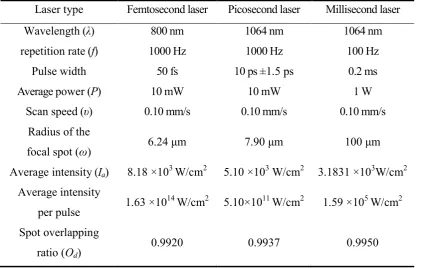
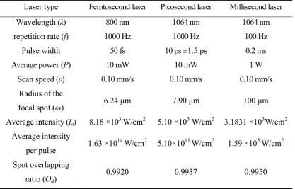
at standard atmospheric pressure in air. Table 1 has given the important experimental
parameters in detail. It can be seen that the average laser intensities and focal spot
overlapping ratios in femtosecond, picosecond, and millisecond laser ablation
experiments are in the same order of magnitude. Also the wavelengths of these laser
pulses are all within the range of near infrared laser wavelength. Therefore, the small
differences of average laser intensities, focal spot overlapping ratios and wavelengths can
[image:10.595.90.513.440.708.2]be ignored in this study.
Table 1. Parameters used in laser ablation experiments
Laser type Femtosecond laser Picosecond laser Millisecond laser
Wavelength (λ) 800 nm 1064 nm 1064 nm
repetition rate (f) 1000 Hz 1000 Hz 100 Hz
Pulse width 50 fs 10 ps ±1.5 ps 0.2 ms
Average power (P) 10 mW 10 mW 1 W
Scan speed (υ) 0.10 mm/s 0.10 mm/s 0.10 mm/s
Radius of the
focal spot (ω) 6.24 μm 7.90 μm 100 μm
Average intensity (Ia) 8.18 ×10³ W/cm2 5.10 ×10³ W/cm2 3.1831 ×10³W/cm2
Average intensity
per pulse 1.63 ×10
14
W/cm2 5.10×10¹¹ W/cm2 1.59 ×10⁵ W/cm2
Spot overlapping
ratio (Od)
3. Results and discussion
3.1 Influence of laser pulse duration variation on the surface morphology and
aspect ratio of titanium by LSCM
[Figure 1 near here]
[Figure 2 near here]
[Figure 3 near here]
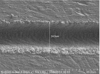
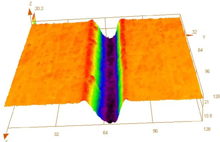
LSCM (OLS4000 from Olympus) was used to observe the 2-D surface morphology and
3-D topography of the ablated region on titanium plates’ surface after femtosecond,
picosecond and millisecond laser ablation. From figs. 1, 2, and 3, it can be seen that
surface morphology quality of the ablated titanium is overall getting worse as laser pulse
duration increases. During the ultrashort pulse duration of femtosecond laser, ultrahigh
laser intensity per pulse is the major reason for the good surface morphology quality of
ablated microgroove. Compared with the extremely high energy input during
femtosecond laser irradiation, the increase of laser pulse duration from femtosecond to
picosecond and then to millisecond scale not only reduces the laser intensity per pulse of
picosecond and millisecond laser, but also provides the absorbed laser energy enough
time to transfer outward as heat and so the heat conduction effect in the irradiated
material is significantly enhanced, which results in more serious thermal and mechanical
damages including melting and solidification of irradiated material, redeposition of
ablation debris, and so on. Therefore, picosecond and millisecond laser produce the
microgrooves with much poorer surface morphology quality, such as worse surface
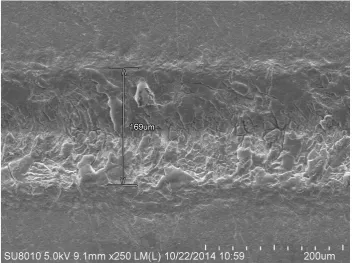
In addition, with the increase of laser pulse duration from femtosecond to
millisecond scale, the widths and depths of microgrooves ablated by femtosecond,
picosecond and millisecond-pulsed laser are 24.5μm and 10.5μm, 69.8μm and 10μm,
169μm and 18.5μm, respectively, so the corresponding aspect ratios of these
microgrooves are 0.4286, 0.1433, and 0.1095, respectively. It can be found that the
microgroove’ aspect ratio decreases with the laser pulse duration increasing. The main
reason can be attributed to the drop of laser intensity per pulse and the enhanced heat
conduction effect as the laser pulse duration increases.
3.2 Influence of laser pulse duration variation on the chemical composition of
titanium by XPS
[Figure 4 near here]
[Figure 5 near here]
[Figure 6 near here]
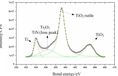
Based on the XPS analysis results shown in figs. 4, 5, and 6, femtosecond laser ablation
product mainly include TiO2, TiO2 rutile, Ti2O3, TiN (loss peak), Ti, the major
compositions of picosecond laser ablation product are TiO2, TiO2 rutile, Ti2O3, TiN (loss
peak), Ti (C, N), TiO0.73, and millisecond laser ablation mainly produces TiO2 (in oxide
films) and TiO2 rutile. Replacing Ti displayed in fig. 4, the additional Ti (C, N) and
TiO0.73 appearing in fig. 5 mainly result from the relatively full reaction between air and
titanium during picosecond pulse duration compared with the femtosecond laser ablation.
So it can be found that, as the laser pulse duration increases from femtosecond to
new generated titanium compounds becomes varied, the composition of compounds
themselves gets more complex, and the content of residual elemental titanium (Ti) is
gradually reduced. While for millisecond laser ablation, because of much longer pulse
duration and significantly enhanced heat conduction effect, millisecond laser pulses melt
the solid sample material to overheated liquid titanium, which reacts with air violently,
then solidify and redeposit to produce a layer of oxide film on the material surface. This
oxide film prevents air from further reacting with inside material in turn. Therefore, the
oxidation reaction dominates in the interaction between millisecond laser and titanium,
TiO2 (in oxide films) and TiO2 rutile then become the major compositions of millisecond
laser ablation product.
A synthesis of above XPS results shows that the proportion of titanium oxide in
final ablation product increases with the laser pulse duration increasing, which becomes a
criterion to reflect the intensity of the interaction between laser and titanium, and to
mirror the degree of thermal and mechanical damages in ablated titanium after laser
irradiation.
3.3 Influence of laser pulse duration variation on the final microstructural states
of titanium by TEM
A TEM (Philips: CM200, acceleration voltage: 2000KV) was used to analyze the
microstructural state of femtosecond, picosecond and millisecond laser ablation products.
Based on the principle of TEM electron diffraction pattern analysis, the TEM electron
diffraction pattern of monocrystal structure is made up of many neatly arranged light
with different radius and only a diffuse center light spot surrounded by one or more very
diffuse diffraction rings, respectively (25). TEM electron diffraction patterns of other
microstructural states can be derived from above three typical patterns.
From the TEM analysis results shown in fig. 7, it can be determined that the final
product of femtosecond laser and titanium ablation mainly contains the deformation
structure, nanocrystalline structure, and amorphous structure. Among them, because the
TEM electron diffraction pattern in fig. 7 (a) is composed of many neatly arranged light
spots, the deformation structure is deduced to contain a lot of regular monocrystal
structure. While for the TEM electron diffraction patterns in figs. 7 (b) and (c), the
appearence of intermittent light rings indicates that the polycrystal and amorphous
structures are their main structural components, in which the proportion of polycrystal
structure is larger. Therefore, the final product of femtosecond laser and titanium ablation
mainly consists of regular monocrystal structure, polycrystal structure and amorphous
structure, and the proportion of amorphous structure is the least among these three
structures.
[Figure 7 (a) near here]
[Figure 7 (b) near here]
[Figure 7 (c) near here]
Based on the TEM analysis results as shown in fig. 8, it can be confirmed that the
final product of picosecond laser and titanium ablation also mainly consists of the
compared with the TEM results in fig. 7, the continuity of TEM diffraction rings in figs.
8 (a), (b) becomes worse and the diffusion phenomenon of TEM diffraction rings gets
more obvious. Therefore, it can be deduced that those deformation structure and
nanocrystalline structure in the picosecond laser ablation product are both made up of
polycrystal and amorphous structures, in which the proportion of amorphous structure is
higher. For the TEM electron diffraction pattern in fig. 8 (c), the occurrence of the diffuse
center light spot surrounded by several very diffuse diffraction rings indicates that the
amorphous structure is the major structural component of the nanocrystalline and
amorphous structures. So the polycrystal structure and amorphous structure are thought to
be main structural components of picosecond laser and titanium ablation product, and the
proportion of amorphous structure is higher between the two structures.
[Figure 8 (a) near here]
[Figure 8 (b) near here]
[Figure 8 (c) near here]
[Figure 9 (a) near here]
[Figure 9 (b) near here]
[Figure 9 (c) near here]
[Figure 9 (d) near here]
TEM analysis results in fig. 9 display that the twin crystal, nanocrystalline
laser and titanium ablation. Similar with the TEM electron diffraction pattern in fig. 7 (a),
the neatly arranged light spots in fig. 9 (a) indicate that twin crystal is made up of some
regular monocrystal structures. The poor continuity and diffusion phenomenon of TEM
diffraction rings in fig. 9 (b) illustrate that the nanocrystalline structure is mainly
composed of polycrystal and amorphous structures, in which the proportion of
amorphous structure is higher. While for the TEM electron diffraction pattern in figs. 9 (c)
and (d), the obvious diffuse center light spot and its surrounding diffuse diffraction rings
show that the amorphous structure is the major structural component for both
nanocrystalline and amorphous (particle) structures. Therefore, the regular monocrystal
structure, polycrystal structure and amorphous structure are the main structural
components of millisecond laser ablation product, however, the proportion of amorphous
structure have the absolute predominance in content.
A comprehensive analysis on the TEM results of femtosecond, picosecond,
millisecond laser and titanium ablation products reveals that polycrystal and amorphous
structures are the common microstructural components of above three ablation products.
Difference of the content proportion of amorphous structure in those ablation products
indicates that the amorphization degree of ablated titanium intensifies as the laser pulse
duration increases from femtosecond to millisecond scale, the main reason of which
could be ascribed to the more and more serious thermal and mechanical damages in
titanium upon the laser irradiation with longer pulse duration. Consequently, together
with the proportion of titanium oxide in final ablation product, amorphization degree of
ablated titanium becomes another criterion to describe the intensity of the interaction
damages in ablated titanium after laser irradiation.
4. Conclusions
An ultrafast melting or vaporing occurs when the sample material is heated by pulsed
lasers, which rapidly results in a significant temperature gradient in the transitional region
between the molten or vaporized material and its surrounding solid region. Then, an
ultrafast cooling happens at the end of laser pulses irradiation. After above melting and
solidification processes, sample material is endowed with some new characteristics of
surface morphology, chemical composition and microstructural state. Based on the
obtained analysis results by LSCM, XPS and TEM, the influence of laser pulse duration
variation on the surface morphology, aspect ratio, chemical composition, and
microstructural state of titanium were analyzed systematically in this study. Finally,
several conclusions can be summarized as follows:
(1) With the laser pulse duration increasing from femtosecond to millisecond
scale, the occurence of worsening surface morphology quality of ablated titanium and
decreased aspect ratios of microgrooves can be attributed to the drop of laser intensity per
pulse and the enhanced heat conduction effect which results in more serious thermal and
mechanical damages in titanium.
(2) A synthetical study on the chemical composition and microstructural state of
femtosecond, picoseond, millisecond laser and titanium ablation products indicates that
the proportion of titanium oxide and amorphization degree of these final ablation
products become the main criteria to evaluate the intensity of the interaction between
ablated material after laser irradiation. Therefore, through the investigation on oxidization
and amorphization of final laser ablation products, it is possible to realize the qualitative
control of laser-induced changes trend, and the quantitative control of laser-induced
changes range in titanium, finally, to improve the controllability of ablation process and
the quality of final processing results.
Acknowledgements, This work was supported by National Natural Science Foundations of China
[Nos. 51405333, 51175372, 51275337]; Independent Innovation Fundation of Tianjin University
[No. 1405]; and Open Foundation of Key Laboratory of Mechanism Theory and Equipment
Design of Ministry of Education in Tianjin University.
References
(1) Oshida, Y. Bioscience and bioengineering of titanium materials; Elsevier: Oxford,
UK, 2007.
(2) Kulka, M.; Makuch, N.; Dziarski, P.; Piasecki, A.; Miklaszewski, A. Microstructure
and Properties of Laser-borided Composite Layers Formed on Commercially Pure
Titanium. Opt. Laser Technol. 2014, 56, 409–424.
(3) Zhang, E.L.; Li, F.B.; Wang, H.Y.; Liu, J.; Wang, C.M.; Li, M.Q.; Yang, K. A New
Antibacterial Titanium–copper Sintered Alloy: Preparation and Antibacterial Property.
Mat. Sci. Eng. C-Mater. 2013, 33 (7), 4280–4287.
(4) Taghavi, A.; Behfrooz, M.R. Production and Investigation About Structural
Properties of Titanium Very Thin Films. J. Appl. Sci. & Agric. 2013, 8 (3), 133-139.
(5) Montealegre, M.A.; Castro, G.; Rey, P.; Arias, J.L.; Vázquez, P.; González, M.
Surface Treatments by Laser Technology. Contemp. Mater. 2010, I (1), 19–30.
(6) Yun, H.G.; Bae, B.S.; Kang, M.G. A Simple and Highly Efficient Method for Surface
Treatment of Ti Substrates for Use in Dye-sensitized Solar Cells. Adv. Energy Mater.
2011, 1 (3), 337–342.
(7) Schuöcker, D., 1st Ed. Handbook of the EuroLaser Academy; Springer: Chapman &
Hall, London, 1998.
(8) Steen, W.M., Watkins, K.G., 3rd Eds.; Laser Material Processing; Springer: New
York, 2003.
Modification of Titanium Alloys. Appl. Surf. Sci. 2005, 242 (1-2), 177–184.
(10)Noh, J.H.; Park, J.H.; Han, H.S.; Lee, S.; Kim, D.H.; Jung, H.S.; Hong, K.S.
Synthesis of Hierarchically Organized Nanostructured TiO2 by Pulsed Laser
Deposition and Its Application to Dye Sensitized Solar Cells, 2010 IEEE 3rd
International Nanoelectronics Conference (INEC 2010), Hong Kong, China, Jan 3–8,
2010, 1056.
(11)Bonis De, A.; Galasso, A.; Ibris, N.; Laurita, A.; Santagata, A.; Teghil, R. Rutile
Microtubes Assembly From Nanostructures Obtained by Ultra-short Laser Ablation
of Titanium in Liquid. Appl. Surf. Sci. 2013, 268, 571–578.
(12)Medina-Valtierra, J.; Frausto-Reyes, C.; Ortiz-Morales, M. Phase Transformation in
Semi-transparent TiO2 Films Irradiated With CO2 Laser. Mater. Lett. 2012, 66 (1),
172–175.
(13)Vorobyev, A.Y.; Guo, C. Femtosecond Laser Structuring of Titanium Implants. Appl.
Surf. Sci. 2007, 253 (17), 7272–7280.
(14)Bonse, J.; Höhm, S.; Rosenfeld, A.; Krüger, J. Sub-100-nm Laser-induced Periodic
Surface Structures Upon Irradiation of Titanium by Ti:sapphire Femtosecond Laser
Pulses in Air, Appl. Phys. A 2013, 110, 547–551.
(15)Nayak, B.K.; Gupta, M.C.; Kolasinski, K.W. Formation of Nano-textured Conical
Microstructures in Titanium Metal Surface by Femtosecond Laser Irradiation. Appl.
Phys. A- Mater. 2008, 90 (3), 399–402.
(16)Shazia Bashir; Shahid Rafique, M.; Chandra Sekher Nathala; Wolfgang Husinsky,
Surface and Structural Modifications of Titanium Induced by Various Pulse Energies
of a Femtosecond Laser in Liquid and Dry Environment, Appl. Phys. A 2014, 114,
243–251.
(17)Zheng, B.X.; Jiang, G.D.; Wang, W.J.; Wang, K.D.; Mei, X.S. Ablation Experiment
and Threshold Calculation of Titanium Alloy Irradiated by Ultra-fast Pulse Laser,
AIP Adv. 2014, 4, 031310.
(18)Nisar Ali; Shazia Bashir; Umm-i-Kalsoom; Mahreen Akram; Khaliq Mahmood,
Effect of Dry and Wet Ambient Environment on the Pulsed Laser Ablation of
Titanium, Appl. Surf. Sci. 2013, 270, 49–57.
on Titanium Through Pulsed Nd:YAG Laser Irradiation in Vacuum. Appl. Surf. Sci.
2002, 197-198, 851–855.
(20)György, E.; Pérez del Pino, A.; Serra, P.; Morenza, J.L. Depth Profiling
Characterisation of the Surface Layer Obtained by Pulsed Nd: YAG Laser Irradiation
of Titanium in Nitrogen. Surf. Coat. Tech. 2003, 173 (2-3), 265–270.
(21)Chen, S.Y.; Shen, P. Laser Ablation Condensation and Transformation of
Baddeleyite-type Related TiO2. Jpn. J. Appl. Phys. 2004, 43 (4A), 1519–1524.
(22)Hamedi, M.; Torkamany, M.; Sabbaghzadeh, J. Effect of Pulsed Laser Parameters on
In-situ TiC Synthesis in Laser Surface Treatment. Opt. Laser. Eng. 2011, 49 (4),
557–563.
(23)Barreca, F.; Acacia, N.; Barletta, E.; Spadaro, D.; Currò, G.; Neri, F. Titanium Oxide
Nanoparticles Prepared by Laser Ablation in Water. Radiat. Eff. Defect. S. 2010, 165
(6-10), 573–578.
(24)Palmieri, F.L.; Watson, K.A.; Morales, G.; Thomas, W.; Robert, H.; Wohl, C.J.;
Hopkins, J.W.; Connell, J.W. Laser Ablative Surface Treatment for Enhanced
Bonding of Ti-6Al-4V Alloy. ACS Appl. Mater. Inter. 2013, 5 (4), 1254–1261.
(25)http://www.photomachining.com/laser-micromachining-products-dpss-laser.html
(26)Milovanović, Dubravka S.; Petrović, Suzana M.; Shulepov, Mikhail A.; Tarasenko,
Victor F.; Radak, Bojan B.; Miljanić, Šćepan S.; Trtica, Milan S. Titanium Alloy
Surface Modification by Excimer Laser Irradiation. Opt. Laser Technol. 2013, 54,
419–427.
(27)Pérez del Pino, A.; Serra, P.; Morenza, J. Coloring of Titanium by Pulsed Laser
Processing in Air. Thin Solid Films 2002, 415 (1), 201–205.
(28)Krishnan, R.; Amirthapandian, S.; Mangamma, G.; Ramaseshan, R.; Dash, S.; Tyagi,
A.K.; Jayaram, V.; Raj, B. Implantation Induced Hardening of Nanocrystalline
Titanium Thin Films. J. Nanosci. Nanotechno. 2009, 9 (9), 5461–5466.
(29)Medina-Valtierra, J.; Frausto-Reyes, C.; Ortiz-Morales, M. Phase Transformation in
Semi-transparent TiO2 Films Irradiated With CO2 Laser. Mater. Lett. 2012, 66 (1),
Tables with captions
Table 1. Parameters used in laser ablation experiments
Laser type Femtosecond laser Picosecond laser Millisecond laser
Wavelength (λ) 800 nm 1064 nm 1064 nm
repetition rate (f) 1000 Hz 1000 Hz 100 Hz
Pulse width 50 fs 10 ps ±1.5 ps 0.2 ms
Average power (P) 10 mW 10 mW 1 W
Scan speed (υ) 0.10 mm/s 0.10 mm/s 0.10 mm/s
Radius of the
focal spot (ω) 6.24 μm 7.90 μm 100 μm
Average intensity (Ia) 8.18 ×10³ W/cm2 5.10 ×10³ W/cm2 3.1831 ×10³W/cm2
Average intensity
per pulse 1.63 ×10
14
W/cm2 5.10×10¹¹ W/cm2 1.59 ×10⁵ W/cm2
Spot overlapping
ratio (Od)
Figure captions
Figure 1. LSCM results of femtosecond laser ablation product
Figure 1 (a). 2-dimensional surface morphology
Figure 1 (b). 3-dimensional topography
Figure 2. LSCM results of picosesecond laser ablation product
Figure 2 (a). 2-dimensional surface morphology
Figure 2 (b). 3-dimensional topography
Figure 3. LSCM results of millisecond laser ablation product
Figure 3 (a). 2-dimensional surface morphology
Figure 3 (b). 3-dimensional topography
Figure 4. XPS result of femtosecond laser ablation product
Figure 5. XPS result of picosecond laser ablation product
Figure 6. XPS result of millisecond laser ablation product
Figure 7. TEM result of femtosecond laser ablation product
Figure 7 (a). Deformation structure and its diffraction pattern
Figure 7 (b). Nanocrystalline structure and its diffraction pattern
Figure 7 (c). Nanocrystalline and amorphous structures and their diffraction patterns
Figure 8. TEM result of picosecond laser ablation product
Figure 8 (a). Deformation structure and its diffraction pattern
Figure 8 (b). Deformation and nanocrystalline structures and their diffraction pattern
Figure 8 (c). Nanocrystalline and amorphous structures and their diffraction pattern
Figure 9. TEM result of millisecond laser ablation product
Figure 9 (a). Twin crystal and its diffraction pattern
Figure 9 (b). Nanocrystalline structure and its diffraction pattern
Figure 9 (c). Nanocrystalline and amorphous structures and their diffraction patterns
Figure 1 (b). 3-dimensional topography
[image:24.595.147.503.126.357.2]Figure 2 (b). 3-dimensional topography
[image:26.595.112.476.174.304.2]Figure 3 (b). 3-dimensional topography
[image:28.595.96.489.173.315.2]Figure 7 (c). Nanocrystalline and amorphous structures and their diffraction patterns
[image:34.595.208.390.102.292.2]Figure 8 (c). Nanocrystalline and amorphous structures and their diffraction pattern
[image:37.595.208.390.102.292.2]Figure 9 (d). Amorphous particles and its diffraction pattern
[image:41.595.207.391.103.291.2]