OPTIMIZATION OF CALLUS BIOMASS YIELD AND EXTRACTION
OF ACTIVE COMPOUND (PHYLLANTHIN) FROM LEAF AND
CALLUS EXTRACT OF PHYLLANTHUS VIRGATUS G. FORST
M. R. Ramachandra Kumar1, S. Ravi Kumar1* and B. Janarthanam2
1
PG and Research Department of Botany, Presidency College, Triplicane, Chennai 600 005.
Tamil Nadu, India.
2
Poonga Biotech Research Centre, Plant Biotechnology Division, Choolaimedu, Chennai -
600 094. Tamil Nadu, India
ABSTRACT
The objective of the present study was to evaluate the optimization of
callus biomass yield and high-performance liquid chromatography
(HPLC) analysis of phyllanthin compound in the leaf and callus culture
extract of Phyllanthus virgatus. Callus cultures were established from
nodal and leaf explants. Leaf explants showed better callus initiation
than nodal explants. Maximum callus induction was observed in
Murashige and Skoog (MS) medium containing 4.52µM 2,4-D. Further
screening of callus culture was carried out on MS medium
supplemented with different concentrations and combinations of 2,
4-D, NAA, IAA, (BA) and KN individually and in combinations.
Optimum callus biomass of 14.90 ± 0.30g/L dry weight (163.4 ±
0.20g/L fresh weight) was developed on MS media containing 4.52µM 2, 4-D, 4.65µM KN
and 1.11µM BA. The harvested callus biomass was subjected to extraction and purification of
phyllanthin compound. In this study, cell biomass extracts were compared with extracts from
leaves of mother plants of Phyllanthus virgatus. HPLC analysis of these extracts showed that
the main components of the active principles namely phyllanthin were present in sufficiently
large amounts in the undifferentiated cultured cells.
KEYWORDS: Phyllanthus virgatus, Callus biomass, phyllanthin, HPLC analysis.
Volume 6, Issue 16, 765-776. Research Article ISSN 2277– 7105
Article Received on 04 October 2017,
Revised on 25 October 2017, Accepted on 15 Nov. 2017
DOI: 10.20959/wjpr201716-10212
*Corresponding Author
S. Ravi Kumar
PG and Research
Department of Botany,
Presidency College,
Triplicane, Chennai 600
INTRODUCTION
Medicinal plants are the most exclusive source of life-saving drugs for majority of the world's
population. The utilization of plant cells for the production of natural or recombinant
compounds of commercial interest has gained increasing attention over past decades.[1] The
secondary metabolites are known to play a major role in the adaptation of plants to their
environment and also represent an important source of pharmaceuticals.[2]
Plants are the tremendous source for the discovery of new products with medicinal
importance in drug development. Today several distinct chemicals derived from plants are
important drugs, which are currently used in one or more countries in the world. Secondary
metabolites are economically important as drugs, flavor and fragrances, dye and pigments,
pesticides, and food additives. Many of the drugs sold today are simple synthetic
modifications or copies of the naturally obtained substances. The evolving commercial
importance of secondary metabolites has in recent years resulted in a great interest in
secondary metabolism, particularly in the possibility of altering the production of bioactive
plant metabolites by means of tissue culture technology. Plant cell and tissue culture
technologies can be established routinely under sterile conditions from explants, such as plant
leaves, stems, roots, and meristems for both the ways for multiplication and extraction of
secondary metabolites.[3]
In the last few decades, plants belong to the genus Phyllanthus (Euphorbiaceae) came in
focus due to their wide distribution, diversity in the genus, broad therapeutic potential and
variety in their secondary metabolites. Substantial amount of the genus are used widely in
traditional medicine for the treatment of flu, dropsy, diabetes, jaundice, gall bladder calculus,
liver disease.[4] Phyllanthus virgatus is rich in polyphenols and is known traditionally for its
antioxidant.[5] Antimicrobial, antiseptic, anti-inflammatory agent, anticancer activity and
antidiabetic properties of various Phyllanthus species have been investigated in experimental
models.[6]
In vitro cell culture techniques are one of the innovative and effective methods to produce
medicinal and aromatic plants for commercial exploitation of valuable plant-derived
pharmaceuticals. With the above mentioned difficulties, callus culture has been an alternative
and efficient source for the production of secondary metabolites. Hence, the present
investigation was carried out for the Production of phyllanthin compound from standardised
MATERIALS AND METHODS Collection of plant material
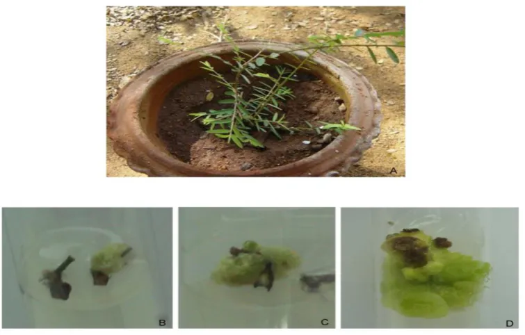
The healthy wild Phyllanthus virgatus plants (figure 1a) were collected during the middle of
October 2015 from Chengalpattu, Tamilnadu, India and were raised in pots containing soil
and farm yard manure (1:1) under greenhouse conditions at PG & Research Department of
Botany, Presidency College, Chennai and heathy leaves and nodal explants used for further
experimental studies.
Explant Preparation
Leaf and nodal explants (nine month old mature plants) were surface sterilized by cleaning
thoroughly under running tap water for 20 min, washed with a solution of labolene (2-3 drops
in 100 ml of water) for 5 min, and again washed with sterile distilled water. The cleaned
explants were then treated with 70% ethanol for 1 min followed by 0.1% mercuric chloride
(HgCl2) treatment for 5 min under aseptic conditions and washed six times with sterile
distilled water to remove traces of HgCl2.[7]
Initiation of callus
The in vitro leaf and nodal explants were cultured on MS basal media containing various
concentrations of 2, 4-D (2.26, 4.52, 6.78 and 9.04 µM); NAA (1.342, 2.68, 5.37, and 8.05
µM) and BA (1.10, 2.22, 4.44 and 8.88µM) for callus induction. Primary callus was
established from cotyledonary leaf explants. For secondary callus production, a small portion
of primary callus was excised using sterile knife holder and was sub-cultured periodically
once in three weeks. The secondary callus was used for all the experimental studies.
A standard approach of Latin square method (Collin and Edwards)[8] was followed for
screening of media to establish optimum culturing of callus by manipulating the
concentration of auxins (2, 4-D, IAA and NAA) and cytokinins (BA and KN) alone and in
combinations. A range of seven concentrations of auxins and cytokinins (0.1, 0.25, 0.5, 1.0,
2.5, 5.0 and 10 mg/L) were used in this study.
Callus growth
The growth measurement of callus was determined by standard method.[9] Growth of the
callus and its biomass was measured in terms of fresh (FW g/L) and dry weight (DW g/L).
the callus surface using blotting paper. Dry weight of callus was determined by drying the
callus in hot air oven at 60º C for 24 h and was expressed in g/L DW culture.
Age of callus culture
The age of callus of Phyllanthus virgatus was determined by standard method.[10] About
0.5gm of secondary callus culture from actively growing callus was inoculated in 250mL
Erlenmeyer flask containing 50 ml of MS solid medium supplemented with 24.52µM 2, 4-D,
1.11µM BA and 4.65µM KN. The culture was incubated under 16/8h photoperiod at 25±1ºC.
Initial weight of the callus biomass was measured in terms of fresh (FW g/L) and dry weight
(DW g/L). Observations were made from the 9th day after incubation up to 36th day with three
days intervals and recorded. On the 9th day the first Erlenmeyer flask containing callus was
used and it was dried at 50oC in the dark for 24 h. The same procedure was adopted for the
callus harvested at different days of intervals 9th, 12th, 15th, 18th, 21st, 24th, 27th, 30th, 33rd and
36th days.
Culture medium and conditions
MS basal medium supplemented with 3% (w/v) sucrose was used for all in vitro culture
studies. The pH of the medium was adjusted to 5.6 0.2 prior to adding 0.9% (w/v) agar, and
autoclaved at 121°C for 15 min. Cultures were maintained at 22 1°C under 16/8h
photoperiod by cool white fluorescent tubes (50µmol m-2s-2) with 55–60% relative humidity.
The plant growth regulators were filter-sterilized using 0.2 m filter (Minisart®, Sartorius)
prior to addition to culture media.
HPLC analysis of phyllanthin compound
The phyllanthin content in Phyllanthus virgatus leaf and callus were determined by HPLC
analysis. One gram of dried leaf powder and callus of Phyllanthus virgatus were used.
Ethanolic extracts of the leaf and callus were prepared by using soxhlet extraction method.
The extracts were incubated at 4ºC for 24 hrs, then filtered and evaporated to dryness vacuum
and re-dissolved in a small amount of the same solvent before separation using HPLC. The
extract was filtered through Sartorius RC membrane syringe filter (0.2mm) and 30µl of the
sample used for the HPLC analysis. Chromatography was performed using Shimadzu HPLC
(Model SPD-10A UV-VIS Detector) and supelcosil LC-18 column (25 cm × 4.6 mm, 5µm)
with mobile phase, linear gradient elution profile started with methanol: water (75:25) and
pressure of 250 psi and the compounds were read at 210 nm using a UV detector. The total
run time was 20 minutes, but preferably it was extended up to 40 minutes.[11] The results were
compared with standard.
RESULT AND DISCUSSION
The growth of callus development varied from leaf and nodal explants. Explants inoculated
on MS medium supplemented with individual concentration of 2, 4-D, NAA and BA. Leaf
explants inoculated on MS medium containing 452µM 2.4-D was noticed to be significantly
higher than nodal explants (Table 1; Fig 1b, c). Callus produced from leaf explants were
friable, creamy and light green in colour. The above findings related to the nature of the
callus was confirmed with the reports of Falcaria vulgaris[12] and Cornukaempferia
aurantiflora.[13] These findings were seen to be in harmony with those reported in Stevia
rebaudiana[14] and also, in accordance with the reports of Cardiospermum halicacabum[15], in
which callus induction was highest (90%) in the leaf explants of C. halicacabum on MS
medium supplemented with 2, 4-D. Calli were transferred to MS medium supplemented with
different concentrations and combination of auxins and cytokinins. In auxins maximum
growth of callus was obtained in MS medium supplemented with 2,4-D 4.52µM followed by
IAA at 5.71µM, NAA at 2.69µM (Table 2; Fig. 2 a, b, c) whereas in cytokinins 1.11µM BAP
was followed by 4.65µM KN(Table 3; Fig. 2 d, e). Callus grown on medium supplemented
with 2, 4-D and IAA was pale in colour. Callus grown on medium supplemented with BA
and KN was green in colour, hard and granular. The maximum callus growth was found with
cytokinins such as KN and BA and auxin (2,4D). Maximum callus growth using auxins and
cytokinins has been reported in G. Sylvestre[16], Eurycoma longifolia[17] and Rosa
bourboniana.[18]
A total of 58 combinations of auxin and cytokinins were tried for optimum callus biomass
production. The hormone combination for optimum callus biomass production was
standardized and the callus biomass was 163.4 g/L fresh weight and 14.91 g/L dry weight in
MS solid medium supplemented with 4.52 µM2, 4-D, 4.65µM KN and 1.11 µM BA after 21st
days of culture (Table.4,5; Fig. 3c). The 24th day callus (Solid medium) extract was used for
HPLC analysis. The HPLC analysis of phyllanthin compound from Phyllanthus virgatus leaf
and callus extract along with the standard phyllanthin has been represented in Fig. 4.
Phyllanthin compound eluted through HPLC analysis and based on standard retention time
used for HPLC analysis recorded a Rt of 13.73 minutes (leaf) and Rt of 13.79 minutes
(Callus) and standard phyllanthin compound recorded a Rt of 13.67 minutes, thus confirming
the presence of phyllanthin compound in leaf and callus extract of Phyllanthus virgatus (Fig.
4a,b,c). In earlier reports considerable evidences are available on the endogenous
accumulation of phyllanthin in cells of Phyllanthus virgatus grown in vitro.[19-20]
In conclusion, a Cell culture methodology for selective metabolite production is found to be
highly useful for commercial production of medicinally important compounds. The increased
use of plant cell culture systems in recent years is perhaps due to an improved understanding
of the secondary metabolite pathway in economically important plants. The present study
developed an efficient and optimum callus biomass using synergetic combination of auxins
and cytokinins and synthesis of phyllanthin content in callus biomass. The HPLC analysis
revealed the identification of active compound namely phyllanthin present in the callus
extract of Phyllanthus virgatus. Further studies will be directed towards large scale
production, testing the efficacy of secondary metabolites through animal cell lines and
exploring market potential.
Table 1: Effect of plant growth regulators (2, 4-D, NAA, BA, 2iP) on callus induction from different explants of Phyllanthus virgatus.
PGR Concentration (µM) Leaf explant Nodal explant
2,4-D NAA BA
0.45 - - 29.97 ± 0.15 20.00 ± 0.30
2.26 - - 40.00 ± 0.10 20.00 ± 0.20
4.52 - - 70.00 ± 0.30 20.00 ± 0.40
9.04 - - 55.00 ± 0.20 30.07 ± 0.50
- 1.34 - 25.00 ± 0.10 30.00 ± 0.50
- 2.68 - 30.00 ± 0.20 40.00 ± 0.50
- 5.37 - 40.00 ± 0.30 25.00 ± 0.10
- 8.05 - 25.00 ± 0.30 20.00 ± 0.30
- - 1.11 35.00 ± 0.20 10.00 ± 0.30
- - 2.22 25.00 ± 0.20 15.00 ± 0.20
- - 4.44 20.00 ± 0.40 10.00 ± 0.20
- - 8.88 20.00 ± 0.20 10.00 ± 0.40
Data were recorded after 40 days of culture. Results represent mean ± SD of six replicated
Table 2: Individual effect of auxins on callus culture of Phyllanthus virgatus.
PGR Concentration (µM) FW(g/L) DW(g/L)
2,4-D 0.45 14.16 ± 0.25 1.55 ± 0. 25
1.13 29.10 ± 0.10 2.54 ± 0. 15 2.26 39.75 ± 0.25 2.74 ± 0. 25
4.52 44.65 ± 0.25 3.18 ± 0. 91
11.31 31.26 ± 0.25 2.19 ± 0. 30 22.62 22.55 ± 0.25 1.84 ± 0. 25 45.25 11.54 ± 0.15 0.90 ± 0. 20
IAA 0.57 24.65 ± 0. 15 1.50 ± 0. 20
1.43 26.54 ± 0. 25 1.45 ± 0. 15 2.85 33.25 ± 0. 25 1.51 ± 0. 12
5.71 39.60 ± 0. 20 2.99 ± 0. 40
14.27 22.39 ± 0. 30 2.67 ± 0. 20 28.54 15.55 ± 0. 15 0.76 ± 0. 15 57.08 12.85 ± 0. 25 0.75 ± 0. 25
NAA 0.54 15.59 ± 0. 20 2.25 ± 0.15
1.34 21.15 ± 0. 15 1.35 ± 0.25
2.69 35.44 ± 0. 25 2.29 ± 0.30
5.37 38.20 ± 0. 20 2.95 ± 0.25 13.43 12.29 ± 0. 20 0.75 ± 0.25 26.85 10.96 ± 0. 25 0.65 ± 0.15 53.71 10.35 ± 0. 25 0.64 ± 0.25
Data were recorded after 40 days of culture. Results represent mean ± SD of six replicated
experiments. FW-Fresh weight; DW-Dry weight.
Table 3: Individual effect of cytokinins on callus culture of Phyllanthus virgatus. PGR Concentration (µM) FW(g/L) DW(g/L)
BA 0.44 11.34 ± 0. 25 2.50 ± 0.10
1.11 48.55 ± 0. 15 3.59 ± 0.30
2.22 32.36 ± 0. 35 2.84 ± 0.25
4.44 23.35 ± 0. 25 2.50 ± 0.20 11.1 21.99 ± 0. 30 2.20 ± 0.20 22.2 13.25 ± 0. 25 1.26 ± 0.15 44.2 10.29 ± 0. 20 0.85 ± 0.25
KN 0.58 11.50 ± 0.20 0.45 ± 0.15
1.16 15.70 ± 0.15 0.65 ± 0.15 2.32 17.40 ± 0.20 1.04 ± 0.15
4.65 39.69 ± 0.15 2.66 ± 0.25
11.62 26.60 ± 0.20 1.30 ± 0.10 22.00 22.49 ± 0.40 1.99 ± 0.20 46.5 15.55 ± 0.25 0.80 ± 0.10
Data were recorded after 40 days of culture. Results represent mean ± SD of six replicated
[image:7.595.136.497.485.703.2]Table 4: Standardization of plant growth regulators for callus culture of Phyllanthus virgatus.
PGR Concentration (µM)
2,4-D NAA BA FW(g/L) DW(g/L) 2,4-D KN BA FW(g/L) DW(g/L)
4.52 2.69 1.11 73.44±0.35 6.35±0.35 4.52 4.65 1.11 163.4±0.30 14.9±0.20
4.52 2.69 2.22 62.74±0.15 4.74±0.25 4.52 4.65 2.22 132.4±0.10 9.30±0.30 4.52 5.37 1.11 83.23±0.25 5.30±0.30 4.52 11.62 1.11 117.6±0.10 8.30±0.10 4.52 5.37 2.22 93.05±0.25 7.36±0.20 4.52 11.62 2.22 93.4±0.20 7.60±0.20 Data were recorded after 40 days of culture. Results represent mean ± SD of six replicated
experiments. FW-Fresh weight; DW-Dry weight.
Table 5: Callus biomass in solid medium at different age culture of Phyllanthus virgatus
Incubation (Days)
Callus Biomass (Solid medium) FW (g/L) DW (g/L)
9 86.10 ±0.30 5.00 ± 0.20
12 93.40 ±0.20 7.30 ± 0.20
15 129.30 ± 0.30 11.75 ± 0.25 18 145.70 ± 0.10 13.25 ± 0.25
21 163.40 ± 0.20 14.90 ± 0.30
24 151.07 ± 0.12 12.75 ± 0.35 27 146.90 ± 0.30 11.50 ± 0.40 30 143.10 ± 0.30 10.84 ± 0.35 33 139.50 ± 0.30 9.35 ± 0.25 36 136.70 ± 0.20 9.11 ± 0.20
Data were recorded after 40 days of culture. Results represent mean ± SD of six replicated
[image:8.595.109.487.503.744.2]experiments. FW-Fresh weight; DW-Dry weight.
(a) Mother plant of Phyllanthus virgatus collected from Chengalpattu, (b) Callus initiation
from nodal explants of Phyllanthus virgatus, (c) Callus initiation from leaf explants of
Phyllanthus virgatus, (d) Secondary callus developed from leaf explants of Phyllanthus
virgatus.
Fig. 2: Individual effect of auxins and cytokinin on callus culture of Phyllanthus virgatus.
(a) Murashige and Skoog (MS) basal medium with 2,4-dichlorophenoxyacetic acid (2,4-D)
(4.52 µM). (b) MS basal medium with Indole-3-acetic acid (IAA) (5.71 µM). (c) MS basal
medium with 1-Naphthaleneacetic acid (NAA) (2.69 µM). (d) MS basal medium with
6-Benzyladenine (BA) (2.22 µM). (e) MS basal medium with Kinetin (KN) (4.65 µM).
Fig. 3: Standardization of plant growth regulators for callus culture of Phyllanthus
[image:9.595.108.488.167.382.2] [image:9.595.148.454.528.715.2](a) Optimum callus biomass developed on MS medium 2,4-D (4.52 µM), KN (11.62 µM) and
BA (1.11 µM), (b) Optimum callus biomass developed on MS medium 2,4-D (4.52 µM), KN
(4.65 µM) and BA (2.22 µM), (c) Optimum callus biomass developed on MS medium 2,4-D
(4.52 µM), KN (4.65 µM) and BA (1.11 µM).
Fig. 4: High-performance liquid chromatography analysis of Phyllanthin compound from Phyllanthus virgatus leaf and callus extract.
(a) Phyllanthin standard (1 mg/1 mL) - Sigma-Aldrich. (b) The leaf extract of Phyllanthus
[image:10.595.112.483.170.671.2]REFERENCE
1. Canter, PH., Thomas, H., Ernst, E. Bringing medicinal plants into cultivation:
opportunities and challenges for biotechnology. Trends Biotechnol, 2005; 23: 180-185.
2. Ramachandra Rao, S and Ravishankar, G.A. Plant cell cultures: chemical factories of
secondary metabolites. Biotechnol. Adv, 2002; 20: 101-153.
3. Hussain, S., Fareed, S., Ansari, S., Rahman, A., Iffat-Zareen, A and Saeed M. Current
approaches toward production of secondary plant metabolites. J. Pharm. Bioall. Sci.,
2012; 4: 10-20.
4. Calixto, JB., Santos, AR., Cechinel, VF and Yunes, RA. A review of the plants of the
genus Phyllanthus: their chemistry, pharmacology, and therapeutic potential. Med. Res.
Rev, 1998; 18(4): 225–258.
5. Kumaran, A and Karunakaran, RJ. In vitro antioxidant activities of methanol extracts of
five Phyllanthus species from India. LWT, Food Science and Technology, 2007; 40:
344-352.
6. Hashim, A., Khan, MS., Khan, MS., Baig, MH and Ahmad, S. Antioxidant and Alpha
Amylase Inhibitory Property of Phyllanthus virgatus L.: An In Vitro and Molecular
Interaction Study. Bio Med Research International, 2013; Article ID 729393, 12 pages,
doi:10.1155/2013/729393
7. Janarthanam, B and Sumathi, E. Rapid and efficient plant regeneration from shoot tip
explants of Orthosiphon spiralis Murr- an important medicinal plant. International
Journal of Current Research, 2013; 5(10): 2718-2720.
8. Collin, HA and Edwards, S. The plant Cell Tissue Culture. In: D. Rickwood and C. Howe
(ed.). Bios scientific publishers Limited, Oxford, UK), 1998; 32.
9. Janarthanam, B., Gopalkrishnan, M and Sekar T. Secondary metabolite production in
callus cultures of Stevia rebaudiana. Bangaldesh journal of science industrial research,
2010; 45(3): 243-248.
10.Janarthanam, B and Seshadri S. In vitro manipulations of Rosa bourboniana L. Acta
Hort. 2008; 357-370.
11.Selvaraj, S., Chittibabu, CV and Janarthanam B. Studies on phytochemical screening,
antioxidant activity and extraction of active compound (swertiamarin) from leaf extract of
Enicostemma littorale. Asian J Pharm Clin Res., 2014; 7(4): 240-244.
12.Hamideh, J., Piri, K and Mohammad Javad, ND. Callus induction and plant regeneration
from leaf explants of Falcaria vulgaris an important medicinal plant. Journal of
13.Saensouk, P. Callus induction and plant regeneration from leaf explant of
Cornukaempferia aurantiflora Mood & Larsen. Pak. J. Bot., 2011; 43(5): 2415-2418.
14.Janarthanam, B., Gopalkrishnan, M, Sai, GL and Sekar, T. Plant regeneration from leaf
derived callus of Stevia rebaudiana Bertoni. Plant Tiss. Cult. Biotech, 2009; 19(2):
133-141.
15.Thomas, TD., Maseena, EA. Callus and plant regeneration in Cardiospermum
halicacabum Linn. An important medicinal plant, Sci. Hortic, 2006; 108: 332 -336.
16.Gopi, C and Vatsala, TM. In vitro studies in effects of plant growth regulators on callus
and suspension culture biomass yield from Gymnema sylvestre R. Br. Afr J Biotechnol,
2006; 5(12): 1215–1219.
17.Siregar, LAM., Keng, LC., Lim, PB. Selection of Cell Source and the effect of pH and
MS Macronutrients on Biomass Production in cell cultures of Tongkat Ali (Euryocoma
Longifoli Jack). J. Plant Biotechnology, 2003; 5: 131-135.
18.Janarthanam B, Seshadri S. In vitro manipulations of Rosa bourboniana L. Acta Hort.
2008; 357-370.
19.Arvind, KT., Verma, RK., Gupta, AK., Gupta, MM and Suman P. S. Khanuja
Quantitative Determination of Phyllanthin and Hypophyllanthin in Phyllanthus Species
by High-performance Thin Layer Chromatography Phytochem. Anal, 2006; 17: 394–397.
20.Prashanth Kumar, S., Mandahasan, A., Vijaya Kumar, S., Dhirendra, BS., Shreedhara, CS
and Manjunath Setty M. Production of Secondary Plant Metabolite Phyllanthin in