SERUM MAGNESIUM LEVELS IN ACUTE MYOCARDIAL
INFARCTION
Dr. Sunanda Amonkar1, Dr. Lavita D’costa2* and Dr. Chitra Y. Dhume3
1
Associate Professor, Department of Internal Medicine, Goa Medical College,
Bambolim-Goa.
2
Assistant Lecturer, Goa Medical College, Bambolim-Goa.
3
Professor and Head, Department of Biochemistry, Goa Medical College, Bambolim-Goa.
ABSTRACT
It has long been known that for normal growth and function inorganic
salts must be supplied to all biological forms. In the human body there
is a tendency to maintain the proper fluid balance, not only as a whole
but between the three compartments of intracellular, interstitial and
intravascular spaces. This is maintained by an intricate play of
hemodynamic, electrolyte and other forces. The field of mineral
metabolism is at present in a phase of rapid expansion. It has become
apparent that not only proteins, fats and carbohydrates, but also
minerals are essential to life. Now the significance of traces not only of
vitamins and other active organic substances, but also of minerals is
under intensive investigation. In this cross sectional study, the mean serum Magnesium level
on day 1 in all 50 patients was 1.86 meq/L (+/- 0.42) and the mean serum Magnesium level
on day 5 was 2.26 meq/L (+/- 0.5) and the mean serum Magnesium level in 26 patients with
arrhythmias was 1.738 meq/L on day 1 and 2.145 meq/L on day 5. In this study group, mean
serum Magnesium level in 24 patients without arrhythmias was 2.004 meq/L on day 1 and
2.390 meq/L on day 5. The difference between the serum Magnesium levels in patients with
and without arrhythmias is statistically significant on day 1. The difference between mean
serum magnesium levels in patients who expired was 1.43 meq/L and in patients who
survived was 1.92 meq/L. Statistically it was significant.
KEYWORDS: Myocardial infarction, serum Magnesium levels, arrhythmic and non
Volume 5, Issue 12, 959-974. Research Article ISSN 2277– 7105
*Corresponding Author
Dr. Lavita D’costa
Assistant Lecturer, Goa
Medical College,
Bambolim-Goa.
Article Received on 14 Oct. 2016,
Revised on 04 Nov. 2016, Accepted on 25 Nov. 2016
INTRODUCTION
Magnesium has been implicated in the pathogenesis of acute myocardial infarction and its
complications like arrhythmias. It plays a significant role in other cardiovascular diseases as
well. Magnesium ions are considered essential for the maintenance of the functional integrity
of the myocardium.[1]
In 1936, Greenberg and associates described myocardial degeneration with fibrosis and
polyplastic infiltration in rats that were fed on low magnesium diet from birth.[1]
Magnesium is the 4th most abundant cation in the body and the second most prevalent
intra-cellular cation, next to potassium [2]. Normal body content in the adult is approximately 2000
meq or 24 gms. Magnesium is distributed unevenly, with greatest concentration in tissues
having the higher metabolic activity such as brain, heart and kidneys. Approximately 60% of
the body Magnesium is in the bone, 1/3rd of the skeletal Magnesium has been shown to be
exchangeable and this fraction may serve as reservoir for maintaining a normal extracellular
Magnesium concentration.
Extracellular Magnesium accounts for only about 1% of total body content. The normal
serum Magnesium concentration is approximately 1.8-2.9 mg/dL.[3] About 70-75% of the
plasma Magnesium is ultra-filtrable of which a major portion is ionised. The non filterable
portion is bound to plasma proteins chiefly albumin. The remaining of the body Magnesium
is intracellular.
The concentration of total Magnesium in cells varies with tissues, but is of order
1-3mmol/L.[4] In general, higher the metabolic activity of the cell, higher the Magnesium
content. Low serum Magnesium concentration usually implies Magnesium deficiency. The
serum Magnesium however may not represent intracellular Magnesium. Intracellular
Magnesium depletion may exist despite normal serum Magnesium concentration.[5] Because
tissue and cellular assays are difficult to perform, and not widely available, the serum
Magnesium determination is the method by which Magnesium deficiency is identified in
clinical practice.
Cardiac muscle has a significant amount of Magnesium (17.4-19.8 meq/L). Higher
differences between Magnesium concentrations in the left and right ventricles or
inter-ventricular septum.[1]
Magnesium has been shown to be involved in ATP hydrolysis by myofibrils, and binding and
release of calcium ions by sacro-tubule reactions, which are essential to the contraction of
heart muscle. Magnesium also stimulates oxidative phosphorylation in heart mitochondria,
and affect sodium potassium ATPase of heart membranes, and activates adenyl-cyclase and
probably phosphorylase-kinase in the heart. Magnesium may have an influence on muscle
tone and conducting system, though the myocardium may be less sensitive to Magnesium
than nervous tissue.[2]
The kidney is the principle organ involved in the regulation of Magnesium homeostasis.
Approximately 8 meq is excreted in the urine each day. During Magnesium deprivation,
Magnesium is retained by the kidneys and <1 meq is excreted into the urine per 24 hours
period. When dietary Magnesium is plentiful, or is administered parenterally so that filtered
load exceed the normal plasma concentration, the excess Magnesium is excreted rapidly.[6]
Investigations revealed that magnesium level in the blood is decreased in the first 48 hours
following a acute myocardial infarction and then increased steadily to reach the normal level
in about three weeks time. The heart muscle subjected to myocardial infarction was found to
contain a low magnesium concentration. These findings directly correlated with the resultant
complications of myocardial infarction, such as arrhythmias.
Myocardial magnesium concentration in patients with sudden death due to ischemic heart
disease was found to be very low.[7] It has been pointed out that magnesium has a vital role in
ventricular fibrillation, which causes sudden death in IHD. The coronary vasospasm resulting
from magnesium deficiency has been suggested as another important factor in the sudden
death of IHD.
Magnesium deficiency was also postulated to have role in the genesis of atheromatous
plaques in that it leads to hyperlipidemia. Also myocardial infarction is one of the common
causes of death at present where prognosis depends on multiple factor of which many still
remain unexplained. This study is designed to know the relationship between serum
MATERIALS AND METHODS
By using simple random method, 50 cases of acute myocardial infarction, admitted over a
period of 1 ½ years between March 2009 to August 2010.
Inclusion criteria for patients
Those patients presenting to the hospital within 12 hours of onset of symptoms were selected.
Patients were considered to have myocardial infarction, only if they had two of the following
criteria.
1. History of chest pain.
2. ECG changes of acute myocardial infarction.
3. Rise of cardiac enzymes.
EXCLUSION CRITERIA
Patients with hypokalemia
Cases selected were subjected to a detailed history and thorough physical examination,
routine investigations like Haemoglobin, blood counts, urine examination, blood sugar, blood
urea, serum creatinine, serum electrolytes, fasting lipid profiles, cardiac enzymes, arterial
blood gas analysis and Echocardiography.
Serum Magnesium levels were estimated on day 1 and day 5.
Method of serum magnesium estimation
Methodology: Arsenazo
Principles of procedure
The method utilizes an arsenzo dye which binds preferentially with magnesium. The
absorbance of the arsenazo-magnesium complex is measured at 572 nm and is proportional to
the concentration of magnesium present in the sample. Calcium interference is prevented by
incorporation of calcium chelating agent.[8]
Specimen
Non-hemolysed serum or lithium heparin plasma may be analysed since the magnesium
concentration inside erythrocytes is 10 times greater than that in the ECF, hemolysis should
be avoided and serum should be separated from the cell as soon as possible.
RESULTS AND DISCUSSION
Table No. 1: Age and sex distribution of the study group.
Age (Years)
Sex
Total F (Female) M (Male)
20 – 30 0 1 1
30 – 40 0 6 6
40 – 50 0 11 11
50 – 60 1 9 10
60 – 70 6 8 14
70 – 80 2 3 5
80 – 90 0 1 1
90 – 100 2 0 2
Total 11 39 50
In this study group of 50 cases, 39 were males and 11 were females. Male Female ratio was
3.5:1. Maximum incidence of acute myocardial infarction was seen in 4th to 6th decades of
life. In males it was more common in 4th and 5th decades and in females it was more common
after the 5th decade. Also there were 7 cases of myocardial infarction, who were <40 years of
age. Probably the incidence of myocardial infarction is slowly increasing in the young.
Figure No: 1 Age and sex distribution of the study group.
Table No: 2 Religion wise distribution of cases.
Religion No. of cases Percentage
Hindus 34 68%
Christians 11 22%
[image:5.596.143.454.404.600.2]In the study of 50 patients, 34 (68%) were Hindus, 11 (22%) were Christians and 5 (10%)
were Muslims. The higher incidence of myocardial infarction is not factual one but it reflects
the difference in population.
[image:6.596.143.454.148.352.2]Figure No. 2: Religion wise distribution of cases.
Table No: 3 Time of presentation
Time No. of cases Percentage
0 – 5 hours 29 58%
5 – 10 hours 21 42%
Is the present study, chest pain was the commonest symptom, and was present in all cases. 29
(58%) cases presented to the hospital in between 0-5 hours of onset of symptoms and 21
(42%) cases presented between 5-10 hours.
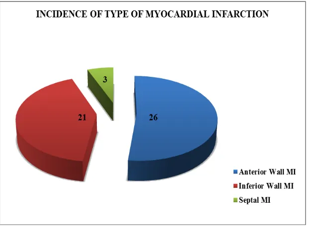
[image:6.596.188.411.413.457.2] [image:6.596.143.454.547.754.2]Table No. 4: Incidence of type of myocardial infarction
Type of Myocardial Infarction (MI) No. of cases Percentage
Anterior Wall MI 26 52%
Inferior Wall MI 21 42%
Septal MI 3 6%
In this present study, 26 (52%) cases were anterior wall MI, 21 (42%) cases were inferior
wall MI and 3 (6%) were septal MI. Right Ventricular MI was seen in 3 patients with
association.
Figure No: 4 Incidence of type of myocardial infarction
Table No: 5 Risk Factors
Risk Factors No. of Cases
Smoking 24
Diabetes Mellitus 20
Hypertension 19
Hyperlipidemia 9
Previous Ischemic Heart Disease 3
Hyper Homocysteinemia 1
Hypothyroidism 1
In this study, smoking is the most common risk factor found in the patients with acute
myocardial infarction (24 cases). Cigarette smoking increases the risk of coronary
atherosclerosis in both sexes and all ages and increases the risk of thrombosis, plaque
[image:7.596.172.427.527.646.2]Diabetes and hypertension were also important risk factors seen in 20 and 19 cases
respectively.
Other risk factors are hyperlipidemia and previous Ischemic Heart Disease (IHD). In this
study, hyper homocysteinemia was found to be a risk factor in 1 patient.
Serum Magnesium Results
In this cross sectional study of 50 patients, the mean serum Magnesium level on day 1 in all
50 patients was 1.86 meq/L (+/- 0.42) and the mean serum Magnesium level on day 5 was
[image:8.596.127.471.294.568.2]2.26 meq/L (+/- 0.5).
Table No: 6 Serum Magnesium Levels (in meq/L) in patients with Arrhythmias.
N Minimum Maximum Mean Std. Deviation
Mg_Day1 50 1.1 2.8 1.866 .4236
Mg_Day5 43 1.5 3.3 2.265 .5056
Figure No: 5 Serum Magnesium Levels (in meq/L) in patients with Arrhythmias.
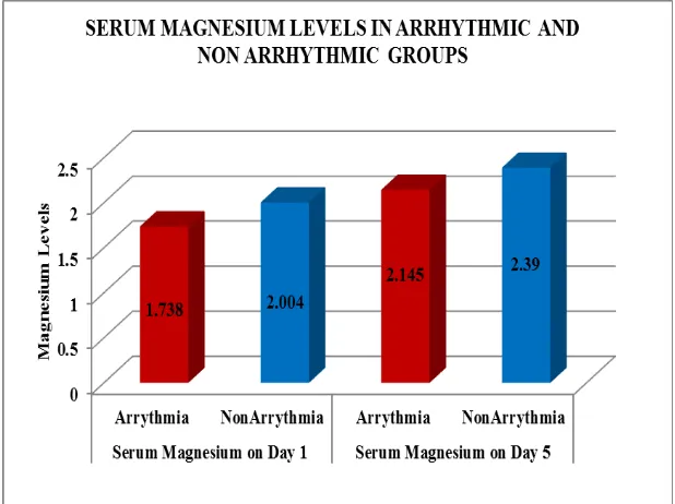
Table No: 7 Serum Magnesium levels in arrhythmic and non arrhythmic groups.
Outcome N Mean Std.
Deviation
Mg_Day1 Arrythmia 26 1.738 .4090
NonArrythmia 24 2.004 .4027
Mg_Day5 Arrythmia 22 2.145 .5068
Mean serum Magnesium levels in patients who had arrhythmias was 1.738 meq/L on day 1
and 2.145 meq/L on day 5. Whereas in patients who did not have arrhythmias, the Mean
serum Magnesium levels were 2.004 meq/L on day 1 and 2.390 meq/L on day 5.
Figure No: 6 Serum Magnesium levels in arrhythmic and non arrhythmic groups.
Table No: 8 Comparison of serum Magnesium levels in patients with arrhythmias and
without arrhythmias on day 1.
No. of cases
Serum Magnesium day 1 (meq/L)
t-value
p-value
Mean Serum Magnesium level
in patients with arrhythmias 26 1.738
2.3 <0.05
(0.025) Mean Serum Magnesium level
in patients without arrhythmias 24 2.004
The above table shows that out of 50 patients, 26 patients had arrhythmias. The Mean value
of serum Magnesium on day 1 in those with arrhythmias is 1.738 meq/L (+/- 0.40) and in
those without arrhythmias is 2.004 meq/L (+/-0.4). p value is < 0.05. There is significant
[image:9.596.143.452.148.379.2]Figure No. 7: Comparison of serum Magnesium levels in patients with arrhythmias and
without arrhythmias on day 1.
Table No. 9: Comparison of serum Magnesium levels in patients with arrhythmias and
without arrhythmias on day 5.
No. of cases
Serum Magnesium day 5 (meq/L)
t-value
p-value
Mean Serum Magnesium level
in patients with arrhythmias 22 2.145
1.6 0.1
Mean Serum Magnesium level
in patients without arrhythmias 21 2.390
The above table shows that serum Magnesium in patients with arrhythmias on day 5 is 2.145
meq/L (+/-0.5) and those without arrhythmias is 2.39 meq/L (+/-0.4). Statistically there is no
significant difference in between the two. Probably because serum Magnesium normalises by
5 days.
Figure No. 8: Comparison of serum Magnesium levels in patients with arrhythmias and
[image:10.596.143.456.70.248.2] [image:10.596.145.453.550.725.2]Mortality
Table No: 10 Comparison of mean serum Magnesium levels in patients who expired and
patients who survived on day 1.
No. of cases
Serum Magnesium
day 5 (meq/L) t-value p-value
Mean Serum Magnesium level
in patients who expired 7 1.43
2.7 <.05
Mean Serum Magnesium level
in patients who survived 43 1.92
In the above study of 50 patients, 7 patients died during 5 days of hospital stay. 4 patients
died of Cardiogenic shock, 2 of Ventricular Tachycardia and 1 of Ventricular Fibrillation.
Mortality percentage was 14%. Mean serum magnesium levels in patients who survived was
1.92 and who expired was 1.43. There was statistically significant difference in values in
these two groups.
Figure No. 9: Comparison of mean serum Magnesium levels in patients who expired
and patients who survived on day 1.
DISCUSSION
Magnesium ion has emerged as a premier cardiovascular cation during the decade. It has
been implicated in the pathogenesis of acute myocardial infarction and complication like
arrhythmias. Magnesium is essential for activation of ATP, which maintains the
[image:11.596.59.542.134.221.2] [image:11.596.145.451.352.571.2]In the study group of 50 patients, 39 were males and 11 were females with a male-female
ratio of 3.5:1. The maximum incidence of acute myocardial infarction was seen in the 4th and
5th decades.
In the present study of 50 patients, the mean serum magnesium level on day-1 in all 50
patients was 1.86 meq/L (+/- 0.42) and the mean serum magnesium level on day-5 was 2.26
meq/L (+/- 0.5).
In this study, mean value of serum Magnesium on day 1 in those with arrhythmias is 1.738
meq/L (+/- 0.40) and in those without arrhythmias is 2.004 meq/L (+/-0.4). p value is < 0.05.
There is significant difference in Magnesium levels in patients with arrhythmias and without
arrhythmias.
Abraham et al[9] reviewed magnesium level of 65 consecutive patients with an admission
diagnosis of acute myocardial infarction. Serum magnesium concentration were low in
patient who had AMI (mean 1.70 mg/dl, p<0.001) or acute coronary insufficiency (mean 1.61
mg/dl, p<0.01), but not in the control group or patients with non-cardiac chest pain (mean
1.91 mg/dl).
Singh A et al[10] checked serum magnesium levels of twenty patients of acute myocardial
infarction on the 1st, 7th and 12th day of admission. In all the cases, there was a significant fall
of serum magnesium on the first day.
Dimtruk[11] in his series of 67 patients of ischemic heart disease showed a distinct reduction
of plasma magnesium during the first 3 days following onset of disease, the level normalized
by 15-25 days from onset of the disease.
Sachdev et al[12] (1978) in 30 patients of myocardial infarction determine the magnesium
levels within 24 hours, 5th and 8th day and reported as 1.83±0.087 mgm%, 1.91±0.149 and
1.97±0.089 as against control of 2.44±0.162 mgm%. The values were statistically lower on
all the three days showing a progressive rise.
In the present study, the serum magnesium level on day-1 was significant lower in patients
with arrhythmias than those without arrhythmia (p<0.001). There was an increase in serum
magnesium from Day-1 to Day-5 in both those with arrhythmias and those without
Ceremuzynski et al[13] assigned 48 patients with acute myocardial infarction over 24 hours
infusion of magnesium or placebo. The incidence of ventricular tachycardia (3 or more
consecutive premature ventricular contraction at a rate faster than 1207 min) recorded by
Holier monitoring was significantly reduced (p<0.001), but the incidence of other ventricular
arrhythmias was not statistically different.
Raismusen et al[14] randomized 273 patients with suspected acute myocardial infarction to
intravenous magnesium or placebo. There is a significant decrease in the ventricular
arrhythmia in the magnesium group compared to placebo (p<0.05).
Shecter et al[15] randomized 103 patients with documented acute myocardial infarction to 48
hours infusion of magnesium or placebo. There is a significant decrease in mortality
(p<0.01). There was also a non-significant decrease in the number of tachyarrhythmias
requiring treatment (10/50) in the magnesium group compared to control (24/53).
Smith et al[16] randomized 400 patients with suspected AMI to a 24 hours infusion of
magnesium sulphate or placebo. Two hundred patients had confirmed acute myocardial
infarction. The difference in mortality and incidence of ventricular dysarrhtymia requiring
treatment between magnesium and placebo groups were not statistically significant.
Abraham et al[17] randomly assigned 94 patients with acute myocardial infarction to receive a
daily magnesium bolus of 30 mmol or placebo for 3-days. There was no significant
difference in mortality or lethal arrhythmias between patients treated with magnesium and
those treated with placebo.
Felstedt et al[18] randomized 298 patients with suspected acute myocardial infarction to 24
hours infusion of magnesium or placebo. Myocardial infarction was documented in 162
patients. During the mean observation period of 245 days, there was no difference in the
incidence of tachyarrhythmias, magnesium infusion was associated with a significant
increase in bradyarrhythmias.
Singh et al[19] randomized 264 patients with suspected acute myocardial infarction to
magnesium, potassium, 10% glucose or 2% glucose infusion. Myocardial infarction was
Morton et al[20] randomized 76 patients to receive either magnesium infusion 0.38 mmol/1 per
kg every 12 hour or placebo over the first 36 hours of hospital, there was no difference in the
incidence of ventricular tachycardia.
Dyckner T et al[7] during their 1½ years, 905 admission, 342 with acute myocardial
infarction, 563 other diagnoses are treated in the CCU on admission both acute myocardial
infarction and non AMI group had significantly lower serum magnesium level than as
reference group. The incidence of serious ventricular premature beats, ventricular tachycardia
and ventricular fibrillation on admission was significantly higher in the hypomagnesemic
patients with acute myocardial infarction.
In this present study, serum magnesium level on day-1 significant lower in patients with
arrythmias and statistically it was significant. But mean serum magnesium on day 5 was not
statistically between arrythmias and non-arrythmias group probably can be explained by
normalization of serum magnesium post myocardial infarction.
Mean serum magnesium in patients who had hyperlipidemia was low compared to normal
lipid profile group statistically there was no difference found.
CONCLUSION
This study was carried out in 50 patients of myocardial infarction who were admitted in the
CCU of Goa Medical College, Goa.
Male to female ratio in the study group was 3.5:1 and maximum incidence of acute
myocardial infarction was seen in the 4th and 5th decade.
In the study Hindus were 68%, Christians were 22% and Muslims were 10%.
In this study, the most common presenting symptom was chest pain. All the patients in this
study had chest pain. 58% of patients presented within 5 hours of onset of symptoms and
42% presented between 5 to 10 hours of onset of symptoms.
In the study, the most common risk factor was smoking followed by Diabetes and
In this cross sectional study, the mean serum Magnesium level on day 1 in all 50 patients was
1.86 meq/L (+/- 0.42) and the mean serum Magnesium level on day 5 was 2.26 meq/L (+/-
0.5).
In this study group, mean serum Magnesium level in 26 patients with arrhythmias was 1.738
meq/L on day 1 and 2.145 meq/L on day 5.
In this study group, mean serum Magnesium level in 24 patients without arrhythmias was
2.004 meq/L on day 1 and 2.390 meq/L on day 5.
The difference between the serum Magnesium levels in patients with and without arrhythmias
is statistically significant on day 1.
The difference between mean serum magnesium levels in patients who expired was 1.43
meq/L and in patients who survive was 1.92 meq/L. Statistically it was significant
Coronary artery disease is a major cause of morbidity and mortality throughout the world.
Major cause of death in coronary artery disease are due to complications like arrythmias.
In the present study, patients with acute myocardial infarction with low magnesium level are
more prone to develop ventricular arrythmias compared to those who are having normal
magnesium levels. Low serum magnesium levels were found in patients who expired
compared to patients who survived. Magnesium replacement therapy in patients with acute
myocardial infarction who is having low serum magnesium level may reduce the incidence of
arrythmias.
REFERENCES
1. Burch GE, Gibs TD. Importance of magnesium deficiency in cardiovascular disease.
American Heart Journal, 1977; 94: 649.
2. Wecker Wee, Parisi AF. Magnesium metabolism. N Engl J Med. 1968; 278: 658-663.
3. Dyckner T. Serum magnesium in acute myocardial infarction: Relation to Arrhythmias.
Acta med scan, 1980; 207: 59-66.
4. Grubbs R, Maguire M. Magnesium as a regulatory cation: Criteria and Evaluation.
Magnesium, 1987; 6: 113.
6. Rude R, Rhyzem E TM. Mg and renal Mg threshold in normal man in certain
pathophysiologic conditions. Magnesium, 1986; 47: 800.
7. Crawford T et al. Prevalence and pathological changes of ischemic heart disease in a hard
water and in a soft water area. Lancet, 1967; 1: 229.
8. Classen HG. Magnesium and potassium deprivation and supplementation in animals and
man - aspects in view of intestinal absorption. Magnesium, 1984; 3: 257-264.
9. Burtis CA, Ashwood ER. Teitz Textbook Of Clinical Chemistry and molecular
diagnostics.2nd ed. Philadelphia: Saunders, 1994.
10.Singh RB et al. Hypomagnesemia in relation to digoxin intoxication in children.
American Heart Journal, 1976; 92: 144.
11.Dmitruk. Magnesium and calcium blood plasma content in patients with ischemic heart
disease. Vrach Delo, 1977; 2(14): 7.
12.Sachadeva et al. Serum magnesium and platelet adhesiveness in acute myocardial
infarction. JIMA, 1978; 71: 165.
13.Ceremuzynski L, Jurgiel R et al. Threatening arrhythmias in acute myocardial infarction
are prevented by intravenous magnesium sulphate. Am Heart J., 1989; 118: 1333-1334.
14.Rasmussen H, McMair P, Norregard P. et al. Effects of IV. magnesium in acute
myocardial infarction. Lancet, 1986; 1: 234.
15.Shechter M, Mark N et al. Beneficial effects of magnesium sulphate in acute myocardial
infarction. Am J Cardiol, 1990; 66: 271-274.
16.Smith LF, Heagerty AM. Intravenous infusion of magnesium sulphate after acute
myocardial infarction: Effects on arrhythmias and mortality. Int J Cardiol, 1986; 12:
175-180.
17.Abraham AS, Rosenmann D. Magnesium in the prevention of lethal arrhythmias in acute
myocardial infarction. Arch. Intern Med., 1987; 147: 753-755.
18.Felstedt M, Boesgarurd et al. Magnesium substitution in acute ischaemic heart syndrome.
Eur Heart J., 1991; 12: 1215-1218.
19.Singh RB, Sircar AR et al. Magnesium and potassium administration in acute myocardial
infarction. Magnesium Trace Elem, 1990; 9: 198-204.
20.Morton BC, Nair RC et al. Magnesium therapy in acute myocardial infarction: A double