www.wjpr.net Vol 4, Issue 05, 2015.
1697
DETECTION OF
BREVUNDIMONAS DIMINUTA
BY POLYMERASE
CHAIN REACTION IN MEMBRANE FILTER VALIDATION
Bhushan.B*1, Uday.D1, Mohan.C1, Abhay.H1, Kakasaheb.M1
1
Department of Quality assurance Techniques, Poona College of Pharmacy, Bharati Vidyapeeth Deemed University, Erandwane, Pune-411 038.
ABSTRACT
The aim of present work was to develop the PCR based microbial testing method for detection of Brevundimonas diminuta (ATCC 19146) for Bacterial challenge test and compare the results of the conventional microbial testing method and the PCR based microbial testing method. It involved the preparation of the sample for the conventional and PCR based microbial testing, microbial testing by conventional method, PCR based microbial testing (extraction of DNA from bacterial cell and PCR thermal cycling, visualization of PCR product by agarose gel electrophoresis and comparison of results of conventional microbial testing method with PCR based microbial testing method. This technical report is intended to provide a systematic approach to selecting and validating the most appropriate filter for a sterilizing filtration application. Early, careful screening of potential filter types and configurations can result in fewer technical and regulatory problems, fewer delays, more efficient product processing and greater sterility assurance.
KEYWORDS: PCR, Brevundimonas diminuta, PCR thermal cycling, agarose gel electrophoresis, sterilizing filtration.
INTRODUCTION
Sterilization is complete eradication of all forms of life along with microbial spores. Various methods of sterilization are used in the pharmaceutical industry. Sterilization filtration is one of the several methods that are widely used. Sterilizing filtration is the process of removing all microorganisms, excluding viruses, from a fluid stream. A sterilizing grade filter must
Volume 4, Issue 5, 1697-1711. Research Article ISSN 2277– 7105
Article Received on 07 March 2015,
Revised on 30 March 2015, Accepted on 20 April 2015
*Correspondence for Author
Bhushan.B
www.wjpr.net Vol 4, Issue 05, 2015.
1698 remove all microorganisms present in a fluid stream without adversely affecting product quality.
In the early 1900s, the first parenteral drugs were manufactured on an industrial scale. The need arose to find a suitable sterilization method for heat-sensitive products that could not be autoclaved in the final container, i.e., had to be aseptically processed. Later, filtration to remove sub visible particulates from parenteral preparations, particularly solutions introduced intravenously, was found to be important.[1]
www.wjpr.net Vol 4, Issue 05, 2015.
1699 Until the late 1960s, 0.45 m-rated membranes were considered "sterilizing grade" filters, and were used successfully in the sterilizing filtration of parenteral. Such filters were qualified using 0.6x1 m Serratia maracescens, a standard bacterium for qualifying analytical membranes used for water quality testing (ASTM, 1980). In the mid-1960s, however, Dr. Frances Bowman of the FDA observed a 0.45 m "sterile-filtered" culture medium to be contaminated with an organism, subsequently shown to penetrate 0.45 m-rated membranes repeatedly in small numbers at challenge levels above 104-106 per cm² (Bowman et al., 1967). Bowman also observed that the next finer grade commercial membrane (nominally 0.22 m-rated) effectively retained this organism at similar challenge levels. This 0.3x0.6-0.8 m contaminant was identified as Pseudomonas diminuta (currently reclassified as Brevundimonas diminuta), and registered with the American Type Culture Collection
(ATCC) as Culture No. 19146. This strain has been accepted widely by filter manufacturers and industry as the standard challenge organism for qualifying sterilizing grade membrane filters (ASTM, 1983; ISO, 1995).[3-4]
Following the broad acceptance of B. diminuta, FDA incorporated demonstration of its retention in the definition of a sterilizing filter. Selecting a sterilizing grade filter requires consideration of many important issues, such as materials of construction and their compatibility with the product. The selection also should consider the processing characteristics, including the volume of product filtered, flow rate, pressure differential, temperature and the chemical characteristics of the product.
www.wjpr.net Vol 4, Issue 05, 2015.
1700 challenging a membrane filter assembly with a suspension of a known organism. The titer reduction is the ratio of influent colony forming units (CFU) to effluent bacteria colonies found downstream of the membrane.[5-8]
During filter evaluation, it is best to consider all processing variables so the appropriate filter surface area and configuration can be selected. There are three basic types of filter configurations generally accepted for sterilizing products: flat stock membranes, preassembled capsules and membrane cartridge assemblies.
TYPES OF FILTRATION
Membrane filtration has become a key element for use in the sterilization of fluids (both gases and liquids) during aseptic processing of pharmaceutical solutions.
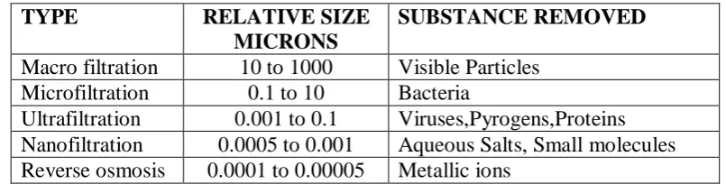
[image:4.595.95.498.381.483.2]Particle removal by membrane filtration may be simply classified as follows: Table 1
Classification of filtration on basis of size.
TYPE RELATIVE SIZE
MICRONS
SUBSTANCE REMOVED
Macro filtration 10 to 1000 Visible Particles Microfiltration 0.1 to 10 Bacteria
Ultrafiltration 0.001 to 0.1 Viruses,Pyrogens,Proteins Nanofiltration 0.0005 to 0.001 Aqueous Salts, Small molecules Reverse osmosis 0.0001 to 0.00005 Metallic ions
Directionally, there are two kinds of membrane filtration.
1) Vertical or direct flow filtration: Microfiltration of pharmaceutical solutions is primarily direct flow filtration. It is also called as a depth filtration.
2) Horizontal or cross flow filtration: Cross flow filtration primarily an Ultrafiltration tech. used in protein and biological material separations.
The various types of membranes that can be used are spiral, ceramic, stainless steel, tubular, hollow fibre, plate and frame.[9-12]
MATERIALS AND METHODS
www.wjpr.net Vol 4, Issue 05, 2015.
1701 Research Laboratories Pvt. Ltd. The strain of Brevundimonas diminuta (MTCC No.1287) was procured from MTCC chandigarh.
(i) Method of isolation of DNA for positive control
For the extraction of the DNA, GeNeiTM Spin Genomic DNA Prep Kit (From Bacteria) from the Bangalore Genei was used. A nutrient broth strain of Brevundimonas diminuta was prepared. 5 ml broth strain of Brevundimonas diminuta was centrifuged at 5000 rpm to form pellet. To these pellet 1 ml of PBS was added and then spined followed by draining, repeated three times. Bacterial pellet of appropriate culture volume was resuspended in 180 μl of lysis buffer I. Proteinase K 20 μl was added and mixed thoroughly by vortexing and then
incubated at 55oC for 1 hour. The sample was vortexed at intervals for better lysis. The sample was spined at maximum speed in a microcentrifuge for 5 minutes and the supernatant decanted carefully to a fresh vial. To the supernatant 200 μl of Lysis buffer II was added and mixed thoroughly by vortexing and incubating at 70oC for 20 minutes. 4 μl of RNase (100
mg/ml) was added, mixed by vortexing and incubated at room temperature for 5 minutes. The sample was spined at maximum speed in a microcentrifuge for 5 minutes and the supernatant decanted carefully to a fresh vial. To the supernatant 200 μl of distilled ethanol was added
and mixed thoroughly by vortexing. The spin column was kept in a 2 ml collection tube and the sample-ethanol mixture was added. It was then centrifuge at maximum speed for 1 minute and the collection tube with flow through was discarded. The spin column was kept in a fresh 2 ml collection tube and 500 μl of wash buffer I was added and spined at maximum speed for
1 minute and collection tube with wash sample was discarded. The spin column was kept in a fresh 2 ml collection tube and 500 μl of wash buffer II was added and spined at maximum speed for 3 minute and wash fraction discarded and collection tube retained. The empty column was spin for two minutes at maximum speed to ensure the removal of wash buffer. The spin column was placed in a fresh 1.5 ml tube and 200 μl of pre warmed elution buffer was added and incubated at room temperature for 5 minutes and centrifuged for 2 minutes to elute the DNA.
(ii) Isolated DNA confirmation by Agarose Gel Electrophoresis.
1 % Agarose with ethidium bromide in 0.5 X TAE was prepared and transferred to submerged Agarose gel electrophoresis apparatus and allow to cool to form a gel .The comb was removed and then the electrophoresis buffer (0.5 X TAE) was added. Then 10 μl isolated
www.wjpr.net Vol 4, Issue 05, 2015.
1702 (100 volt.) for 45 minutes and monitored by the tracer present in the gel loading buffer. The isolated DNA bands were observed in Gel documentation instrument.
(iii) Quantitation of DNA by UV Spectrophotometric method
1 μl from each isolated DNA of five different strains was dissolved in 500 μl double distilled
water. The OD values on spectrophotometer were taken at 260 nm. The amount of DNA were quantified using the formula DNA concentration (ng/ml.) = OD260 x 500 (dilution factor) x 50
µg/ml.[13-19]
(iv) PCR Thermal Cycling for positive control
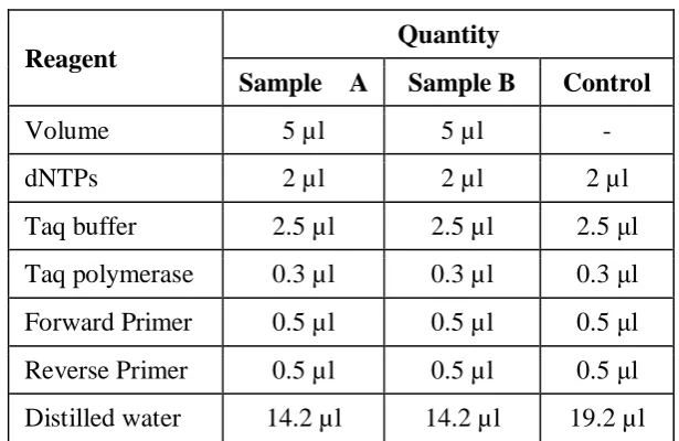
[image:6.595.143.451.337.537.2]The PCR thermal cycling of strain of Brevundimonas diminuta which was previously subjected to the DNA extraction procedure and the negative control was done as follows.
Table 2: A typical 25 µl PCR reaction.
Reagent
Quantity
Sample A Sample B Control
Volume 5 µl 5 µl -
dNTPs 2 µl 2 µl 2 µl
Taq buffer 2.5 µl 2.5 µl 2.5 μl Taq polymerase 0.3 µl 0.3 µl 0.3 μl
Forward Primer 0.5 µl 0.5 µl 0.5 μl
Reverse Primer 0.5 µl 0.5 µl 0.5 μl Distilled water 14.2 µl 14.2 µl 19.2 µl
The above mixture was subjected to the PCR thermal cycling. The above mixture was first of all subjected to 94oC for 5 minutes and then the 30 cycles at different temperatures as shown in the table 6 were run by setting the programmed on the PCR thermal cycler.[20-24]
Table 3: PCR Conditions
Process Temperature oC Time
Initial denaturation 94 oC 5 minutes Denaturation 94 oC 30 second
Annealing 50 oC 30 second
Extension 72 oC 30 second
Incubation 72 oC 7 minutes
www.wjpr.net Vol 4, Issue 05, 2015.
1703 (v) For microbial testing in ophthalmic solutions.
0.1 ml nutrient broth solution A of Brevundimonas diminuta (ATCC NO. 19146.) was transferred in the conical flask containing 100 ml of saline solution (B). 0.1 ml of solution was transferred from B to the other conical flask containing 100 ml of saline solution (C -103 dilutions). Then 0.1 ml of solution was transferred from C to the other conical flask containing 100 ml of saline solution (D -106 dilutions). Again 0.1 ml of solution was transferred from D to the other conical flask containing 100 ml of saline solution (E -109 dilutions). Again 0.1 ml of solution was transferred from E to the other conical flask containing 100 ml of saline solution (F -1012 dilution). In this way the dilutions of Brevundimonas diminuta (ATCC NO. 19146.) in the saline solution were made. The 100 ml solution A was passed on 0.2 um filter at 760 mM Hg subjected to filtration.[25-30]
The membrane filter was washed with PBS solution for the collection of micro-organism (Sample A). Filtrate was again filter and the membrane filter was washed with PBS solution for the collection of micro-organism in the filtrate. (Sample B). Both the samples were subjected to PCR.
[image:7.595.147.450.483.682.2]The PCR thermal cycling of Sample A and Sample B prepared as above and the negative control was done as follows.
Table 4: A typical 25 µl PCR reaction
R
Reeaaggeennt t QQuuaannttiitty y S
SaammpplleeA A SSaammpplleeB B CCoonnttrrool l
V
Voolluumme e 55µµll 55µµll -
-d
dNNTTPPs s 22µµll 22µµll 22µµl l
T
Taaqqbbuuffffeer r 22..55µµll 22..55µµl l 22..55µµll
T
Taaqqppoollyymmeerraasse e 00..33µµll 00..33µµl l 00..33µµll F
FoorrwwaarrddPPrriimmeerr 00..55µµll 00..55µµl l 00..55µµll
R
ReevveerrsseePPrriimmeerr 00..55µµll 00..55µµl l 00..55µµll
D
Diissttiilllleeddwwaatteer r 1144..22µµl l 1144..22µµl l 1199..22µµl l
www.wjpr.net Vol 4, Issue 05, 2015.
[image:8.595.165.438.93.195.2]1704 Table 5: PCR conditions
Process Temperature oC Time
Initial denaturation 94 oC 10 minutes Denaturation 94 oC 30 second
Annealing 50 oC 30 second
Extension 72 oC 30 second
Incubation 72 oC 7 minutes
Cooling 4 oC 5 minutes
(v) Visualization of PCR product by Agarose gel electrophoresis
The method adopted for the visualization of PCR product of both i.e. positive control and microbial testing in ophthalmic solutions remains the same, which is carried out as described- 1 % Agarose with ethidium bromide in 0.5 X TAE was prepared and transferred to submerged Agarose gel electrophoresis apparatus and allow to cool to form a gel .The comb was removed and then the electrophoresis buffer (0.5 X TAE) was added. The standard 100 bp ladder was loaded in first well and then all 25 µl of PCR product in each PCR tubes with gel loading buffer (2 µl) were loaded in subsequent wells. The gel was run at a constant voltage (100 volt.) for 45 minutes and monitored by the tracer present in the gel loading buffer. The isolated DNA bands were observed in Gel documentation instrument.
RESULTS AND DISCUSSION
Conventional Microbial Testing: The CFU of Sample A was observed 4 x 10¹². No colonies were observed in sample B. PCR Thermal Cycling for positive control: The Oligonucleotide sequences which are used in the PCR thermal cycling for the amplification of DNA , the strain of Brevundimonas diminuta, hence PCR based microbial testing developed by us can detect strain of Brevundimonas diminuta in the sample.
[image:8.595.152.447.566.735.2]
www.wjpr.net Vol 4, Issue 05, 2015.
1705 Lane 1- DNA ladder
Lane 2- 1 μl DNA (ATCC.19146) Lane 3 -2 μl DNA (ATCC.19146) Lane 4 -4 μl DNA (ATCC.19146) Lane 5 –Control.
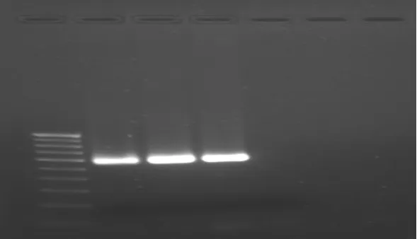
PCR Thermal Cycling for microbial testing of Sample A Sample B: After PCR thermal cycling Sample A and Sample B run on agarose gel electrophoresis.
[image:9.595.144.453.228.443.2]
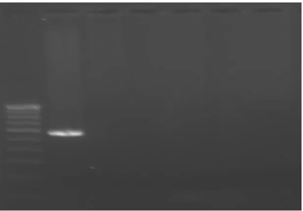
Figure 2 PCR thermal cycling result of Sample A and Sample B after filtration. Lane 1- DNA ladder
Lane 2- Sample A Lane 3 – Sample B Lane 4 -Control
1. Figure 1 showing band of isolated DNA of Brevundimonas diminuta (ATCC No.19146). The third lane on the agarose gel was of isolated DNA band. These resµlts confirmed the DNA of Brevundimonas diminuta isolated from source.
2. The figure 2 Showing different bands of different concentrations of Brevundimonas diminuta positive control. The first lane on the agarose gel was of 100 bp standard DNA
ladder, hence it showed different bands at 100 bp intervals. The second third and fourth lanes were of PCR product having concentrations 1µl, 2 µl, 4 µl respectively. The fifth lane was of negative control. All bands correspond with 600 bp of standard DNA ladder. 3. In fig. 3 showing band of Sample A in second lane of Brevundimonas diminuta at 600 bp
www.wjpr.net Vol 4, Issue 05, 2015.
1706 amplification occurring. This result confirms that 0.2 µm filter is valid for its use in filtration process.
CONCLUSION
Membrane filtration has become a key element for use in the sterilization of fluids (both gases and liquids) during aseptic processing of pharmaceutical solutions. Sterilizing filtration is the process of removing all microorganisms, excluding viruses, from a fluid stream. A sterilizing grade filter must remove all microorganisms present in a fluid stream without adversely affecting product quality. This technical report is intended to provide a systematic Approach to selecting and validating the most appropriate filter for a sterilizing filtration application. Early, careful screening of potential filter types and configurations can result in fewer technical and Regulatory problems, fewer delays, more efficient product processing and greater sterility assurance.
Until the late 1960s, 0.45 µm-rated membranes were considered "sterilizing grade" filters, and were used successfully in the sterilizing filtration of parenteral. Such filters were qualified using 0.6x1 µm Serratia maracescens, a standard bacterium for qualifying analytical membranes used for water quality testing (ASTM, 1980). In the mid-1960s, however, Dr. Frances Bowman of the FDA observed a 0.45 µm "sterile-filtered" culture medium to be contaminated with an organism, subsequently shown to penetrate 0.45 µm-rated membranes repeatedly in small numbers at challenge levels above 104-106 per cm² (Bowman et al., 1967). Bowman also observed that the next finer grade commercial membrane (nominally 0.22 µm-rated) effectively retained this organism at similar challenge levels. This 0.3x0.6-0.8 µm contaminant was identified as Pseudomonas diminuta (currently reclassified as Brevundimonas diminuta), and registered with the American Type Culture Collection (ATCC) as Culture No. 19146. This strain has been accepted widely by filter manufacturers and industry as the standard challenge organism for qualifying sterilizing grade membrane filters (ASTM, 1983; ISO, 1995).
www.wjpr.net Vol 4, Issue 05, 2015.
1707 viable, change from Brevundimonas diminuta use bacteria isolated from formulation, solution or environment are used.
The disadvantage about PCR technique of detecting dead microorganism can be converted in to advantage. By application of PCR procedure for bacterial challenge test.
In the present work by using PCR based microbial detection we can detect the Brevundimonas diminuta species though, they are found in not-viable from in pharmaceutical
formulation or solution. Therefore, the different approaches mentioned above required to be carried out in ase of non- viable from of Brevundimonas diminuta are bypassed.
The study was undertaken to introduce the new method for the microbial testing of the pharmaceuticals, which is based on the Polymerase Chain Reaction hence called as the PCR based microbial testing. On comparison of results it was seen that the PCR based microbial testing has following advantages over conventional microbial testing.
Very rapid : Conventional microbial testing requires the enrichment of the culture but in case of PCR the enrichment of culture is not required hence the time is saved. Again as the incubation of the culture is not required in case of PCR . There are PCR thermal cyclers available which can amplify the 10 to 3000 samples at a time hence no need of repetitively preparing the different media, sterilization of the media and performing the biochemical tests.
Cost effective: Conventional microbial testing requires the more inventory in the form of different equipments e.g. Autoclave, Hot air oven, Incubator etc. and different media for the culturing of the microorganisms, Petri plates, biochemical reagent kits which are different for different microorganisms but in case of PCR based microbial testing all these equipments are not required. The manpower required is also less in case of PCR because 20-3000 samples can be subjected to the PCR based microbial testing at a time hence the labor cost is also less in case of PCR.
www.wjpr.net Vol 4, Issue 05, 2015.
1708 Very less space is required: PCR based microbial testing requires less space as compared to the conventional microbial testing.
Simple: Conventional microbial testing requires the change of reagents, media, chemicals for microbial testing of the different microorganisms but in case of PCR same protocol is followed for the different microorganisms, only change required is the change of primer which is specific for the specific microorganisms hence the PCR based microbial testing is simple as compared to the conventional microbial testing which is very tedious.
Sensitive: For PCR based microbial testing the presence of a single DNA, even one single stranded DNA in the sample is sufficient. The sample requirement in case of PCR is 20-100microlitre but in case of conventional microbial testing the sample required is more.
Selective: The PCR based microbial testing is very very selective because only that sequence will get amplified for which the primer is added, but in case of conventional microbial testing the selectivity is less.
Time required is less: Conventional microbial testing requires about three to four days to confirm the presence of Pseudomonas aeruginosa but in case of PCR based microbial testing the time required is very less i.e. For the extraction of DNA from the sample, five hours are required, for PCR thermal cycling three hours are required and for the agarose gel electrophoresis two hours are required. Thus it means that we get the confirm result within nine days hence the time required for the PCR based microbial testing is very less.Sometimes due to the incomplete sterilization the microorganism get injured but not completely killed and in case of conventional microbial testing during incubation in the media for the prescribed period of time the microorganism recovers from the injury but do not show the growth and hence false negative result is obtained but in case of PCR based microbial testing no need of incubation and we are concerned with the DNA of that microorganism only hence this disadvantage is absent in case of PCR based microbial testing.
www.wjpr.net Vol 4, Issue 05, 2015.
1709 Hence the conclusion is
By conventional microbial method false positive result may be possible as it can not
detect dead organism, but PCR can be detect even dead organism and this is biggest advantage.
There is a need of fast and accurate method to screen microbes from pharmaceuticals; we
have developed PCR based method for quicker and selective microbial testing. PCR analysis is selective, simple and cost effective.
Quantitative analysis will be able to calculate microbial load, which is usually expressed
as colony forming units in conventional microbial testing methods.
REFERENCES
1. Sterilizing filtration of liquids, PDA journal of science and technology, 1997.
2. Robert A. Nash et al, Membrane filters for Microfiltration of pharmaceutical solution, Journal of Validation Technology.
3. FDA, “Guidelines on sterile drug products produced by aseptic processing”. Centre for Drugs and Biologics, Rockville. MD, 1987.
4. Validation Guide for Pall® Emflon® PFR Filter Cartridges, 2003. 5. Pharmaceutical Journal, Oct. 23: 400-402.
6. S.J. Bloomfield, R. Baird, R. E. Leak. R. Leech, Microbiological Quality assurance in Pharmaceuticals, Cosmetics and toilatories, Ellis harwood Ltd. Chichester, 18.
7. The Pharmacopoeia of India, Vol. 1, 4th Edn, Published by the controller of publications of India.
8. The United State Pharmacopoeia, 22nd Edn, United states pharmacopoeial Convention, 1990,1943,1896,1945,1897,1585.
9. Breakthroughs in bioscience, The Polymerase Chain Reaction, Tabitha M. Powledge. 10. Dr. U.Satyanarayana, Biochemistry, 2nd edition, Books and Allied (P) Ltd., 1999; 571. 11. Sambrook and Russell, Molecular Cloning, A Laboratory Manual, 3rd edition, 2,
8.4-8.9,8.14-8.15.
12. http://www.fermentas.com/index.html (accessed on 10/01/2008)
13. Carl W. Dieffenbach, Gabriela S. Dveksler, PCR Primer- A laboratory manual, 7-16. 14. R. W. Old, S. B. Primrose, Principles Of Gene Manipulation: An introduction to Genetic
www.wjpr.net Vol 4, Issue 05, 2015.
1710 15. James A. Watson, Nancy H. Hopkins, Jeffery W. Roberts, Joan Argetsinger Steitz, Alan M. Wener, Molecular Biology Of The Gene, 3rd edition, The Benzamin/ Cummings Publishing Company. Inc., 175.
16. http://en.wikipedia.org/wiki/Applications_of_PCR" (accessed on 25/01/2008)
17. Doane A., Standard Operating Procedure: Gel Documentation system, College of Biological Science, University of Guelph, 2007.
18. http://www.pcrstation.com/pcrtypes (accessed on 25/01/2008)
19. Praful B. Godkar, Textbook of Medical Laboratory Technology, Bhalani Publishing House, 1994; 340.
20. John C. Sherris et.al, Medical Microbiology: An introduction to infectious diseases, Elsevier, 2nd edition, 394.
21. Duguid J. P., Marmion B. P., Swain R. H., Mackie McCartney Medical Microbiology, English Language Book Society,13th edition, 1973; vol.I: 341.
22. Xiang Y. Han1 and Roberto A. Andrade, Brevundimonas diminuta infections and its resistance to fluoroquinolones, Journal of Antimicrobial Chemotherapy., 2005; 55: 853–859.
23. Elizabeth Wheeler Alm et al, The Oligonucleotide Probe Database, APPLIED AND ENVIRONMENTAL MICROBIOLOGY, Oct. 1996; 3557–3559
24. Singh B.; Biotechnology, 1st edition, Kalyani Publishers, Ludhiana, New Dehli, 2002; 32-40.
25. Kohler R., Wheat L., White A.; Detection of pseudomonas aeruginosa antigenemia in granulocytopenic rabbits by radiommunoassay. J. Clin. Microbiol., 1980; 39–43.
26. Ludwig,W.et al, Comparative sequence analysis of 23S rRNA from Proteobacteria, Syst. Appl. Microbiol., 1995; 18: 164-188.
27. Kingsford N., Raadsma H.; Detection of pseudomonas aeruginosa from ovine fleece washings by PCR amplification of 16S ribosomal RNA. Vet. Microbiol., 1995; 47(1-2): 61-70.
28. Kevin G., Sarah E., Linda S.; Method for detection of pseudomonas aeruginosa using polymerase chain reaction. US Patent Issued on April 11, 2000.
29. Brodsky M., Nixon M.; Rapid Method for Detection of pseudomonas aeruginosa on MacConkey Agar under ultraviolet light. Appl. Microbiol., 1973; 26(2): 219–220.
www.wjpr.net Vol 4, Issue 05, 2015.
1711 31. Fundamentals of microbiology, 6th edition, Edited by Hugo W. B., Russel A. D.,
Blackwell Science, 283.
32. Rosamund M. Baird, R.A. Shooter, British Medical Journal, 1976; 2: 49-350.
33. Richard C.B., John F.P., Medical Microbiology, A guide to microbial infections: pathogenesis, immunity, laboratory diagnosis and control, Churchill Livinnstone, 16th edition, 2002; 282.