Regulatory T cells in embryo implantation and
the immune response to pregnancy
Sarah A. Robertson, … , Alison S. Care, Lachlan M.
Moldenhauer
J Clin Invest.
2018;
128(10)
:4224-4235.
https://doi.org/10.1172/JCI122182
.
At implantation, the embryo expresses paternally derived alloantigens and evokes
inflammation that can threaten reproductive success. To ensure a robust placenta and
sustainable pregnancy, an active state of maternal immune tolerance mediated by CD4
+regulatory T cells (Tregs) is essential. Tregs operate to inhibit effector immunity, contain
inflammation, and support maternal vascular adaptations, thereby facilitating trophoblast
invasion and placental access to the maternal blood supply. Insufficient Treg numbers or
inadequate functional competence are implicated in idiopathic infertility and recurrent
miscarriage as well as later-onset pregnancy complications stemming from placental
insufficiency, including preeclampsia and fetal growth restriction. In this Review, we
summarize the mechanisms acting in the conception environment to drive the Treg
response and discuss prospects for targeting the T cell compartment to alleviate
immune-based reproductive disorders.
Review
Find the latest version:
Introduction
Within days of conception, the embryo attaches to the uterine lining, and trophoblast cells invade into the uterine decidua (1). A finely controlled developmental program then unfolds, with successive waves of trophoblast invasion, proliferation, and dif-ferentiation to form a mature placenta that sustains fetal growth throughout gestation.
This remarkable feat occurs in apparent defiance of the moth-er’s immune response. Abundant immune cells reside in the decid-ua in close contact with infiltrating trophoblasts, and paternally derived alloantigens are expressed in the developing placental and fetal tissues. Far from the immune evasion or systemic immune suppression historically invoked to explain maternal- fetal toler-ance (2), maternal immune cells exhibit priming toward fetal allo-antigens (3–5) and actively participate in many aspects of estab-lishing, sustaining, and terminating pregnancy (6).
Mammalian pregnancy cannot be readily reconciled with the classical self/non-self discrimination theory or alternative models of immune regulation (7). Central tolerance, where-in self-reactive lymphocytes are deleted where-in the thymus (8), is not relevant, since fetal alloantigens are not encountered outside of the reproductive context. Instead, a range of spe-cialized mechanisms in both the innate and adaptive immune compartments mediate an active state of functional tolerance that permits fetal and maternal cells to coexist. Key suppres-sive mechanisms include attenuated placental expression of polymorphic MHC molecules (9, 10); placental release of anti-inflammatory and protolerogenic hormones, cytokines, and immune modulatory molecules (11–13); and specialized
decid-ual regulation of immune cell access and egress (refs. 14, 15, and for additional information, see refs. 6, 16–18).
There is a strong imperative to define how pregnancy toler-ance is established, as an immune etiology is implicated in com-mon reproductive conditions including recurrent implantation failure and miscarriage (19–21), as well as later-onset gestational disorders that arise as a result of disturbed implantation and pla-cental morphogenesis (22–24). Recurrent implantation failure occurs when overtly healthy embryos fail to implant normally and is the cause of infertility in approximately 10% of women seeking in vitro fertilization (IVF) treatment (25). Recurrent pregnancy loss, defined as loss of three or more karyotypically normal embryos before 20 weeks’ gestation, occurs in approxi-mately 1% to 2% of women (26). Preeclampsia affects 3% to 5% of pregnancies (27) and is a major cause of morbidity and mor-tality for women and infants, particularly in low- and middle- income countries, and it is often accompanied by fetal growth restriction and preterm birth (28, 29).
Tolerance arises in the preimplantation phase of early preg-nancy and appears to require a unique dialog involving mater-nal-, patermater-nal-, and conceptus-derived signals and specialized anatomical elements of the reproductive tissues (30, 31). Their interaction drives a cascade of immune changes that initiate prior to conception, persist through gestation, and culminate with birth (6, 18). Innate immune cells, particularly macro-phages (32), DCs (33), and a unique population of NK cells with a CD56hiCD57lo phenotype (uterine NK cells, or uNK cells) (34), are abundant in the decidua in the luteal phase of the menstrual cycle when implantation commences. These cells influence pla-cental development through immune regulation, provision of growth factors, and facilitation of adaptations in the uterine vas-culature to support trophoblast invasion. Innate immune cells exhibit altered phenotypes and contribute to pathophysiological processes in many gestational conditions (refs. 35, 36, and for more information, see refs. 17, 37, 38).
At implantation, the embryo expresses paternally derived alloantigens and evokes inflammation that can threaten reproductive success. To ensure a robust placenta and sustainable pregnancy, an active state of maternal immune tolerance mediated by CD4+ regulatory T cells (Tregs) is essential. Tregs operate to inhibit effector immunity, contain inflammation, and
support maternal vascular adaptations, thereby facilitating trophoblast invasion and placental access to the maternal blood supply. Insufficient Treg numbers or inadequate functional competence are implicated in idiopathic infertility and recurrent miscarriage as well as later-onset pregnancy complications stemming from placental insufficiency, including preeclampsia and fetal growth restriction. In this Review, we summarize the mechanisms acting in the conception environment to drive the Treg response and discuss prospects for targeting the T cell compartment to alleviate immune-based reproductive disorders.
Regulatory T cells in embryo implantation and the
immune response to pregnancy
Sarah A. Robertson, Alison S. Care, and Lachlan M. Moldenhauer
Robinson Research Institute and Adelaide Medical School, University of Adelaide, Adelaide, South Australia, Australia.
Conflict of interest: SAR is an inventor on international patent PCT/AU99/00499 and receives royalty income from Origio A/S. SAR and LMM receive research support from Ferring Pharmaceuticals (US).
events surrounding conception that critically impact the availabil-ity and function of Tregs for implantation and later gestation. We explain the evidence for Tregs in the pathophysiology of infertility and obstetric disorders, discuss the origins of Treg deficiency in some women, and speculate on the prospect of targeting Tregs to address common reproductive and obstetric conditions.
The immune response and embryo implantation
Normal fetal growth depends on establishing a robust placenta to nurture the fetus; remarkably, two-thirds of genetic mutations identified as embryonically lethal in mice affect placental mor-phogenesis (69). Even minor derangement of the developmental program of the placenta can cause later miscarriage or set a tra-jectory toward preeclampsia (22, 27, 70). These conditions can be traced back to aberrant interactions between trophoblasts and the uterine decidua at conception and implantation (70–72).
Critical to implantation is an adequate decidual response. Immune cells are instrumental, with reciprocal interactions between DCs, uNK cells, and invading trophoblasts (73, 74) in response to hormonal triggers to transform the uterine lining in the luteal phase (1, 71, 72). Extensive contact between trophoblasts and immune cells in the decidua is common to all placental mam-mals (75) but is most conspicuous with invasive hemochorial pla-centation, as occurs in mice and humans (17). A sufficient number of decidual immune cells must acquire appropriate phenotypes to support the decidual response and remodel the local vascular net-work for embryo attachment and implantation (71, 76).
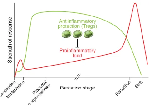
Leukocyte recruitment builds during the estrogen-dom-inated periovulatory phase, and a threshold level of inflam-matory activation may facilitate the generation of a receptive endometrium (77, 78). However, within days of conception, inflammation must be contained and controlled in order for decidualization and implantation to progress (72). The capacity to resolve decidual inflammation has evolved as a key feature underpinning placentation in viviparous mammals (79), and disturbance to the dynamic balance between pro- and antiin-flammatory mediators is a hallmark of impaired implantation (21, 80). Through their potent antiinflammatory actions, Tregs appear to be critical for controlling inflammation in early preg-nancy and establishing a receptive decidual environment (refs. 18, 81, and Figure 1).
The adaptive immune response is also critical to pregnancy tolerance (refs. 39–41 and for more information, see refs. 18, 42), and an imbalance of T regulatory cells (Tregs) and effector T cells (Teffs) is emerging as a key underpinning factor in common fer-tility and obstetric disorders (19–21, 23, 24). Tregs are well known for their capacity to limit excessive inflammation and recalibrate tissue homeostasis after insult or injury, as well as to suppress Teff reactions to self or non-self antigens (43, 44). Tregs exert potent antiinflammatory, immune-regulatory, and vaso-regulatory func-tions (43–45) relevant to establishing pregnancy. Also significant is their distinct phenotypic plasticity, or capacity to transdifferenti-ate into Th17 cells (46, 47), which provides a potential mechanism for strategic female reproductive investment (48).
In women, T cells comprise 10% to 20% of decidual immune cells in the first trimester (49). Many of these are CD8+ T cells, including regulatory subsets (50, 51). Among the CD4+ T cells, approximate-ly 10% to 30% express the Treg transcription factor FOXP3, which constitutes a substantial enrichment compared with its expres-sion in peripheral blood (52–54). Decidual Th1 cell frequencies are moderately elevated, while Th17 and Th2 cells are generally not enriched, indicating a mild inflammatory environment controlled by Tregs (53, 55). The Tregs are composed of both thymus-derived Tregs (tTregs) and peripheral Tregs (pTregs) and exhibit phenotyp-ic heterogeneity according to the cycle and pregnancy phase (36, 56, 57). Uterine recruitment of Tregs in preparation for conception commences in the proliferative phase of each cycle, with an estro-gen-driven increase peaking at ovulation (58). After increasing in early pregnancy, decidual Tregs remain elevated through mid-gesta-tion before declining prior to birth (52, 53, 59), with peripheral blood Tregs following a broadly similar pattern (60, 61).
In reproductive disorders, insufficient numbers of Tregs or impaired function is a common feature (19, 20, 23), with a coun-teractive increase in Teffs (21, 24). Compelling evidence that Treg deficiency is causal in pregnancy loss comes from animal models (39, 62–64). An underlying T cell etiology in women is supported by correlations with prior sexual and reproductive history (65) and by couple-specific, HLA-linked dispositions to reproductive condi-tions (66–68), consistent with a protective effect of adaptive immune “memory” for partner histocompatibility antigens.
[image:3.585.48.287.54.223.2]In this Review, we describe the current understanding of Tregs as master regulators of pregnancy tolerance, focusing on
increases at implantation are consistently greater with alloge-neic fetuses, indicating fetal antigen–driven expansion, and are accompanied by specific suppression of anti-paternal alloantigen responses (83, 85). Male-specific minor antigens such as H-Y con-tribute to driving the Treg response, with male fetuses being more vulnerable to Treg depletion (86).
Studies in which Tregs are depleted at various time points show that the pre- and peri-implantation phase is the most vulnerable. Anti-CD25 Ab administered shortly after mating causes complete implantation failure (62, 87). Depletion of FOXP3+ cells from
Foxp3-Dtr mice during early placentation increases later fetal resorption
(41, 63), but mid-gestation depletion increases fetal death only mod-erately (40), unless mice receive a second-hit inflammatory chal-lenge (64, 88, 89). Mice deficient in T cells due to Rag1-null mutation
Tregs as essential mediators of pregnancy
tolerance
[image:4.585.39.546.56.425.2]Experiments in mice provide compelling evidence that Tregs are essential for the antiinflammatory transition accompanying implan-tation and placental development. Initially, this was shown by trans-ferring T cells depleted of CD4+CD25+ Tregs into pregnant T cell– deficient mice (39). In the absence of Tregs, allogeneic fetuses were uniformly rejected, but Tregs were not essential when fetuses shared maternal MHC (39). Likewise, depletion of CD25+ T cells using the PC21 mAb on the day of mating caused a dramatic increase in activat-ed CD8+ and CD4+ T cells in para-aortic lymph nodes (PALNs) drain-ing the uterus, and few fetuses survived in allogeneic pregnancies (82). Treg dynamics in mice mirror those in human pregnancy, providing a useful model for key regulatory features (83, 84). Treg
responses, reveals a key function of Tregs in preventing destruc-tive Teff responses to fetal alloantigens (41, 92–94). Paternal antigen–reactive CD8+ Teffs arise in uterus-draining lymph nodes in early pregnancy but normally don’t exhibit cytotoxicity in mice (95) or women (5). However, excessive IFN-γ and IL-2 at priming can promote the generation of cytotoxic CD8+ T cells that later drive fetal loss (96), consistent with evidence of CD8+ T cell–associated trophoblast damage in early-onset preeclamp-sia (59). Unrestrained Teffs adversely affect placental devel-opment in a fetal antigen–independent manner, presumably through inflammatory cytokine release (96), as well as through antigen-dependent trophoblast cytotoxicity (97, 98). Decidual Tregs secrete IL-10 and TGF-β and express CD25, CTLA4, and PD-L1 (53, 88, 99, 100), all of which are hallmark mediators of Treg suppression that probably contribute to Teff constraint in early pregnancy (88, 101).
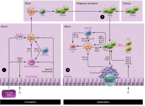
Second, Tregs regulate other leukocytes and nonhemopoi-etic cell lineages to influence decidual support of implantation (Figure 2 and ref. 42). Notably, Tregs promote antiinflammatory and tolerogenic phenotypes in alternatively activated (M2) mac-rophages and tolerogenic DCs (tDCs) through TGF-β, IL-10, and CTLA4-mediated mechanisms. Indoleamine 2,3-dioxygen-ase (IDO) produced by tDCs impairs Th1 cell survival (102, 103). Additionally, Tregs release heme oxygenase-1 (HO-1), which are susceptible to inflammation-induced fetal loss, which is
mitigat-ed by CD4+ T cells that differentiate into Tregs after transfer (64). Tregs also protect against fetal loss elicited by activated invariant NK T cells (iNKT cells) (88) or after IL-10 depletion from NOD mice (89). mAb-mediated depletion of CD25+ cells in mid-gestation has a less severe impact, but this may be because Teffs as well as Tregs express CD25 and are removed by PC61 mAb treatment (62, 90).
Mouse models with a high rate of spontaneous fetal loss rein-force the critical importance of Tregs for implantation. CBA/J females mated with DBA/2J males have fewer Tregs and elevat-ed decidual Th1 cell numbers, attributable to DBA/2J expression of the superantigen MIs (87, 91). Adoptive transfer of Tregs from CBA/J females mated with BALB/c males elevates decidual Tregs to restore fetal viability (87), but only if Tregs are transferred before embryo implantation (87). These findings confirm that Tregs have essential roles in the uterus, particularly in the peri- implantation period, consistent with managing the antiinflammatory transition required for embryo receptivity (Figure 1).
Mechanisms by which Tregs mediate
implantation success
[image:5.585.43.551.57.332.2]Mouse studies indicate at least three mechanisms by which Tregs facilitate implantation and placental development (Figure 2). Selective depletion of Tregs, or induction of overwhelming Teff
The preeclampsia symptoms are T cell dependent: they cannot be induced in T cell–deficient athymic rats, but can be induced by transfer of Th17 cells (120), and are mitigated by Tregs transferred from pregnant controls (119). Treatments that boost endogenous Tregs, including IL-10 (121) or low-dose CD28 superagonist (119), also reduce hypertension and fetal growth restriction in the RUPP model. In a rat model of preeclampsia, induction of Tregs using a CD28 superagonist treatment alleviates maternal and fetal disease, most effectively in the preimplantation phase (122).
Treg origins and antigen specificity in early
pregnancy
The two lineages of Tregs required for implantation have differ-ent ontogenies: tTregs emerge from the thymus fully competdiffer-ent to suppress responses to self and alloantigens, while pTregs differen-tiate from conventional naive CD4+ precursors in peripheral lymph nodes or tissues (123) following presentation of cognate antigen by tDCs in the presence of IL-2 and TGF-β (124). Both pTregs and recent thymic emigrant (RTE) tTreg populations require antigen contact to activate full suppressive function and memory (125).
Expansion of CD4+Helios+FOXP3+ pTregs predominantly accounts for the Treg expansion in blood and decidua in early human pregnancy (36). This is consistent with nonredundant functions for pTregs shown in CNS1-null mice, wherein elevated fetal loss is attributable to pTreg deficiency (41). Helios+ tTregs, as opposed to pTregs, may be preferentially recruited into first trimester decidua (56). Among peripheral blood tTregs, the CD45RA+CD31+ RTE population expands in the first trimester and differentiates into CD45RA–CD31– memory Tregs (57).
By using tetramer-based enrichment, selective stimulation and expansion of endogenous Tregs with fetal alloantigen speci-ficity can be demonstrated initially in the PALNs and then decidua in mice (40), explaining why fetal-maternal MHC disparity is an important determinant of Treg numbers (126). Antigen-experi-enced Tregs retain protective memory for fetal MHC antigen and rapidly reaccumulate during second pregnancies (40). Although antigen is required for Treg generation, the anti gen independence of the effector functions of Tregs confers bystander suppression and infectious tolerance that protect a wider array of fetal and placental antigens than the antigens against which the Tregs were initially primed (127).
Contact with conceptus alloantigens must occur under con-ditions that favor stable Treg (not Teff) development. pTregs require the support of tDCs to differentiate from naive Th0 cells. A tolerogenic phenotype is imposed on uterine tDCs by TGF-β, granulocyte-macrophage–CSF (GM-CSF), IL-10, galectin-1 (gal-1), and prostaglandin E (refs. 38, 100, 128, and Figure 2). Treg- derived IL-10, TGF-β, and HO-1 induce tDC and M2 macrophages to express IDO and sustain pTreg generation (102–104). Decidual Tregs also express CTLA4 (53, 105), which downregulates the DC costimulatory molecules CD80 and CD86 needed for Teff activa-tion (43). uNK cells contribute to the reinforcement of protolero-genic M2 through IFN-γ–mediated promotion of IDO expression in decidual M2 macrophages (105) and IL-10–mediated stabi-lization of tDCs (76). A PD-1–PD-L1 interaction is essential for Treg-mediated protection of alloantigenic fetuses, as shown by fetal death after PD-L1 blockade (129) or in PD-L1–null mutant targets uterine DCs and maintains their immature state (104).
In turn, these M2 and tDC phenotypes promote further Treg generation (104, 105).
In mice, DCs are key regulators of decidual transformation (73, 76), and through regulation of the uterine DC phenotype (104), Tregs may influence the extent and quality of the decidual response (Figure 2). uNK cells also promote decidualization (106) and regu-late decidual artery remodeling (107). Tregs may be important reg-ulators of the uNK phenotype and function at implantation (108), since Tregs control IL-15 release from DCs (109) and suppress uNK cytolytic activity (110). Invading trophoblasts engage with Tregs in a reciprocal interaction that modulates the secretory phenotype of both lineages (111). These coordinated interactions allow Tregs to constrain and limit the inflammatory damage and oxidative stress associated with trophoblast invasion (112).
Third, Tregs are emerging as important regulators of the maternal vascular changes that are essential for normal pla-cental development and adequate plapla-cental access to mater-nal blood. Recent studies have shown that Tregs are critical for modulating cardiovascular function and vascular homeostasis (45). How this relates to the vascular changes required for preg-nancy is detailed below.
Tregs and maternal vascular adaptation
for placental development
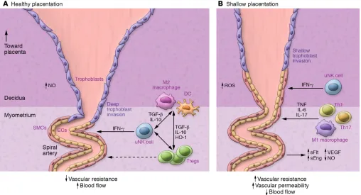
uNK cells regulate decidual blood vessel remodeling to enable ves-sel dilatation, trophoblast invasion, and secure placental access to maternal blood (ref. 107 and Figure 3). Mouse models show that T cells can interact with uNK cells to influence the maternal hemo-dynamic response to pregnancy (113, 114), and adverse effects of uNK deficiency on decidual vessel remodeling are exacerbated when T cells are also deficient (115).
Treg-deficient mice show consistent impairment in uterine spiral arterial modification, reduced placental blood flow, and fetal growth restriction (Figure 3 and refs. 41, 63, 116). Acute depletion of FOXP3+ Tregs in early pregnancy causes later uter-ine artery dysfunction associated with increased production of the vasoconstrictor endothelin-1 (ET-1) (63). A particular function for pTregs is indicated by CNS1-null mice, in which deficiency for the
Foxp3 enhancer impairs remodeling of uterine spiral arteries and
placental development (41). Poor trophoblast invasion and failure to transform spiral arteries is also seen in mice after depletion of proangiogenic, neutrophil-induced Tregs (116).
mice, which can be reversed by WT Tregs (130). Trophoblasts reinforce the tDC phenotype and drive local Treg differentiation by inducing DC production of the cytokine thymic stromal lymph-opoietin (TSLP) (111).
The significance of antigen exposure for Treg effector function at implantation is evidenced by studies in abortion-prone mice. Tregs are only effective in rescuing fetal loss when transferred from donors carrying MHC-matched fetuses, whereas antigen-inexpe-rienced donor Tregs from nonpregnant mice are ineffective (87). Consistent with a role for fetal alloantigens in facilitating uNK and Treg responses, studies in congenic mice show that fetal-maternal MHC mismatch promotes decidual vascular adaptations and facil-itates fetal growth (9). Nevertheless, endogenous antigens such as hyaluronidase or microbial antigens might also influence the acti-vation and expansion of Tregs in early pregnancy (131).
The majority of T cells in the human decidua have a memory phenotype (CD45RA– or CD45RO+) (51, 132). HLA-C is the only poly-morphic HLA expressed in human trophoblasts, and fetal- maternal HLA-C mismatch is associated with elevated decidual Tregs (4) and maternal protection from preeclampsia (68). Many decidual Tregs show fetal HLA-C antigen specificity (54, 133), but specificity to reproductive and other antigens has not been evaluated.
Seminal fluid priming of Tregs
In mice, appropriate conditions for T cell antigen priming occur in two waves during the reproductive process. A first exposure to paternal alloantigens that will later be expressed by the conceptus occurs following contact with seminal fluid at coitus, which primes the activation of antigen-specific Tregs (84). Once placental mor-phogenesis is complete in mid-gestation and maternal blood con-tacts the syncytiotrophoblast surface, fetal antigens in the form of placental exosomes are released into the maternal blood, provid-ing a second surge of antigen exposure (134).
The two stages of T cell activation can be tracked in mice expressing transgenic T cell receptors (TCRs) that are reactive with surrogate paternal antigens. Mating with male mice engineered to constitutively express OVA in semen elicit a pulse in the prolifer-ation of OVA-reactive CD4+ and CD8+ T cells within the PALNs, followed by a progressive gain in the postimplantation phase once OVA+ placental cells contact maternal blood (90). Release of pla-cental microparticles containing OVA sustains a progressively sys-temic T cell response until the postpartum phase (90). A similar pattern of T cell responsiveness is seen in T cell–transgenic models tracking female responses to paternally derived H-Y antigen (135). Seminal fluid contains high levels of TGF-β as well as TLR4 ligands, which provoke an inflammation-like response in the female reproductive tract (136), eliciting DCs and macrophages that take up male antigens, traffic to the PALNs, and present antigen to naive T cells (ref. 90 and Figure 2A). This provokes a wave of T cell activation corresponding with a 2-fold expansion in Tregs detected within days of conception in the PALNs and then the spleen and peripheral blood (refs. 39, 84, 137, and Figure 2B). Circulating Tregs then accumulate in the uterus (39) in response to CCL3, CCL4, CCL5, and CCL19 secreted by epithelial cells (85, 137) and may undergo further rounds of proliferation (39) to induce a state of hypo-responsiveness to paternal alloantigens in time for embryo implantation (refs. 84, 135, and Figure 2C).
Mouse studies imply that seminal fluid priming is a key step for expansion of the pTreg population in early pregnancy (41). Consistent with this, pregnancies sired in the absence of seminal fluid contact have poor outcomes (138). The size and suppressive competence of the pTreg pool are determined by the strength of the antigenic challenge and the cytokine context in which antigen contact occurs, parameters determined by seminal fluid composi-tion as well as female tract factors. The plasma fraccomposi-tion of seminal fluid is instrumental in Treg activation, as surgical excision of the seminal vesicle glands ablates the maternal response (135, 137) and abrogates the paternal alloantigen hyporesponsiveness seen after mating with intact males (84).
A population of tTregs also expands systemically prior to con-ception and embryo implantation (83, 139). These cells accumu-late in the uterus and PALNs during the estrous stage of the repro-ductive cycle in mice in response to increases in estradiol (E2) at ovulation (139). The antigen specificity of tTregs and whether they can be further stimulated by tDC presentation of seminal fluid or trophoblast antigens is not clear.
In women, vaginal intercourse elicits an immune response to seminal fluid, with elevated cytokine expression, immune cell recruitment, and T cell activation in the cervix (140, 141). Human seminal fluid contains soluble HLA antigens and high levels of TGF-β among an array of immune regulatory cytokines (141). Analysis of peripheral blood T cells provides evidence that prior seminal fluid contact contributes to the priming of paternal anti-gen–specific Tregs (54) to build on the follicular-phase Treg pool expansion (58). Although it is yet to be proven that seminal fluid induces pTregs in vivo, seminal fluid has been demonstrated to skew DCs to a tDC phenotype and induce Tregs in vitro (141–143). A priming effect of seminal fluid explains the benefit of seminal fluid contact for implantation success in IVF treatment cycles (144) and the protective effect in preeclampsia of sexual cohab-itation with the conceiving partner (145, 146), particularly when maternal-fetal HLA sharing is greater (68).
T cell imbalance in unexplained infertility,
miscarriage, and preeclampsia
and reduced expression of CTLA4 (153) and Ubc13, an enzyme important for Treg stability (158, 159). Conversely decidual Th17 cell numbers are elevated (155), and elevated susceptibility to IL-6
trans-signaling, which can confer abnormal suppressive function
and disposition to Th17 conversion, may contribute to this (160). In primary unexplained infertility, IL-1A, IFN-γ, and CCL11 are elevated in uterine fluid (161), and increased IL-12, IL-15, and IL-18 levels are linked with elevated uNK numbers in the decidua (80). Reduced endometrial expression of FOXP3 indicates fewer Tregs (19) potentially due to a systemic defect, as IVF success cor-relates with circulating levels of CD4+CD25+FOXP3+ cells (162, 163). Primate studies suggest that systemic and endometrial Treg deficiency is exacerbated by endometriosis, a common condition in infertile women (164). The association between reduced fer-tility and fewer Tregs in asthma (165), allergy (166), and autoim-mune disease (167) supports the view of Treg dysfunction as an underlying cause (168).
In preeclampsia, Tregs in the maternal peripheral blood and decidual tissue are reduced (23, 36, 59), and suppressive func-tion is impaired (169, 170), with an accompanying rise in proin-flammatory Th17 cells (24), CD8+ effector cells, and trophoblast apoptosis (59). This is linked with pTreg deficiency (36), most notable in early-onset severe preeclampsia (59), as well as with CD45RA+CD31+ RTE tTregs being less able to acquire a memo-ry phenotype (171). Dysfunctional DCs with reduced HLA-G and ILT4 expression (36) and/or insufficient expression of PD-L1 (172) may amplify Treg deficiency.
The clinical features of preeclampsia point to immune prim-ing and memory, invokprim-ing a causal role for the adaptive immune response. Preeclampsia is more common in first pregnancies, especially when sexual contact with the conceiving partner has been limited because of a short duration of sexual cohabitation or barrier contraceptive use — and protection afforded by prior preg-nancies is lost with a change of partner (65, 145, 173). Use of donor oocytes in assisted reproduction, where there is no prior contact with the donor’s alloantigens, leads to a striking 4.3-fold increase in preeclampsia compared with natural conception (174). With donor sperm, the risk is also increased, but, remarkably, this is mitigated by multiple exposures to the same donor’s semen (175).
Inadequate Treg priming and “shallow”
placentation
Considering the extant evidence, it is reasonable to infer that insufficient Tregs in the periconception phase is a key upstream driver of the altered decidual environment and failure to achieve appropriate inflammatory resolution that precede limited invasion of trophoblasts and maternal vessel remodeling, in turn causing “shallow” placentation and ultimately preeclampsia and/or fetal growth impairment in later gestation (refs. 27, 70, 176, and Figure 3). This view fits the emerging paradigm invoking early pregnancy as the origin not just of miscarriage, but also of disorders of pla-centation linked with early-onset, severe preeclampsia, and as a contributing factor to fetal growth restriction, late spontaneous abortion, and preterm labor (27, 176, 177). Direct evidence for this comes from transcriptional analysis of chorionic villous samples, showing evidence of immune changes originating at implantation in women who later developed preeclampsia (178).
This raises the question of why some women have insufficient Treg numbers and function. Because newly generated pTregs are more vulnerable to phenotype switching and lineage instability (47), a secure Treg fate will depend on the conception environment. Treg priming may be dysregulated as a result of seminal fluid composi-tion or responsiveness (112, 141). For example, when CD4+ cells from patients with recurrent miscarriage are cultured with DCs and the partner’s seminal fluid antigens, CD4+IL-17 and CD4+IFN-γ+ cells proliferate excessively, and fewer CD4+CD25+FOXP3+ Tregs are generated compared with fertile controls (158). The composition of seminal fluid immune-regulatory agents, particularly protolerogenic TGF-β, varies among and within men over time (179). The antitolero-genic cytokine IFN-γ, which drives the generation of Th1 immuni-ty, fluctuates substantially in seminal fluid and can be elevated in response to infection (180). IFN-γ is elevated in the seminal fluid of male partners of women with recurrent miscarriage (181, 182). IFN-γ
can interfere with the synthesis of GM-CSF required to drive T cell activation at conception (128, 183), skew Th0 differentiation toward Th17 cells (46, 184), and cause Tregs to transdifferentiate (185).
Bioavailability of cytokines, hormones, and microRNAs and the reproductive tract microbiome in the conception environment all potentially influence the Treg response (186–188). IL-10 defi-ciency elicits an unstable Treg response at implantation, with more rapid phenotype conversion and a reduced capacity to withstand an inflammatory challenge in later gestation (101, 189). Neutrophils also contribute, with neutrophil-depleted mice displaying insuffi-ciency of proangiogenic, neutrophil-induced Tregs (116). Proges-terone bioavailability impacts the Treg phenotype and secure fate commitment (190, 191), in part by inducing gal-1 to reinforce the tDC phenotype (100). Progesterone is commonly administered as luteal-phase support in IVF cycles (192), but whether this elevates Tregs at implantation remains to be determined.
Within hyperinflammatory environments, pTregs exhibit phenotypic plasticity and lineage instability, with a capacity to shift the phenotype and express cytokines that are characteristic of Teff lineages (47, 185). Tregs that undergo transdifferentiation into effector Th1 or Th17 cells, known as exTregs, drive pathology in inflammatory conditions and autoimmune disorders (193, 194). Epigenetic regulation of FOXP3 expression is a key determinant of whether T cells can maintain a suppressive phenotype (195, 196).
There is emerging evidence for Treg phenotype instability in reproductive disorders. Defects in stability would explain the observations of reduced Treg-suppressive competence (60) and evidence of elevated Th1 and Th17 cells in preeclampsia (24, 99). An intrinsic deficiency in peripheral blood Tregs in recurrent mis-carriage is indicated by diminished IL-2 and TGF-β secretion as well as reduced IL-2/STAT5 signaling (197), while decidual Tregs have elevated IFN-γ expression (198). Exploratory studies impli-cate gene polymorphisms in the promoter region of FOXP3 in pre-eclampsia (199, 200). Tregs that express insufficient FOXP3 may be phenotypically plastic and express inflammatory cytokines, and exTregs could directly contribute to the pathology.
Conclusions and therapeutic prospects
1. Schatz F, Guzeloglu-Kayisli O, Arlier S, Kayisli UA, Lockwood CJ. The role of decidual cells in uterine hemostasis, menstruation, inflam-mation, adverse pregnancy outcomes and abnormal uterine bleeding. Hum Reprod Update. 2016;22(4):497–515.
2. Medawar PB. Some immunological and endo-crinological problems raised by the evolution of viviparity in vertebrates. Symp Soc Exp Biol. 1953;7:320–338.
3. Tafuri A, Alferink J, Möller P, Hämmerling GJ, Arnold B. T cell awareness of paternal alloantigens during pregnancy. Science. 1995;270(5236):630–633.
4. Tilburgs T, et al. Fetal-maternal HLA-C mis-match is associated with decidual T cell activa-tion and inducactiva-tion of funcactiva-tional T regulatory cells. J Reprod Immunol. 2009;82(2):148–157. 5. Lissauer D, Piper K, Goodyear O, Kilby MD, Moss
PA. Fetal-specific CD8+ cytotoxic T cell respons-es develop during normal human pregnancy and exhibit broad functional capacity. J Immunol. 2012;189(2):1072–1080.
6. Robertson SA, Petroff MG, Hunt JS. Immunology of pregnancy. In: Plant TM, Zeleznik AJ, eds.
Knobil and Neill’s Physiology of Reproduction.
London, United Kingdom: Academic Press;
2015:1835–1874.
7. Bonney EA. Alternative theories: Pregnancy and immune tolerance. J Reprod Immunol. 2017;123:65–71.
8. Anderson MS, Venanzi ES, Chen Z, Berzins SP, Benoist C, Mathis D. The cellular mechanism of Aire control of T cell tolerance. Immunity. 2005;23(2):227–239.
9. Madeja Z, et al. Paternal MHC expression on mouse trophoblast affects uterine vasculariza-tion and fetal growth. Proc Natl Acad Sci U S A. 2011;108(10):4012–4017.
10. Apps R, Murphy SP, Fernando R, Gardner L, Ahad T, Moffett A. Human leucocyte antigen (HLA) expression of primary trophoblast cells and placental cell lines, determined using single antigen beads to characterize allotype speci-ficities of anti-HLA antibodies. Immunology. 2009;127(1):26–39.
11. Kshirsagar SK, et al. Immunomodulatory mol-ecules are released from the first trimester and term placenta via exosomes. Placenta. 2012;33(12):982–990.
12. Tilburgs T, et al. Human HLA-G+ extravillous tro-phoblasts: Immune-activating cells that interact with decidual leukocytes. Proc Natl Acad Sci U S A. 2015;112(23):7219–7224.
13. Taglauer ES, Trikhacheva AS, Slusser JG, Petroff MG. Expression and function of PDCD1 at the human maternal-fetal interface. Biol Reprod. 2008;79(3):562–569.
14. Collins MK, Tay CS, Erlebacher A. Dendritic cell entrapment within the pregnant uterus inhibits immune surveillance of the maternal/fetal inter-face in mice. J Clin Invest. 2009;119(7):2062–2073. 15. Nancy P, Tagliani E, Tay CS, Asp P, Levy DE,
Erle-bacher A. Chemokine gene silencing in decidual stromal cells limits T cell access to the maternal- fetal interface. Science. 2012;336(6086):1317–1321. 16. Trowsdale J, Betz AG. Mother’s little helpers:
mechanisms of maternal-fetal tolerance. Nat
Immunol. 2006;7(3):241–246.
17. Moffett A, Loke C. Immunology of placenta-tion in eutherian mammals. Nat Rev Immunol. 2006;6(8):584–594.
18. Erlebacher A. Immunology of the maternal-fetal interface. Annu Rev Immunol. 2013;31:387–411. 19. Jasper MJ, Tremellen KP, Robertson SA. Primary
unexplained infertility is associated with reduced expression of the T-regulatory cell transcription factor Foxp3 in endometrial tissue. Mol Hum
Reprod. 2006;12(5):301–308.
20. Yang H, Qiu L, Chen G, Ye Z, Lü C, Lin Q. Propor-tional change of CD4+CD25+ regulatory T cells
based disorders in reproductive and obstetric medicine. Ques-tions to first resolve include the antigen specificity of decidual Tregs, the key female and male partner determinants of optimal Treg generation and phenotype commitment, and the relevance of different Treg subsets including CD8+ Tregs.
Treg therapies relevant to pregnancy disorders could take one of three approaches: (a) advice on sexual activity, contraception, and lifestyle modifications to promote endogenous Treg gener-ation; (b) in vivo interventions to increase Treg numbers and/or function in an antigen-nonspecific, tissue-targeted, or systemic manner; or (c) cell therapy methods that could include ex vivo generation and/or expansion of Treg numbers in vitro. Candi-date biological agents to boost Treg numbers and stability using PF-L1 Fc (172) or CD28 superligand (122) already show a promis-ing proof of concept in mouse models of preeclampsia. It will be important to focus on interventions and matching diagnostics that can be applied sufficiently early in pregnancy to have a reasonable chance of diverting the course of disease development. It is critical that any experimental evaluation of these approaches in a human reproductive setting take a highly cautious approach and be based on robust clinical trial design principles, as the risk-to-benefit ratio in reproductive and obstetric conditions is different than that for life-threatening immune diseases.
Acknowledgments
SAR has received funding from the National Health and Medical Research Council of Australia (NHMRC) (APP1099461). ASC has received funding from the NHMRC (APP1092191).
Address correspondence to: Sarah A. Robertson, Robinson Research Institute, Adelaide Medical School, University of Ade-laide, North Tce, Adelaide 5005, South Australia, Australia. Phone: 618.8313.4094; Email: sarah.robertson@adelaide.edu.au. environment conducive to implantation receptivity and robust
pla-cental formation. It seems likely that in a substantial proportion of cases of infertility, recurrent miscarriage, and preeclampsia, Tregs are central and causal in disease development (21, 80). Immune imbalance or maladaptation causing incompetent phenotypes or insufficient Tregs provides a point of intersection channeling envi-ronmental, metabolic, and genetic factors (187) that likely interact with clinical factors such as prior pregnancy and male partner his-tocompatibility, which are identified as important in the prepreg-nancy antecedents of adverse pregprepreg-nancy outcomes (22).
The biological properties of Tregs, particularly their respon-siveness to environmental context and capacity to undergo phenotype switching (47, 187), may confer a maternal ability to distinguish and differentially invest in reproductive oppor-tunities. The capacity of Tregs to transdifferentiate into Teffs in the event of infection, excessive inflammation, or disrupted fetal development (129) confers the capacity to terminate preg-nancy and ensure maternal survival. The plasticity of Tregs may thereby provide an evolutionary benefit by contributing to maternal “quality control” that ensures optimal reproductive investment and maximizes offspring fitness (48). Vulnerability to reproductive disorders due to Treg instability (56, 57, 170) may be the biological trade-off (48).
immune-in decidua and peripheral blood immune-in unexplaimmune-ined recurrent spontaneous abortion patients. Fertil
Steril. 2008;89(3):656–661.
21. Lee SK, et al. An imbalance in interleukin-17-pro-ducing T and Foxp3+ regulatory T cells in women with idiopathic recurrent pregnancy loss. Hum
Reprod. 2011;26(11):2964–2971.
22. Roberts JM, et al. Global Pregnancy Collabora-tion Symposium: Prepregnancy and very early pregnancy antecedents of adverse pregnancy outcomes: overview and recommendations.
Placenta. 2017;60:103–109.
23. Sasaki Y, et al. Proportion of peripheral blood and decidual CD4(+) CD25(bright) regulatory T cells in pre-eclampsia. Clin Exp Immunol. 2007;149(1):139–145.
24. Santner-Nanan B, et al. Systemic increase in the ratio between Foxp3+ and IL-17-producing CD4+ T cells in healthy pregnancy but not in pre-eclampsia. J Immunol. 2009;183(11):7023–7030. 25. Coughlan C, et al. Recurrent implantation failure:
definition and management. Reprod Biomed
Online. 2014;28(1):14–38.
26. Ford HB, Schust DJ. Recurrent pregnancy loss: etiology, diagnosis, and therapy. Rev Obstet
Gyne-col. 2009;2(2):76–83.
27. Redman CW, Sargent IL. Immunology of pre-eclampsia. Am J Reprod Immunol. 2010;63(6):534–543.
28. Bilano VL, Ota E, Ganchimeg T, Mori R, Souza JP. Risk factors of pre-eclampsia/eclampsia and its adverse outcomes in low- and middle-income countries: a WHO secondary analysis. PLoS One. 2014;9(3):e91198.
29. Stevens W, et al. Short-term costs of preeclamp-sia to the United States health care system. Am J
Obstet Gynecol. 2017;217(3):237–248.e16.
30. Beer AE, Billingham RE. Maternal immunolog-ical recognition mechanisms during pregnancy.
Ciba Found Symp. 1978;(64):293–322.
31. Robertson SA, Mau VJ, Hudson SN, Tremellen KP. Cytokine-leukocyte networks and the estab-lishment of pregnancy. Am J Reprod Immunol. 1997;37(6):438–442.
32. Houser BL, Tilburgs T, Hill J, Nicotra ML, Strominger JL. Two unique human decidual macrophage popu-lations. J Immunol. 2011;186(4):2633–2642. 33. Gardner L, Moffett A. Dendritic cells in the human
decidua. Biol Reprod. 2003;69(4):1438–1446. 34. Koopman LA, et al. Human decidual natural
killer cells are a unique NK cell subset with immunomodulatory potential. J Exp Med. 2003;198(8):1201–1212.
35. Higuma-Myojo S, et al. Cytokine profile of nat-ural killer cells in early human pregnancy. Am J
Reprod Immunol. 2005;54(1):21–29.
36. Hsu P, et al. Altered decidual DC-SIGN+ anti-gen-presenting cells and impaired regulatory T-cell induction in preeclampsia. Am J Pathol. 2012;181(6):2149–2160.
37. Lash GE, Pitman H, Morgan HL, Innes BA, Agwu CN, Bulmer JN. Decidual macrophages: key regulators of vascular remodeling in human pregnancy. J Leukoc Biol. 2016;100(2):315–325. 38. Blois SM, et al. Dendritic cells: key to fetal
toler-ance? Biol Reprod. 2007;77(4):590–598. 39. Aluvihare VR, Kallikourdis M, Betz AG.
Regulato-ry T cells mediate maternal tolerance to the fetus.
Nat Immunol. 2004;5(3):266–271.
40. Rowe JH, Ertelt JM, Xin L, Way SS. Pregnancy imprints regulatory memory that sustains anergy to fetal antigen. Nature. 2012;490(7418):102–106. 41. Samstein RM, Josefowicz SZ, Arvey A, Treuting
PM, Rudensky AY. Extrathymic generation of regulatory T cells in placental mam-mals mitigates maternal-fetal conflict. Cell. 2012;150(1):29–38.
42. Guerin LR, Prins JR, Robertson SA. Regulatory T-cells and immune tolerance in pregnancy: a new target for infertility treatment? Hum Reprod
Update. 2009;15(5):517–535.
43. Sakaguchi S, Yamaguchi T, Nomura T, Ono M. Regulatory T cells and immune tolerance. Cell. 2008;133(5):775–787.
44. Rudensky AY. Regulatory T cells and Foxp3.
Immunol Rev. 2011;241(1):260–268.
45. Yamashita T, Sasaki N, Kasahara K, Hirata K. Anti-inflammatory and immune-modulatory therapies for preventing atherosclerotic cardio-vascular disease. J Cardiol. 2015;66(1):1–8. 46. Bettelli E, et al. Reciprocal developmental pathways for the generation of pathogenic effector TH17 and regulatory T cells. Nature. 2006;441(7090):235–238.
47. Hori S. Lineage stability and phenotypic plas-ticity of Foxp3+ regulatory T cells. Immunol Rev. 2014;259(1):159–172.
48. Robertson SA. Immune regulation of conception and embryo implantation — all about quality con-trol? J Reprod Immunol. 2010;85(1):51–57. 49. Williams PJ, Searle RF, Robson SC, Innes BA,
Bulmer JN. Decidual leucocyte populations in early to late gestation normal human pregnancy.
J Reprod Immunol. 2009;82(1):24–31.
50. Shao L, Jacobs AR, Johnson VV, Mayer L. Activa-tion of CD8+ regulatory T cells by human placental trophoblasts. J Immunol. 2005;174(12):7539–7547. 51. Tilburgs T, et al. Human decidual tissue
con-tains differentiated CD8+ effector-memory T cells with unique properties. J Immunol. 2010;185(7):4470–4477.
52. Dimova T, et al. Maternal Foxp3 expressing CD4+ CD25+ and CD4+ CD25– regulatory T-cell populations are enriched in human early normal pregnancy decidua: a phenotypic study of paired decidual and peripheral blood samples. Am J
Reprod Immunol. 2011;66(suppl 1):44–56.
53. Mjösberg J, Berg G, Jenmalm MC, Ernerudh J. FOXP3+ regulatory T cells and T helper 1, T help-er 2, and T helphelp-er 17 cells in human early preg-nancy decidua. Biol Reprod. 2010;82(4):698–705. 54. Tilburgs T, et al. Evidence for a selective migration of fetus-specific CD4+CD25bright regulatory T cells from the peripheral blood to the decidua in human pregnancy. J Immunol. 2008;180(8):5737–5745. 55. Nakashima A, Ito M, Yoneda S, Shiozaki A,
Hida-ka T, Saito S. Circulating and decidual Th17 cell levels in healthy pregnancy. Am J Reprod
Immu-nol. 2010;63(2):104–109.
56. Inada K, Shima T, Ito M, Ushijima A, Saito S. Helios-positive functional regulatory T cells are decreased in decidua of miscarriage cases with normal fetal chromosomal content. J Reprod
Immunol. 2015;107:10–19.
57. Wagner MI, et al. Differentiation of ICOS+ and ICOS– recent thymic emigrant regulatory T cells
(RTE T regs) during normal pregnancy, pre-ec-lampsia and HELLP syndrome. Clin Exp
Immu-nol. 2016;183(1):129–142.
58. Arruvito L, Sanz M, Banham AH, Fainboim L. Expansion of CD4+CD25+ and FOXP3+ regu-latory T cells during the follicular phase of the menstrual cycle: implications for human repro-duction. J Immunol. 2007;178(4):2572–2578. 59. Quinn KH, Lacoursiere DY, Cui L, Bui J, Parast
MM. The unique pathophysiology of early-onset severe preeclampsia: role of decidual T regulato-ry cells. J Reprod Immunol. 2011;91(1–2):76–82. 60. Steinborn A, et al. Distinct subsets of regulatory
T cells during pregnancy: is the imbalance of these subsets involved in the pathogenesis of pre-eclampsia? Clin Immunol. 2008;129(3):401–412. 61. Mjösberg J, et al. Systemic reduction of function-ally suppressive CD4dimCD25highFoxp3+ Tregs in human second trimester pregnancy is induced by progesterone and 17β-estradiol. J Immunol. 2009;183(1):759–769.
62. Shima T, et al. Regulatory T cells are necessary for implantation and maintenance of early preg-nancy but not late pregpreg-nancy in allogeneic mice.
J Reprod Immunol. 2010;85(2):121–129.
63. Care AS, Bourque SL, Morton JS, Hjartarson EP, Robertson SA, Davidge ST. Reduction in regulatory T cells in early pregnancy causes uterine artery dysfunction in mice. Hypertension. 2018;72(1):177–187.
64. Bizargity P, Del Rio R, Phillippe M, Teuscher C, Bonney EA. Resistance to lipopolysaccha-ride-induced preterm delivery mediated by regulatory T cell function in mice. Biol Reprod. 2009;80(5):874–881.
65. Kho EM, et al. Duration of sexual relationship and its effect on preeclampsia and small for ges-tational age perinatal outcome. J Reprod
Immu-nol. 2009;82(1):66–73.
66. Johnsen GM, et al. The combination of maternal KIR-B and fetal HLA-C2 is associated with decid-ua basalis acute atherosis in pregnancies with preeclampsia. J Reprod Immunol. 2018;129:23–29. 67. Meuleman T, Lashley LE, Dekkers OM, van Lith
JM, Claas FH, Bloemenkamp KW. HLA associa-tions and HLA sharing in recurrent miscarriage: a systematic review and meta-analysis. Hum
Immunol. 2015;76(5):362–373.
68. Triche EW, Harland KK, Field EH, Rubenstein LM, Saftlas AF. Maternal-fetal HLA sharing and preeclampsia: variation in effects by sem-inal fluid exposure in a case-control study of nulliparous women in Iowa. J Reprod Immunol. 2014;101–102:111–119.
69. Perez-Garcia V, et al. Placentation defects are highly prevalent in embryonic lethal mouse mutants. Nature. 2018;555(7697):463–468. 70. Huppertz B. Placental origins of preeclampsia:
challenging the current hypothesis. Hypertension. 2008;51(4):970–975.
71. Evans J, et al. Fertile ground: human endometrial programming and lessons in health and disease.
Nat Rev Endocrinol. 2016;12(11):654–667.
73. Plaks V, et al. Uterine DCs are crucial for decidua formation during embryo implantation in mice.
J Clin Invest. 2008;118(12):3954–3965.
74. Blois SM, et al. Interaction between dendritic cells and natural killer cells during pregnancy in mice. J Mol Med. 2008;86(7):837–852. 75. Croy BA, Wessels J, Linton N, Tayade C.
Compar-ison of immune cell recruitment and function in endometrium during development of epithelio-chorial (pig) and hemoepithelio-chorial (mouse and human) placentas. Placenta. 2009;30(suppl A):S26–S31. 76. Tirado-González I, et al. Uterine NK cells are
critical in shaping DC immunogenic functions compatible with pregnancy progression. PLoS
One. 2012;7(10):e46755.
77. Dekel N, Gnainsky Y, Granot I, Racicot K, Mor G. The role of inflammation for a successful implan-tation. Am J Reprod Immunol. 2014;72(2):141–147. 78. Gnainsky Y, et al. Local injury of the
endome-trium induces an inflammatory response that promotes successful implantation. Fertil Steril. 2010;94(6):2030–2036.
79. Griffith OW, Chavan AR, Protopapas S, Maziarz J, Romero R, Wagner GP. Embryo implanta-tion evolved from an ancestral inflammatory attachment reaction. Proc Natl Acad Sci U S A. 2017;114(32):E6566–E6575.
80. Lédée N, et al. The Uterine Immune profile may help women with repeated unexplained embryo implantation failure after in vitro fertilization.
Am J Reprod Immunol. 2016;75(3):388–401.
81. Robertson SA, Moldenhauer LM. Immunological determinants of implantation success. Int J Dev
Biol. 2014;58(2-4):205–217.
82. Darrasse-Jèze G, Darasse-Jèze G, Klatzmann D, Charlotte F, Salomon BL, Cohen JL. CD4+CD25+ regulatory/suppressor T cells prevent allo-geneic fetus rejection in mice. Immunol Lett. 2006;102(1):106–109.
83. Zhao JX, Zeng YY, Liu Y. Fetal alloantigen is responsible for the expansion of the CD4(+) CD25(+) regulatory T cell pool during pregnancy.
J Reprod Immunol. 2007;75(2):71–81.
84. Robertson SA, Guerin LR, Bromfield JJ, Branson KM, Ahlström AC, Care AS. Seminal fluid drives expansion of the CD4+CD25+ T regulatory cell pool and induces tolerance to paternal alloanti-gens in mice. Biol Reprod. 2009;80(5):1036–1045. 85. Kallikourdis M, Andersen KG, Welch KA,
Betz AG. Alloantigen-enhanced accumula-tion of CCR5+ ‘effector’ regulatory T cells in the gravid uterus. Proc Natl Acad Sci U S A. 2007;104(2):594–599.
86. Kahn DA, Baltimore D. Pregnancy induces a fetal antigen-specific maternal T regulatory cell response that contributes to tolerance. Proc Natl
Acad Sci U S A. 2010;107(20):9299–9304.
87. Zenclussen AC, et al. Abnormal T-cell reactivity against paternal antigens in spontaneous abor-tion: adoptive transfer of pregnancy-induced CD4+CD25+ T regulatory cells prevents fetal rejection in a murine abortion model. Am J
Pathol. 2005;166(3):811–822.
88. Li L, Tu J, Jiang Y, Zhou J, Schust DJ. Regulatory T cells decrease invariant natural killer T cell- mediated pregnancy loss in mice. Mucosal
Immu-nol. 2017;10(3):613–623.
89. Lin Y, Liu X, Shan B, Wu J, Sharma S, Sun Y.
Pre-vention of CpG-induced pregnancy disruption by adoptive transfer of in vitro-induced regulatory T cells. PLoS One. 2014;9(4):e94702.
90. Moldenhauer LM, Diener KR, Thring DM, Brown MP, Hayball JD, Robertson SA. Cross- presentation of male seminal fluid antigens elicits T cell activation to initiate the female immune response to pregnancy. J Immunol. 2009;182(12):8080–8093.
91. Thuere C, et al. Kinetics of regulatory T cells during murine pregnancy. Am J Reprod Immunol. 2007;58(6):514–523.
92. Zenclussen AC, Fest S, Joachim R, Klapp BF, Arck PC. Introducing a mouse model for pre-eclampsia: adoptive transfer of activated Th1 cells leads to pre-eclampsia-like symptoms exclusively in preg-nant mice. Eur J Immunol. 2004;34(2):377–387. 93. Wegorzewska M, et al. Fetal intervention
increas-es maternal T cell awarenincreas-ess of the foreign con-ceptus and can lead to immune-mediated fetal demise. J Immunol. 2014;192(4):1938–1945. 94. Xin L, et al. Cutting edge: committed Th1 CD4+
T cell differentiation blocks pregnancy-induced Foxp3 expression with antigen-specific fetal loss.
J Immunol. 2014;192(7):2970–2974.
95. Moldenhauer LM, Hayball JD, Robertson SA. Utilising T cell receptor transgenic mice to define mechanisms of maternal T cell tolerance in preg-nancy. J Reprod Immunol. 2010;87(1–2):1–13. 96. Moldenhauer LM, Diener KR, Hayball JD,
Robertson SA. An immunogenic phenotype in paternal antigen-specific CD8+ T cells at embryo implantation elicits later fetal loss in mice.
Immu-nol Cell Biol. 2017;95(8):705–715.
97. Chaturvedi V, et al. CXCR3 blockade pro-tects against Listeria monocytogenes infec-tion-induced fetal wastage. J Clin Invest. 2015;125(4):1713–1725.
98. Mellor AL, et al. Prevention of T cell-driven com-plement activation and inflammation by trypto-phan catabolism during pregnancy. Nat Immunol. 2001;2(1):64–68.
99. Zhang Y, Liu Z, Tian M, Hu X, Wang L, Ji J, Liao A. The altered PD-1/PD-L1 pathway delivers the ‘one-two punch’ effects to promote the Treg/Th17 imbalance in pre-eclampsia. Cell Mol Immunol. 2018;15(7):710–723.
100. Blois SM, et al. A pivotal role for galectin-1 in fetoma-ternal tolerance. Nat Med. 2007;13(12):1450–1457. 101. Prins JR, Zhang B, Schjenken JE, Guerin LR,
Barry SC, Robertson SA. Unstable Foxp3+ regula-tory T cells and altered dendritic cells are associ-ated with lipopolysaccharide-induced fetal loss in pregnant interleukin 10-deficient mice. Biol
Reprod. 2015;93(4):95.
102. Fallarino F, et al. Modulation of tryptophan catabolism by regulatory T cells. Nat Immunol. 2003;4(12):1206–1212.
103. Munn DH, et al. Prevention of allogeneic fetal rejection by tryptophan catabolism. Science. 1998;281(5380):1191–1193.
104. Schumacher A, et al. Blockage of heme oxygen-ase-1 abrogates the protective effect of regulatory T cells on murine pregnancy and promotes the maturation of dendritic cells. PLoS One. 2012;7(8):e42301.
105. Vacca P, et al. Crosstalk between decidual NK and CD14+ myelomonocytic cells results in
induction of Tregs and immunosuppression. Proc
Natl Acad Sci U S A. 2010;107(26):11918–11923.
106. Blois SM, Klapp BF, Barrientos G. Decidualiza-tion and angiogenesis in early pregnancy: unrav-elling the functions of DC and NK cells. J Reprod
Immunol. 2011;88(2):86–92.
107. Ashkar AA, Di Santo JP, Croy BA. Interferon gamma contributes to initiation of uterine vascular modifi-cation, decidual integrity, and uterine natural killer cell maturation during normal murine pregnancy.
J Exp Med. 2000;192(2):259–270.
108. Zhang J, Dunk C, Croy AB, Lye SJ. To serve and to protect: the role of decidual innate immune cells on human pregnancy. Cell Tissue Res. 2016;363(1):249–265.
109. Terme M, Chaput N, Combadiere B, Ma A, Ohte-ki T, Zitvogel L. Regulatory T cells control den-dritic cell/NK cell cross-talk in lymph nodes at the steady state by inhibiting CD4+ self-reactive T cells. J Immunol. 2008;180(7):4679–4686. 110. Ghiringhelli F, et al. CD4+CD25+ regulatory T
cells inhibit natural killer cell functions in a trans-forming growth factor-β-dependent manner.
J Exp Med. 2005;202(8):1075–1085.
111. Du MR, et al. Embryonic trophoblasts induce decidual regulatory T cell differentiation and maternal-fetal tolerance through thymic stromal lymphopoietin instructing dendritic cells.
J Immunol. 2014;192(4):1502–1511.
112. Saito S, Sakai M, Sasaki Y, Nakashima A, Shiozaki A. Inadequate tolerance induction may induce pre- eclampsia. J Reprod Immunol. 2007;76(1–2):30–39. 113. Croy BA, et al. Identification of the primary out-comes that result from deficient spiral arterial modification in pregnant mice. Pregnancy
Hyper-tens. 2011;1(1):87–94.
114. Burke SD, Barrette VF, Carter AL, Gravel J, Adams MA, Croy BA. Cardiovascular adapta-tions of pregnancy in T and B cell-deficient mice.
Biol Reprod. 2011;85(3):605–614.
115. Kieckbusch J, Gaynor LM, Moffett A, Colucci F. MHC-dependent inhibition of uterine NK cells impedes fetal growth and decidual vascular remodelling. Nat Commun. 2014;5:3359. 116. Nadkarni S, et al. Neutrophils induce pro-angiogenic T cells with a regulatory pheno-type in pregnancy. Proc Natl Acad Sci U S A. 2016;113(52):E8415–E8424.
117. Matrougui K, et al. Natural regulatory T cells control coronary arteriolar endothelial dys-function in hypertensive mice. Am J Pathol. 2011;178(1):434–441.
118. Maganto-García E, et al. Foxp3+-inducible regulatory T cells suppress endothelial acti-vation and leukocyte recruitment. J Immunol. 2011;187(7):3521–3529.
119. Cornelius DC, et al. An increased population of regulatory T cells improves the pathophysiology of placental ischemia in a rat model of pre-eclampsia. Am J Physiol Regul Integr Comp Physiol. 2015;309(8):R884–R891.
120. Cornelius DC, et al. Reduced uterine perfusion pressure T-helper 17 cells cause pathophysiology associated with preeclampsia during preg-nancy. Am J Physiol Regul Integr Comp Physiol. 2016;311(6):R1192–R1199.
rat model of preeclampsia. Hypertens Pregnancy. 2015;34(3):291–306.
122. Przybyl L, et al. Regulatory T cells ameliorate intrauterine growth retardation in a transgen-ic rat model for preeclampsia. Hypertension. 2015;65(6):1298–1306.
123. Josefowicz SZ, Lu LF, Rudensky AY. Regulatory T cells: mechanisms of differentiation and func-tion. Annu Rev Immunol. 2012;30:531–564. 124. Chen W, et al. Conversion of peripheral
CD4+CD25– naive T cells to CD4+CD25+ regulato-ry T cells by TGF-beta induction of transcription factor Foxp3. J Exp Med. 2003;198(12):1875–1886. 125. Kohler S, Thiel A. Life after the thymus: CD31+
and CD31–human naive CD4+ T-cell subsets.
Blood. 2009;113(4):769–774.
126. Zhao JX, Zeng YY, Liu Y. Fetal alloantigen is responsible for the expansion of the CD4(+) CD25(+) regulatory T cell pool during pregnancy.
J Reprod Immunol. 2007;75(2):71–81.
127. Tang Q, Bluestone JA. The Foxp3+ regulatory T cell: a jack of all trades, master of regulation.
Nat Immunol. 2008;9(3):239–244.
128. Moldenhauer LM, Keenihan SN, Hayball JD, Robertson SA. GM-CSF is an essential regulator of T cell activation competence in uterine den-dritic cells during early pregnancy in mice.
J Immunol. 2010;185(11):7085–7096.
129. D’Addio F, et al. The link between the PDL1 costimulatory pathway and Th17 in fetomaternal tolerance. J Immunol. 2011;187(9):4530–4541. 130. Habicht A, et al. A link between PDL1 and T
regu-latory cells in fetomaternal tolerance. J Immunol. 2007;179(8):5211–5219.
131. Chen T, et al. Self-specific memory regulatory T cells protect embryos at implantation in mice.
J Immunol. 2013;191(5):2273–2281.
132. Saito S, et al. A study of CD45RO, CD45RA and CD29 antigen expression on human decidual T cells in an early stage of pregnancy. Immunol Lett. 1994;40(3):193–197.
133. Mjösberg J, Berg G, Ernerudh J, Ekerfelt C. CD4+ CD25+ regulatory T cells in human pregnancy: development of a Treg-MLC-ELISPOT suppres-sion assay and indications of paternal specific Tregs. Immunology. 2007;120(4):456–466. 134. Chamley LW, Chen Q, Ding J, Stone PR,
Abu-maree M. Trophoblast deportation: just a waste disposal system or antigen sharing? J Reprod
Immunol. 2011;88(2):99–105.
135. Shima T, et al. Paternal antigen-specific pro-liferating regulatory T cells are increased in uterine-draining lymph nodes just before implantation and in pregnant uterus just after implantation by seminal plasma-priming in allogeneic mouse pregnancy. J Reprod Immunol. 2015;108:72–82.
136. Schjenken JE, Glynn DJ, Sharkey DJ, Robertson SA. TLR4 signaling is a major mediator of the female tract response to seminal fluid in mice.
Biol Reprod. 2015;93(3):68.
137. Guerin LR, Moldenhauer LM, Prins JR, Brom-field JJ, Hayball JD, Robertson SA. Seminal fluid regulates accumulation of FOXP3+ regulatory T cells in the preimplantation mouse uterus through expanding the FOXP3+ cell pool and CCL19-mediated recruitment. Biol Reprod. 2011;85(2):397–408.
138. Bromfield JJ, Schjenken JE, Chin PY, Care AS, Jasper MJ, Robertson SA. Maternal tract factors contribute to paternal seminal fluid impact on metabolic phenotype in offspring. Proc Natl Acad
Sci U S A. 2014;111(6):2200–2205.
139. Kallikourdis M, Betz AG. Periodic accumulation of regulatory T cells in the uterus: preparation for the implantation of a semi-allogeneic fetus? PLoS
One. 2007;2(4):e382.
140. Sharkey DJ, Tremellen KP, Jasper MJ, Gem-zell-Danielsson K, Robertson SA. Seminal fluid induces leukocyte recruitment and cytokine and chemokine mRNA expression in the human cervix after coitus. J Immunol. 2012;188(5):2445–2454. 141. Robertson SA, Sharkey DJ. Seminal fluid and
fer-tility in women. Fertil Steril. 2016;106(3):511–519. 142. Balandya E, Wieland-Alter W, Sanders K, Lahey
T. Human seminal plasma fosters CD4(+) regu-latory T-cell phenotype and transforming growth factor-β1 expression. Am J Reprod Immunol. 2012;68(4):322–330.
143. Meuleman T, et al. The immunomodulating effect of seminal plasma on T cells. J Reprod
Immunol. 2015;110:109–116.
144. Crawford G, Ray A, Gudi A, Shah A, Homburg R. The role of seminal plasma for improved outcomes during in vitro fertilization treatment: review of the literature and meta-analysis. Hum
Reprod Update. 2015;21(2):275–284.
145. Dekker G. The partner’s role in the etiology of pre-eclampsia. J Reprod Immunol. 2002;57(1–2):203–215. 146. Saftlas AF, Rubenstein L, Prater K, Harland KK,
Field E, Triche EW. Cumulative exposure to paternal seminal fluid prior to conception and subsequent risk of preeclampsia. J Reprod
Immu-nol. 2014;101-102:104–110.
147. von Wolff M, Thaler CJ, Strowitzki T, Broome J, Stolz W, Tabibzadeh S. Regulated expression of cytokines in human endometrium throughout the menstrual cycle: dysregulation in habitual abortion. Mol Hum Reprod. 2000;6(7):627–634. 148. Banerjee P, Jana SK, Pasricha P, Ghosh S,
Chakra-varty B, Chaudhury K. Proinflammatory cyto-kines induced altered expression of cyclooxygen-ase-2 gene results in unreceptive endometrium in women with idiopathic recurrent spontaneous miscarriage. Fertil Steril. 2013;99(1):179–187. 149. Michimata T, et al. Decrease of T-helper 2 and
T-cytotoxic 2 cells at implantation sites occurs in unexplained recurrent spontaneous abortion with normal chromosomal content. Hum Reprod. 2003;18(7):1523–1528.
150. Lissauer D, Goodyear O, Khanum R, Moss PA, Kilby MD. Profile of maternal CD4 T-cell effec-tor function during normal pregnancy and in women with a history of recurrent miscarriage.
Clin Sci. 2014;126(5):347–354.
151. Lédée N, et al. Specific and extensive endo-metrial deregulation is present before con-ception in IVF/ICSI repeated implantation failures (IF) or recurrent miscarriages. J Pathol. 2011;225(4):554–564.
152. Banerjee P, et al. Identification of key contribu-tory factors responsible for vascular dysfunction in idiopathic recurrent spontaneous miscarriage.
PLoS One. 2013;8(11):e80940.
153. Jin LP, Chen QY, Zhang T, Guo PF, Li DJ. The CD4+CD25 bright regulatory T cells and
CTLA-4 expression in peripheral and decidual lympho-cytes are down-regulated in human miscarriage.
Clin Immunol. 2009;133(3):402–410.
154. Inada K, Shima T, Nakashima A, Aoki K, Ito M, Saito S. Characterization of regulatory T cells in decidua of miscarriage cases with abnormal or normal fetal chromosomal content. J Reprod
Immunol. 2013;97(1):104–111.
155. Wang WJ, et al. Increased prevalence of T helper 17 (Th17) cells in peripheral blood and decidua in unexplained recurrent spontaneous abortion patients. J Reprod Immunol. 2010;84(2):164–170. 156. Sasaki Y, Sakai M, Miyazaki S, Higuma S,
Shioza-ki A, Saito S. Decidual and peripheral blood CD4+CD25+ regulatory T cells in early pregnancy subjects and spontaneous abortion cases. Mol
Hum Reprod. 2004;10(5):347–353.
157. Winger EE, Reed JL. Low circulating CD4(+) CD25(+) Foxp3(+) T regulatory cell levels pre-dict miscarriage risk in newly pregnant women with a history of failure. Am J Reprod Immunol. 2011;66(4):320–328.
158. Liu C, Wang XZ, Sun XB. Assessment of sperm antigen specific T regulatory cells in women with recurrent miscarriage. Early Hum Dev. 2013;89(2):95–100.
159. Chang JH, et al. Ubc13 maintains the suppressive function of regulatory T cells and prevents their conversion into effector-like T cells. Nat
Immu-nol. 2012;13(5):481–490.
160. Arruvito L, Billordo A, Capucchio M, Prada ME, Fainboim L. IL-6 trans-signaling and the frequency of CD4+FOXP3+ cells in women with reproductive failure. J Reprod Immunol. 2009;82(2):158–165.
161. Fitzgerald HC, Salamonsen LA, Rombauts LJ, Vollenhoven BJ, Edgell TA. The proliferative phase underpins endometrial development: altered cytokine profiles in uterine lavage fluid of women with idiopathic infertility. Cytokine. 2016;88:12–19.
162. Zhou J, Wang Z, Zhao X, Wang J, Sun H, Hu Y. An increase of Treg cells in the peripheral blood is associated with a better in vitro fertilization treatment outcome. Am J Reprod Immunol. 2012;68(2):100–106.
163. Lu Y, Zhang F, Zhang Y, Zeng B, Hu L, Liao A. Quantitative reduction of peripheral CD4+ CD25+ FOXP3+ regulatory T cells in reproductive failure after artificial insemination by donor sperm. Am J
Reprod Immunol. 2013;69(2):188–193.
164. Braundmeier A, Jackson K, Hastings J, Koehler J, Nowak R, Fazleabas A. Induction of endometrio-sis alters the peripheral and endometrial regula-tory T cell population in the non-human primate.
Hum Reprod. 2012;27(6):1712–1722.
165. Toldi G, et al. Peripheral T(h)1/T(h)2/T(h)17/ regulatory T-cell balance in asthmatic pregnancy.
Int Immunol. 2011;23(11):669–677.
166. Wegienka G, et al. Within-woman change in reg-ulatory T cells from pregnancy to the postpartum period. J Reprod Immunol. 2011;88(1):58–65. 167. Varghese S, Crocker I, Bruce IN, Tower C.
Systemic lupus erythematosus, regulatory T cells and pregnancy. Expert Rev Clin Immunol. 2011;7(5):635–648.