Thrombosis in Giant Basilar Tip Aneurysms during Coil Embolization
S. A. Renowden and A. J. Molyneux
Summary: Two patients with giant basilar tip aneurysms under-went coil embolization, one with both platinum fiber and plati-num Guglielmi detachable coils and the other with Guglielmi detachable coils only. In both cases, spontaneous intraaneurys-mal thrombosis occurred outside the coil mass, presumably a result of disruption of the intraaneurysmal flow pattern.
Index terms: Aneurysms, embolization; Interventional instru-ments, coils
During the last decade, endovascular treat-ment has been accepted as a therapeutic alter-native to surgery in the management of giant vertebrobasilar aneurysms, which are consid-ered inoperable or pose an unacceptable surgi-cal risk. Surgisurgi-cal treatment of these aneurysms is difficult even in the most experienced hands and is associated with a mortality rate of 25% to 50% (1, 2).
Until recently, endovascular treatment of in-tracranial aneurysms has depended mainly on the use of detachable balloons to obliterate the aneurysm or parent vessel. However, the inabil-ity of the balloons to adapt to the irregular shape of the aneurysm may result in stress and strain on the aneurysm during balloon placement and detachment, with the risk of aneurysm rupture. Recently the placement of soft metallic coils within the lumen of the aneurysm has become possible. With the advent of Guglielmi detach-able coils (an experimental device currently un-der review by the Food and Drug Administra-tion), which use the principle of electro-thrombosis and which allow more controlled coil placement and detachment (3, 4), coil use may increase significantly.
Our experience with coil embolization in two patients with giant basilar tip aneurysms is de-scribed. Thrombosis within the coil mesh work is well recognized, but in both patients, sponta-neous intraaneurysmal thrombosis occurred
re-mote from the coils. This phenomenon is likely to result from alteration of the blood flow pattern within the aneurysm.
Case Reports
Case 1
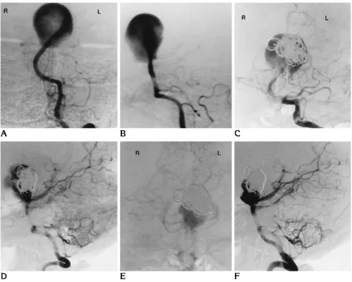
A 30-year-old woman presented with a long history of episodic, severe frontal headache. At the time of admis-sion, the headaches were daily, worse in the morning, lasted up to 10 hours, and lately had been associated with vomiting. Physical examination revealed no abnormality. Contrast-enhanced cranial computed tomography and magnetic resonance revealed a giant basilar tip aneurysm. This was confirmed by vertebral angiography (Fig 1A and B), which demonstrated rapid and rotatory flow within the aneurysm lumen.
It was decided to treat the aneurysm by coil emboliza-tion using Guglielmi detachable coils, in a staged proce-dure. A 6F guiding catheter was introduced into the left vertebral artery; through it was passed a Tracker 18 Guglielmi detachable coil variable-stiffness catheter (3F tapering to 2.5F) with a steerable guide wire (Target Ther-apeutics, San Jose, Calif). The variable-stiffness catheter was introduced into the aneurysm, and through this cath-eter five platinum Guglielmi detachable coils (Tracker 18, 8-mm helix by 40-cm length) were inserted sequentially, without complication. The detachment time for four of the coils ranged from 11 to 25 minutes, but the fifth coil had still not detached at 100 minutes. The delivery wire was torqued, and the fifth coil was successfully released. At this stage the procedure was terminated, and it was estimated that approximately 40% of the aneurysm had been occluded (Fig 1C and D). The patient remained well.
Four days later, a repeat angiogram was performed (Fig 1E and F) with the intent of further coil embolization. It was noted that a significant proportion of the aneurysm had completely thrombosed, not only within the coils but also over the aneurysm dome, anteriorly, and on the right, outside the coil mass.
Five more coils (Tracker 18, one 8-mm helix, 40-cm length, and Tracker 10, three 8-mm helix, 40-cm length,
Received December 16, 1992; accepted after revision April 29, 1993. From the Department of Neuroradiology, Radcliffe Infirmary, Oxford, England.
Address reprint requests to Dr A. J. Molyneux, Department of Neuroradiology, Radcliffe Infirmary, Woodstock Road, Oxford, Oxfordshire 0X2 6HE, England.
AJNR 16:866–871, Apr 1995 0195-6108/95/1604 –0866qAmerican Society of Neuroradiology
and one 6-mm helix, 20-cm length) were successfully inserted, as described above, without complication. De-tachment times ranged from 7 to 20 minutes, and at the end of the procedure, approximately 90% aneurysm oc-clusion was achieved (Fig 1G and H).
Six weeks later, follow-up angiography (Fig 1I) showed that the residual lumen had significantly increased in size. Further Guglielmi detachable coil embolization using two 8-mm340-cm coils, two 6-mm320-cm coils, and two 5-mm315-cm coils resulted in 100% occlusion (Fig 1J).
Case 2
A 47-year-old hypertensive man was admitted in an emergency after a generalized convulsion; he had a history of increasing headache over a period of weeks. Physical examination findings were normal apart from diplopia on right lateral gaze. Cranial computed tomography demon-strated blood in the third and fourth ventricles and basal cisterns.
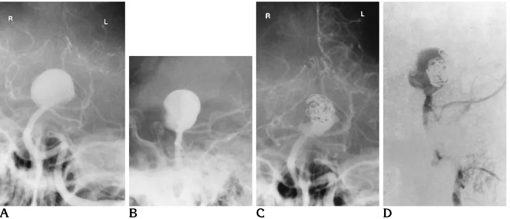
Vertebral angiography revealed a giant basilar tip an-eurysm; again, rapid rotatory blood flow was noted within Fig 1. AandB, Anteroposterior and lateral digital subtraction angiographic image of the posterior fossa arterial circulation confirms the presence of a giant basilar tip aneurysm (a termination-type aneurysm). The basilar artery is angled to the left. The direction of the inflow jet of contrast is upward and to the left, and its passage is predominantly anterior and to the right over the dome of the aneurysm. CandD, Anteroposterior and lateral subtraction left vertebral angiogram performed at the end of the first Guglielmi detachable coil embolization procedure demonstrates approximately 40% aneurysm occlusion. The coils are positioned directly in the path of the high-velocity inflow jet entering the aneurysm neck. Note the appearance of the anterior aspect of the aneurysm, suggesting thrombus formation, remote from the coils.
EandF, Anteroposterior and lateral subtraction left vertebral angiogram taken at the start of the second procedure, 4 days later, demonstrates progressive intraaneurysmal thrombosis, not only in the mesh work of the coils but also significantly over the dome, anteriorly, and on the right, remote from the coils. Comparing this image withCandD, it seems likely that thrombus formation remote from the coils commenced actuallyduringthe first procedure. The percentage of aneurysm occlusion is now approximately 70%.Figure continues.
[image:2.612.57.556.99.498.2]the aneurysm lumen (Fig 2A and B). We decided to per-form coil embolization using 20-mm and 30-mm platinum fiber complex helical coils (Target Therapeutics) (Guglielmi detachable coils were not available at the time). A 6F guiding catheter was introduced into the left ver-tebral artery, and 18 coils were placed within the aneurysm via a Tracker 18 unibody variable-stiffness catheter (3F tapering to 2.5F) achieving 40% to 50% aneurysm occlu-sion (Fig 2C and D). The patient remained well.
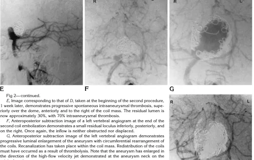
One week later, the patient was reexamined angio-graphically as a prelude to repeat embolization. At the beginning of the second procedure, considerable throm-bosis of the aneurysm had occurred spontaneously, supe-rior, antesupe-rior, and to the right of the coils (Fig 2E and F).
The remaining loculus was embolized with two 3-cm-long coils and five 2-mm-diameter coils. At the end of the procedure, a small loculus still remained (Fig 2G and H). We decided not to embolize this further in case of coil impingement into the basilar termination.
The patient remained well and was reexamined 2 months later when significant enlargement of the residual aneurysm had occurred. An additional three 30-mm and two 20-mm coils were placed into the aneurysm, and once more a small residuum remained. Although the patient remained asymp-tomatic, angiograms performed 4 and 16 months subse-quently again demonstrated progressive enlargement of the lumen without change in the external diameter but with cir-cumferential redistribution of coils (Fig 2G).
Fig 1— continued.
GandH, Anteroposterior and lateral subtraction right vertebral angiogram taken at the end of the second procedure demonstrates approximately 90% aneurysm occlusion. Two small loculi remain inferiorly and anteriorly, one on theleftand a smaller one on theright. The inflow is neither obstructed nor displaced.
I, Anteroposterior left vertebral subtraction angiogram 6 weeks later demonstrates considerable enlargement of the residual aneurysm with recanalization of thrombus out-side the coil mass.
J, Anteroposterior left vertebral subtraction angiogram after the final embolization demonstrates total aneurysm occlusion.
[image:3.612.57.557.95.555.2]Guglielmi detachable coil embolization was then per-formed via a Guglielmi detachable coil Tracker 18 vari-able-stiffness catheter (3F tapering to 2.5F). Six 8-mm3 40-cm coils, one 8-mm340-cm, and two 6-mm320-cm coils were successfully inserted, and 95% or greater aneu-rysm occlusion was achieved (Fig 2H). If the aneuaneu-rysm is not completely occluded at follow-up, then further Guglielmi detachable coil embolization is planned.
Discussion
Coil embolization is described in two patients with giant basilar tip aneurysms. The thrombo-genicity of platinum-fiber coils is enhanced by the attached dacron fibers. These coils are ex-truded through the variable-stiffness catheter using a coil pusher. Guglielmi detachable coils are platinum spiral coils with a circular memory, soldered to a stainless steel delivery system. When this coil is positioned within the variable-stiffness catheter, it adopts a straight shape and may be easily advanced into the aneurysm. When the coil has been placed in a satisfactory position, a low-voltage positive direct electric current (0.5 to 1.0 mA) is applied to the stain-less steel guide wire. The platinum coil, now positively charged, attracts the negatively charged blood particles, resulting in electro-thrombosis. The electric current also induces electrolysis at the solder joint, and the coil is gradually detached (3). The size and weight of
the thrombus is directly proportional to the cou-lombs (milliamperes times minutes) of electric-ity delivered.
In both patients spontaneous thrombosis in the aneurysm occurred outside the coil mass. The responsible mechanism is thought to be altered aneurysm hemodynamics. Hemody-namic forces are known to be important in an-eurysm thrombosis and are also important in terms of the occurrence, growth, enlargement, and rupture of intracranial saccular aneurysms (5–7). Although the precise factors are not known, the geometrical relationship between an aneurysm and its parent artery; the size of neck and aneurysm volume are thought to be impor-tant in determining the intraaneurysmal flow pattern (6 – 8). The aneurysm flow characteris-tics in both patients are of the terminal type, with rapid rotatory flow and a high-velocity jet within the inflow zone located at the side of the ostium nearest an imaginary line passing through the long axis of the parent vessel (9).
In both patients, the coils were deposited di-rectly in the path of the high-velocity jet enter-ing the aneurysm neck, thereby decreasenter-ing an-eurysm volume and obstructing the inflow track and subsequent passage of blood within the aneurysm. It is considered likely that the coil mass, by reflecting the inflowing blood, would initiate stasis within the aneurysm and precipi-Fig 2. AandB, Anteroposterior and lateral left vertebral angiogram before embolization.
CandD, Lateral subtraction image of a left vertebral angiogram shows approximately 40% aneurysm occlusion at the end of the first procedure. The coils are again positioned directly in the path of the high-velocity inflow jet. Thrombosis has already commenced anteriorly away from the coil mass.Figure continues.
[image:4.612.58.558.101.316.2]tate to thrombus formation. In both cases, the direction of the inflow jet was upward and to the left, followed by its passage over the dome, anteriorly and to the right. By impeding the initial flow, the coils inhibited blood making this passage; hence, stasis and subsequent throm-bosis would result.
In the canine model, complete aneurysmal thrombosis has been shown to depend on com-plete obstruction of the inflow zone, displace-ment of the inflow zone, and complete luminal filling (10). None of these factors had been achieved initially in either patient, and it is therefore hardly surprising that aneurysm reca-nalization occurred in each case. Thrombus for-mation and dissolution is the product of an un-stable dynamic balance between coagulation and fibrinolysis. Unfortunately, detailed his-topathologic information about thrombus evo-lution and organization within aneurysms is not
available. Thrombus evolution involves fibrinol-ysis with potential subsequent embolization and organization. Completely organized thrombi are converted to smooth muscle and collagen. Mononuclear cells within the fibrinous matrix differentiate into either macrophages responsi-ble for hematoclasia and hematophagocytosis. Endothelial cells that line autolytic slits in the fibrinous matrix form new capillaries. Last, fi-broblasts and smooth-muscle cell precursors form connective tissue. Persistence of any an-eurysm lumen inhibits complete thrombus or-ganization.
It is interesting to note that in case 1 aneu-rysm recanalization occurred outside the coil mass where Guglielmi detachable coils had been used. This contrasts with case 2, in which platinum-fiber coils were used and recanaliza-tion occurred within the coil mass with circum-ferential redistribution of the coils. The inflow Fig 2— continued.
E, Image corresponding to that ofD, taken at the beginning of the second procedure, 1 week later, demonstrates progressive spontaneous intraaneurysmal thrombosis, supe-riorly over the dome, antesupe-riorly and to the right of the coil mass. The residual lumen is now approximately 30%, with 70% intraaneurysmal thrombosis.
F, Anteroposterior subtraction image of a left vertebral angiogram at the end of the second coil embolization demonstrates a small residual loculus inferiorly, posteriorly, and on the right. Once again, the inflow is neither obstructed nor displaced.
G, Anteroposterior subtraction image of the left vertebral angiogram demonstrates progressive luminal enlargement of the aneurysm with circumferential rearrangement of the coils. Recanalization has taken place within the coil mass. Redistribution of the coils must have occurred as a result of thrombolysis. Note that the aneurysm has enlarged in the direction of the high-flow velocity jet demonstrated at the aneurysm neck on the original angiogram.
H, Anteroposterior subtraction left vertebral angiogram after Guglielmi detachable coil embolization demonstrates 95% or greater aneurysm occlusion. A tiny loculus remains on theright, posteriorly, but the inflow is now obstructed.
[image:5.612.57.558.111.428.2]jets were similar in both aneurysms, and it is postulated that the different sites of recanaliza-tion are related to the type of thrombus and the type of coil. It is suggested that the thrombus formed by the Guglielmi detachable coils differs from that formed in association with the non-Guglielmi detachable coils, and both are differ-ent to the thrombus formed outside the coil mass. The latter is formed secondary to blood stasis and is likely to resemble venous throm-bus rather than arterial thromthrom-bus. Although the initial nidus in venous thrombus is composed of platelets, the bulk of the propagating thrombus comprises largely fibrin and red blood cells. In the case of the non-Guglielmi detachable coils, the thrombus is likely to resemble arterial thrombus, formed in a high-blood-flow location. The platinum-fiber coils act as a foreign body on which fibrinogen is adsorbed and platelets are deposited and aggregate. Thrombin is re-leased by the aggregating platelets, and this enhances further platelet aggregation and fibrin formation, which may be associated with the entrapment of red and white cells. Arterial-type thrombus is, however, composed mostly of fi-brin and platelets. In the case of the Guglielmi detachable coils, electrothrombosis also con-tributes to the thrombotic process, whereby red and white blood cells and fibrinogen in addition to platelets are attracted to and deposited on the foreign-body surface. The Guglielmi detachable coils also provide a larger surface area for this to occur than the small discrete non-Guglielmi de-tachable coils. It is suggested that Guglielmi detachable coils also form a stable framework and mesh to hold a possibly more stable throm-bus and prevent recanalization within the coil mass. The inflow jet was deviated by the Guglielmi detachable coil mass and resulted in thrombolysis and recanalization outside the coil mesh in the “venous-type” thrombus. The dis-crete non-Guglielmi detachable coils may not
form such a stable framework and may not pre-vent lysis and contraction of the thrombus be-tween the coils. Recanalization therefore oc-curred within the coil mass with redistribution of the coils.
In conclusion, we have shown in two patients that by altering aneurysm hemodynamics in gi-ant aneurysms, spontaneous intraaneurysmal thrombosis remote from the coil mass may oc-cur. This observation may be used beneficially by staging coil embolization, but it is recom-mended that there be only a short delay be-tween procedures, because failure to obliterate the aneurysm totally or to obstruct or displace the inflow zone may result in prompt aneurysm recanalization and possible rupture.
References
1. Drake CG. Ligation of the vertebral (unilateral or bilateral) or basilar artery in the treatment of large intracranial aneurysms.J Neurosurg1975;43:255–274
2. Yasergil MG.Microneurosurgery.vol II. Stuttgart: Thieme, 1984 3. Guglielmi G, Vinuela F, Sepetka I, Macellar V. Electrothrombosis
of saccular aneurysms via endovascular approach, 1: electro-chemical basis, technique and experimental results.J Neurosurg 1992;75:1–7
4. Guglielmi G, Vinuela V, Dion J, Duckwiler G. Electrothrombosis of saccular aneurysms via endovascular approach, 2: preliminary clinical experience.J Neurosurg1991;75:8 –14
5. Sekhar LH, Heros RC. Origin, growth and rupture of saccular aneurysms: a review.Neurosurgery1981;18:248 –260
6. Liepsch DW, Steiger HJ, Poll A, Reulen HJ. Hemodynamic stress in lateral saccular aneurysms.Biorheology1987;24:689 –710 7. Steiger HJ, Liepsch DW, Poll A, Reulen HJ. Hemodynamic stress
in terminal saccular aneurysms: a laser-Doppler study.Heart Ves-sels1988;4:162–169
8. Steiger HJ, Reulen HJ. Low frequency fluctuations in saccular aneurysms.Acta Neurochir (Wien)1987;83:131–137
9. Strother CM, Graves VB, Rappe A. Aneurysm hemodynamics: an experimental study.AJNR Am J Neuroradiol1992;13:1089 –1095 10. Graves VB, Strother CM, Partington CR, Rappe A. Flow dynamics of lateral carotid artery aneurysms and their effects on coils and balloons: an experimental study in dogs.AJNR Am J Neuroradiol 1992;13:189 –196