Radiologic-Pathologic Correlation
Diffuse Pontine Astrocytoma
Arthur G. Kane, I Hector A. Robles, James G. Smirniotopoulos, James D. Heironimus, and Melton H. Fish
From the Depart~ents of Radiology (AGK), Nuclear Medicine (JDH), and Pathology (MHF), Brooke Army Medical Center, San Antonio, TX; Department of Radiology Walter Reed Army
Medical Center, Washington, DC (HAR); and Department of
Radiolo~ic Pathology
Armed Forces Institute of Pathology, Washington, DC (JGS) '
Clinical History
A 5-year-old girl presented with a several-month history of slurred speech, attention def-icit, gait abnormalities, and visual problems. Computed tomography (CT) scan without con-trast was obtained (Fig 1 ). A magnetic reso-nance (MR) scan was then obtained; it revealed diffuse enlargement of the pons, which was slightly hypointense on T1 (600/27) (TR/TE)-weighted images and hyperintense on T2-(2500/80) and proton density (2500/30)-weighted images. There was no appreciable enhancement after gadolinium administration. Without a biopsy, the patient underwent cis-platinum chemotherapy and radiation therapy twice a day for 6 weeks, for the presumed diagnosis of brain stem glioma. The patient experienced remission of symptoms and was started on steroid treatment. Serial MR scans at 1, 2, and 4 months after initial therapy revealed a slight reduction in the mass effect of the tumor without any change in signal and without enhancement or development of any other abnormality.
However, an MR scan performed 7 months after therapy revealed marked enhancement in
The opinions or assertions contained herein are the private views of the authors and are not to be construed as reflecting the views of the Department of the Army or the Department of Defense.
1
Address reprint requests to Arthur G. Kane, MD, Captain, [)e.
partment of Radiology, Brooke Army Medical Center, San Antonio, TX 78234.
Index terms: Brain stem, neoplasms; Radiologic-pathologic correla -tions
AJNR 14:941-945, Jul/Aug 1993 0195-6108/93/1404-0941
© American Society of Neuroradiology
941
the central pons. To differentiate between re-growth of tumor and radiation necrosis or non-neoplastic causes of enhancement, thallium-201
e
01T1) single-photon emission CT (SPECT) was performed (Fig 2). This was interpreted as showing increased uptake in the pons consist-ent with regrowth of tumor.The patient improved, but symptoms re-curred 9 months after therapy, to include right-sided extremity weakness, dysphagia, nausea, and vomiting. MR scan at that time revealed enhancement in the pons increased over that noted at 8 months as well as enlarge-ment of the lateral ventricles consistent with hydrocephalus. A ventriculoperitoneal shunt was placed, after which the patient became more alert and regained some function in the right upper and lower extremity. Bone marrow harvest was performed to allow intense
[image:1.617.311.549.510.674.2]942 KANE
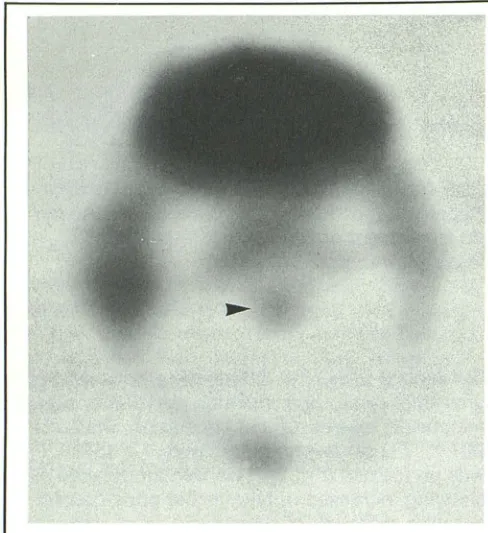
Fig. 2. Axial 201Tl-SPECT image through the brain stem
8 months after initial therapy reveals increased uptake in the region of the pons (arrowhead), consistent with regrowth of tumor.
regimen chemotherapy. One week later, the patient underwent infusion with her autologous bone marrow, after which she showed wors-ening symptoms, which improved with steroid therapy. MR performed at that time (10 months after initial therapy) confirmed reduction of the hydrocephalus but persistent enhancement on postcontrast study. Over the next 3 weeks the patient developed progressive respiratory dif-ficulty with tachypnea, wheezing, and wors-ening of neurologic symptoms. A "
do-not-resuscitate" order was written in light of the patient's poor response to maximal therapy and of the results of an MR scan performed at that time (Figs 3-5). The following day, the patient died.
Autopsy revealed symmetrical nodular en-largement of the pons and the medulla (Fig 48). Sectioning of the pons revealed a central pontine hematoma (Fig 58) surrounded by necrosis and tumor. The neoplasm extended
into the midbrain, medulla, and cerebellar white matter via the middle cerebellar peduncles (Fig 58). Histologic exam revealed diffuse pontine fibrillary astrocytoma (grade II/IV) with exten-sive necrosis and surrounding areas of
hemor-rhage (Fig 6).
AJNR: 14, July/ August 1993
General Discussion
Brain stem tumors comprise approximately 10% to 15% of infratentorial central nervous system neoplasms in the pediatric population (1, 2). The natural history of brain stem glioma is a progression of the disease with a median survival of only 4 to 15 months (3, 4). Most (75%) occur before the age of 10, with a median age of presentation being approxi-mately 6 years, and a predominant number of cases occurring in males (2.5: 1) (2). Even with radiation and chemotherapy, the overall 5-year survival in histologically documented lesions is 5% (5). Although the prognosis remains poor,
early diagnosis may result in improved survival with treatment. Classic presenting symptoms may include headaches, nausea, vomiting, gait disturbance, ataxia, visual deficits, seizures, hemiparesis, and cranial nerve dysfunction (6). Occasionally more bizarre isolated symptoms may lead one to suspect a brain stem lesion (7). The average duration of symptoms at di-agnosis is 3 to 4 months (6).
Location
Brain stem gliomas most commonly origi-nate in or involve the pons (54%), followed in frequency by the medulla (32% ); infrequently they involve the mesencephalon (5). Pontine gliomas and mesencephalic gliomas often ex-tend superiorly into the thalamus, inferiorly into the medulla, or posterolaterally through the cerebellar peduncles into the cerebellar hemisphere (Fig 58) (8, 9).
Pathology
Gross
[image:2.613.58.302.83.350.2]AJNR: 14, July/August 1993 PONTINE ASTROCYTOMA 943
A
B
c
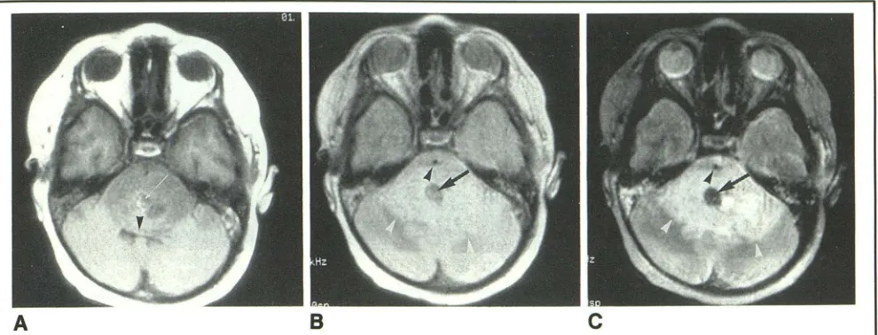
Fig. 3. A, MR axial T1-weighted image (spin echo (SE) 600/27) 1 day before the patient's death demonstrates low signal in the pons with central focal hyperintense signal (small white arrow) in the central pons. There is mass effect of the pontine glioma on the fourth ventricle (black arrowhead). The central focus demonstrates signal loss on (B) (arrow) a proton density (SE 2500/ 30)-weighted image and becomes even darker with increasingly T2-weighted (SE 2500/80) image (C) (arrow). This is consistent
with early subacute hematoma (predominantly deoxyhemoglobin with some intracellular methemoglobin). Note that the
surrounding area of hyperintensity (white arrowhead) in the double-echo images is much larger than the portions of hypointense pons on axial T1-weighted image. The basilar artery (black arrowheads) in Band Cis engulfed by tumor.
ventricle and the pontine tegmentum may
be-come convex posteriorly. However,
hydro-cephalus is rare unless the tumor involves the
midbrain (Fig 3).
Microscopic
Brain stem gliomas are typically low-grade malignancies but can be high grade. Tumor cells tend to infiltrate diffusely in intimate as-sociation with the existing neurons and blood vessels (Figs 6A and 6B). Histologic grade is based on the degree of anaplasia or dediffer-entiation, number of mitoses, and presence of necrosis (12-14).
The delayed effects of radiation and chemo-therapy are varied, but demyelination with a reduction of glial parenchymal elements and damage to peripheral vessels leading to necro-sis and infarction in zones of radiation are characteristic (Fig 6C) (15). Brain stem tumors
that have focal involvement or are exophytic
may have a more favorable prognosis in that
they tend to be low grade and are often
ame-nable to surgical resection ( 12-14).
Imaging
CT
Brain stem tumors almost always appear as a focally expanded hypodense area in the brain
stem on CT (Fig 1). The presence of contrast
enhancement before treatment is variable and
apparently does not predict alteration of
sur-vival time (13). However, two CT features,
hypodensity and involvement of the entire
brain stem, have correlated with a worse
prog-nosis (13). CT may be more sensitive than MR
in the detection of acute hemorrhage within
the brain stem, whereas MR may be more
sensitive in the early subacute to late stage of
hemorrhage (Figs 3-5) (16).
MR
Many of the lesions in the differential
diag-nosis for an intra-axial brain stem lesion have
the effect of lengthening both the T1 and T2
relaxation times of adjacent brain stem tissue.
In brain stem tumors, however, the area of
abnormal signal on T2-weighted images is
usu-ally more extensive than the areas of abnormal
signal on T1-weighted images. Except in cases
of hemorrhage, the advantage of MR in
char-acterizing a brain stem lesion lies in its ability
to demonstrate lesion location, focality, and
[image:3.612.60.555.78.267.2]944 KANE AJNR: 14, July/ August 1993
A
BFig. 4. A, Saggital T1-weighted nonenhanced image 1 day before death (SE 600/27) illustrates hypointense pons. The
enlarged pons obliterates the normal pontomedullary angle (white arrowhead) and the pontine tegmentum has become convex posteriorly (large arrow). The central pontine focus of high signal again suggests hemorrhage (small white arrow). A focal
exophytic component of tumor extends into prepontine cistern just posterior to the clivus (black arrowhead).
B, Gross specimen confirms symmetrical nodular enlargement of the pons which encases the basilar artery (arrow) as seen in Figs 3B and 3C. The focal exophytic component (black arrowhead) is again seen.
A
BFig. 5. A, Axial T1-weighted (SE 600/27) gadolinium-enhanced image through the pons from the same MR scan as Figs 3
and 4 demonstrates marked enhancement in the pons with a central, dumbbell-shaped, nonenhancing region consistent with hemorrhage and necrosis (black arrow) as seen in B.
B, Section through the pons confirms dumbbell-shaped acute central pontine hematoma (black arrow) evident on MR images, as well as extension of the whiteish to pale gray tumor into the medulla, midbrain, and middle cerebellar peduncles (arrowheads).
Fig. 6. A, Low-power (100X, hematoxylin and eosin stain) photomicrograph demonstrates widely dispersed small astrocytic tumor cells tracking along nerve axons and vessels (arrows).
B, High-power (400X) view demonstrating small tumor cells containing enlarged, irregular hyperchromatic nuclei (long black arrows) surrounding a neuron (malignant satellitosis) and its axon (arrowheads).
C, Low-power (1 OOX) view of area of necrosis and hemorrhage. There is dissolution of the walls of small vessels (arrowhead)
[image:4.615.61.562.78.234.2] [image:4.615.63.556.256.456.2] [image:4.615.67.553.527.732.2]AJNR: 14, July/ August 1993
The delayed effects of radiation and
chemo-therapy include injury to the endothelial lining
of small blood vessels, eventually resulting in
ischemia, infarction, necrosis, and damage to
the blood-brain barrier. These effects can occur months after radiation therapy. The late onset of enhancement after gadolinium
administra-tion, therefore, is common and often coincides
with the recurrence of symptoms (15).
Functional Brain Imaging
The onset of enhancement after treatment of an initially nonenhancing brain stem glioma
can pose a diagnostic dilemma. 201T1-SPECT
can be useful in distinguishing between
recur-rent tumor and nonneoplastic lesions such as
radiation necrosis, resolving hematoma, or
postsurgical change. Normal brain and nonneo-plastic lesions within the central nervous
sys-tem show little or no 201T1 uptake. 201
T1-SPECT imaging can thus be used in lieu of biopsy to establish tumor regrowth or to guide biopsy or additional treatment by localizing
areas of greatest radionuclide uptake (19, 20).
Differential Diagnosis
The differential diagnosis of brain stem
as-trocytoma includes encephalitis, tuberculoma,
vascular malformation, and resolving hema-toma. Because of the characteristic signal of blood and blood breakdown products, MR can differentiate brain stem glioma from vascular malformations and hemorrhage. A lack of
mass effect on MR scan combined with an
appropriate clinical history may be more suggestive of an infectious rather than a neo-plastic lesion etiology (21, 22). Stereotactic techniques have reduced the morbidity and mortality of biopsy and improved the accuracy of biopsy in achieving an initial histologic di-agnosis. Biopsy may be indicated in cases in
which the distinction between brain stem
neo-plasm and infection is clinically unclear (23,
24).
Summary
This case demonstrated the classic gross,
pathologic, CT, and MR findings of pontine
astrocytoma. The role of functional brain
im-PONTINE ASTROCYTOMA 945
aging in identifying regrowth of tumor was
illustrated and the differential diagnosis of a
brain stem lesion summarized.
References
1. Segall HD, Zee CS, Naidich TP, et al. Computed tomography in neoplasms of the posterior fossa in children. Radio/ C/in North
Am 1982;20:237-253
2. Farwell JR, Dohrmann GJ, Flannery JT. Central nervous system
tumors in children. Cancer 1977;40:3123-3132
3. Lassman LP, Arjona VE. Pontine gliomas of childhood. Lancet
1967;1:913-915
4. Panitch HS, Berg BO. Brain stem tumors of childhood and adolescence. Am J Dis Child 1970; 119:465-472
5. Fulton OS, Levin VA, Wara WM, et al. Chemotherapy of pediatric
brain-stem tumors. J Neurosurg 1981;54:721-725
6. Flores LE, Williams DL, Bell BA, O'Brien M, Ragab AH. Delay in diagnosis of pediatric brain tumors. Am J Dis Child
1986; 140:684-686
7. Martin RA, Handel SF, Aldama AE. Inability to sneeze as a manifestation of medullary neoplasm. Neurology 1991 ;41:
1675-1676
8. Naidich TP, Zimmerman RA. Primary brain tumors in children.
Semin Roentgenol 1984; 19: 100-114
9. Buckley RC. Pontine gliomas: a pathologic study and classification of twenty-five cases. Arch Pat hoi 1930:9:779-819
I 0. Pilcher C. Spongioblastoma polare of the pons. Arch Neural Psychiatr 1934; 1210-1230
11. Lassiter KRL, Alexander E Jr, David CH Jr, et al. Surgical
treatment of brainstem gliomas. J Neurosurg 1971;34:719-725
12. Stroink AR, Hoffman HJ, Hendrick EB, Humphreys RP, Davidson
G. Transependymal benign dorsally exophytic brain stem gliomas
in childhood: diagnosis and treatment recommendations. Neur
o-surgery 1987;20:439-444
13. Albright AL, Guthkelch AN, Packer RJ, Price RA, Rourke LB.
Prognostic factors in pediatric brainstem gliomas. J Neurosurg
1986;65:751-755
14. Stroink AR, Hoffman HJ, Hendrick EB, Humphreys RP. Diagnosis and management of pediatric brain stem gliomas. J Neurosurg
1986;65:745-750
15. Ball WS Jr, Prenger EC, Ballard ET. Neurotoxicity of radio/ chemotherapy in children: pathologic and MR correlation AJNR:
Am J Neuroradio/1992;13:761-776
16. Byrne JV, Kendall BE, Kingsley DP, Moseley IF. Lesions of the
brain stem: assessment by magnetic resonance imaging. Neur
o-radiology 1989;31: 129-133
17. Lee BCP, Kneeland JB, Walker RW, et al. MR imaging of brainstem tumors. AJNR: Am J Neuroradiol 1985;6: 159-163
18. Hueftle MG, Hans JS, Kaufman B, et al. MR imaging of brainstem gliomas. J Comput Assist Tomogr 1985;9:263-267
19. Kaplan WD, Takvorian T, Morris JH, et al. Thallium-201 brain tumor imaging: a comparative study with pathologic correlation.
J Nucl Med 1987;28:47-52
20. Mountz JM, Stafford-Schuck K, McKeever PE, et al.
Thallium-201 tumor/cardiac ratio estimation of residual astrocytoma. J
Neurosurg 1988;68: 705-709
21. Bradley WG Jr. Hemorrhage and brain iron. In: Stark DO, Bradley WG Jr, eds. Magnetic resonance imaging. 2nd ed. St. Louis:
Mosby, 1992:721-769
22. Barhovich AJ. Pediatric neuroimaging. New York: Raven,
1990:164
23. Abernathy CD, Camacho A, Kelly PJ. Stereotaxic suboccipital transcerebellar biopsy of pontine mass lesions. J Neurosurg
1989; 70:195-200