According to Mitchell’s chemiosmotic hypothesis, the respiratory chain converts redox energy into an electrochemical gradient of protons (proton-motive force, ∆p) which subsequently drives ATP formation from ADP and phosphate by the F1Fo ATP synthase. The two parameters of ∆p are the membrane potential (∆ψ) and the transmembrane proton gradient (∆pH), which are thermodynamically equivalent:
∆p =∆ψ+ 2.3RT/F∆pH ,
where R is the gas constant, T is absolute temperature and F is Faraday’s constant.
The contribution of either parameter to the proton-motive force varies widely in different organisms. In mitochondria, the ∆ψ component significantly exceeds the ∆pH component and is therefore regarded as the main driving force for ATP production. In contrast, chloroplasts appear to operate mainly on ∆pH which, at approximately 3 pH units in the steady state, far exceeds the ∆ψ of ⭐50 mV (Gräber and Witt, 1976). Bacteria are remarkably flexible and are able grow in environments with a wide range of pH values. Extreme species are the alkaliphiles and acidophiles, which have specialised to grow at pH values above 10 or below 4, respectively. For the alkaliphiles, which keep their internal pH at around 8.5, the membrane potential not only serves as the exclusive thermodynamic driving force for ATP synthesis, but it also has to compensate for the counteracting inverse ∆pH. In spite of
these variations, there is a remarkable similarity in the phosphorylation potentials, i.e. the [ATP]/[ADP][phosphate] ratios, in the various organisms (Hoffmann and Dimroth, 1991; Krulwich, 1995).
Hence, thermodynamically, both parameters of the proton-motive force undoubtedly contribute driving forces for the synthesis of ATP according to equation 1. However, this equilibrium equation says nothing about how the two components contribute to the kinetics of ATP formation during normal operation; this depends on the mechanism of energy conversion. Important early studies reported on ATP synthesis by thylakoid membranes energised with either ∆pH or ∆ψ (Jagendorf and Uribe, 1966; Uribe, 1973), and similar results were obtained using submitochondrial particles (Thayer and Hinkle, 1975). These results were corroborated and extended with the reconstituted ATP synthases of chloroplasts or other sources, leading to the conclusion that ∆pH and ∆ψare not only thermodynamically but also kinetically equivalent driving forces for the synthesis of ATP (Junesch and Gräber, 1991; Turina et al., 1991). However, these results have recently been challenged: it was shown that the ‘acid bath procedure’ used in all these experiments not only generates the desired ∆pH but also a ∆ψof significant size (Kaim and Dimroth, 1998a, 1999). Remarkably, the ATP synthase of chloroplasts or Escherichia coli is dependent on the ∆ψ component, because no ATP synthesis can be measured in its absence in spite of considerable ∆pH values. Here, we will give a detailed account of the essential importance Printed in Great Britain © The Company of Biologists Limited 2000
JEB2336
ATP, the universal carrier of cell energy, is manufactured from ADP and phosphate by the enzyme ATP synthase using the free energy of an electrochemical gradient of protons (or Na+). The proton-motive force
consists of two components, the transmembrane proton concentration gradient (∆pH) and the membrane potential. The two components were considered to be not only thermodynamically but also kinetically equivalent, since the chloroplast ATP synthase appeared to operate on ∆pH only. Recent experiments demonstrate, however, that the chloroplast ATP synthase, like those of mitochondria and
bacteria, requires a membrane potential for ATP synthesis. Hence, the membrane potential and proton gradient are not equivalent under normal operating conditions far from equilibrium. These conclusions are corroborated by the finding that only the membrane potential induces a rotary torque that drives the counter-rotation of the a and c subunits in the Fomotor of Propionigenium modestum ATP
synthase.
Key words: F1Fo ATP synthase, Fo motor, intersubunit rotation, membrane potential, torque generation, Propionigenium modestum.
Summary
Introduction
CRUCIAL ROLE OF THE MEMBRANE POTENTIAL FOR ATP SYNTHESIS BY F
1F
oATP SYNTHASES
PETER DIMROTH*, GEORG KAIM ANDULRICH MATTHEY
Institut für Mikrobiologie, Eidgenössische Technische Hochschule, ETH-Zentrum, Schmelzbergstraße 7, CH-8092 Zürich, Switzerland
*e-mail: dimroth@micro.biol.ethz.ch
of the membrane potential in generating rotational torque by the Fomotor. This torque is required to liberate, via rotation of the γ subunit, tightly bound ATP from catalytic sites in F1.
The Na+(Li+or H+)-translocating ATP synthase of Propionigenium modestum
The F1FoATP synthases comprise a huge family of enzymes with members found in the bacterial cytoplasmic membrane, the inner membrane of mitochondria and the thylakoid membrane of chloroplasts. Although most members of this family use H+ as the exclusive coupling ion, the P. modestum enzyme is the prototype of a few ATP synthases that use Na+ as the physiological coupling ion and switch to Li+or H+under certain conditions (Dimroth, 1997). We will see below that just a few amino acid substitutions in the a and c subunits determine changes in coupling ion specificity and that the overall structure and function of the H+- and Na+-translocating ATP synthases are the same. This has been most convincingly demonstrated by combining subunits of the H+-translocating E. coli ATP synthase with those of the Na+-translocating P. modestum ATP synthase to form functional chimeras (Kaim and Dimroth, 1993).
Fig. 1 shows the overall geometry of the P. modestum ATP synthase. The structure is divided into the membrane-integral Fomoiety, comprising subunits ab2c9–12, and the peripheral F1
part, composed of subunits α3β3γδε. For our purposes, we need only recognise that the entire structure can be subdivided into two counter-rotating assemblies denoted by convention as rotor and stator. The rotor consists of a ring of 9–12 c subunits connected to a shaft consisting of the γ and εsubunits. The remainder of the assembly consists of subunits ab2α3β3δand is the stator. During ATP synthesis, the Fomotor converts the energy stored in a transmembrane electrochemical gradient of Na+ (or H+) into rotary torque (Kaim and Dimroth, 1998b). This drives the rotation of the γ subunit, eliciting conformational changes in the catalytic β subunits so that tightly bound ATP is released. In its reverse operation, the engine acts as an ATP-driven Na+or H+pump. Rotation of the
γ, εand c-ring assembly is now driven by the hydrolysis of ATP, and this rotation forces Na+ (or H+) to pass from the c subunit binding site through the a subunit channel to the other side of the membrane.
Structural features of the membrane-bound rotor and stator subunits
As the atomic structure of Fohas not yet been resolved, one is restricted to understanding its function from low-resolution images, from the topological arrangements of the a and c subunits, from solution structures of the c subunit monomer
b
2 aMembrane
Cytoplasm
Rotor
Stator
δ
α
α
α
β
β
β
γ
+
Na+
Na+ Na+
Na+ Na+
Na+ Na+
Na+
Na+
Rotatio n
+
_ _ _ _ _
a
c
9−12ε
b
2 [image:2.609.235.560.67.424.2]Periplasm
and from a variety of biochemical data. The overall structure of Foinferred from electron and atomic force microscopy is that of a ring consisting of 9–12 copies of subunit c flanked at the periphery by one a and two b subunits (Birkenhäger et al., 1995; Singh et al., 1996; Takeyasu et al., 1996). Exceptionally stable c subunit assemblies are present in the ATP synthases from P. modestum (Laubinger and Dimroth, 1988) and Ilyobacter tartaricus (Neumann et al., 1998), in which even boiling with sodium dodecylsulphate (SDS) is not sufficient to dissociate the complex into monomers. The monomeric c subunits are conveniently isolated from whole cells by extraction with chloroform/methanol and fractionation on an ion-exchange column in mixed organic solvents. The c subunit contains only 80–90 amino acid residues and, therefore, structure determination by nuclear magnetic resonance (NMR) is feasible. In chloroform/methanol/H2O (4:4:1), E. coli subunit c packs into two antiparallel α-helices of 30–40 amino acid residues connected by a short hydrophilic loop (Girvin et al., 1998). Fillingame and colleagues proposed that the two extended α-helices span the membrane, which places the H+ -binding D61 residue into an occluded position in the centre of
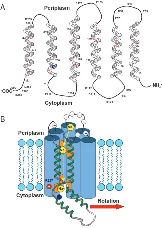
the bilayer (Girvin and Fillingame, 1994). The P. modestum subunit c, which was not stable in the organic solvent mixture, was subjected to NMR structure analysis in SDS micelles (Matthey et al., 1997). It was assumed that the detergent micelles mimic more closely the biphasic character of the membrane than the organic solvent mixture, which is monophasic. In the micelles, P. modestum subunit c folds into four clearly defined α-helices connected by short peptides with non-regular secondary structures (Matthey et al., 1999) (Fig. 2A). The N-terminal and C-terminal helices were proposed to span the membrane, and the additional helices II and III and the intervening hydrophilic loop were proposed to protrude into the cytoplasm. In this structure, the Na+-binding residues Q32, E65 and S66 (Kaim et al., 1997) are located in the helix I→II and helix III→IV connections, probably near the membrane surface on the cytoplasmic side. Hence, direct cytoplasmic access of these sites is readily conceivable, in accord with several lines of experimental evidence obtained with the ATP synthase of P. modestum (Kaim et al., 1998). Since structures have been conserved during evolution, it is not satisfactory that the two subunit c structures determined by
Na+
-COO
– -NH 3 +
3 nm
3
32
44
52 69
88
α I
α II
α III
α IV
26
66
Na+
A64
T67
G33
G31
E65
O
C
O
N
Q32
H
H
O
S66
H
O
C
CH
2CH
2CH
2CH
2CH
2B
A
[image:3.609.68.540.72.387.2]NMR are different. A reasonable explanation would be different folding patterns of the two proteins in the organic solvent mixture and in detergent micelles. It has been argued that the structure of E. coli subunit c in organic solvents closely mimics that in the native Focomplex because the specificity of D61 for dicyclohexylcarbodiimide (DCCD) was retained (Girvin and Fillingame, 1994). However, reduction of the modification rate by about four orders of magnitude makes this conclusion rather doubtful. P. modestum subunit c in dodecylsulphate micelles also retained its specificity for modification by DCCD (Matthey et al., 1997). The modification rate was approximately 10-fold lower than in the F1Fo complex, and Na+ provided partial protection from DCCD labelling, indicating that the Na+-binding site is retained in the detergent-solubilised c subunit monomer. However, even with these results, additional information is
needed to make a confident judgement about the structure of subunit c in its native folding pattern.
Probably the most important function of subunit c is to bind the coupling ions during their translocation across the membrane. For this purpose, each subunit c contains a binding site that specifically recognises the coupling ion(s). These features were first discovered for P. modestum subunit c, which may bind Na+, Li+ or H+ (Kluge and Dimroth, 1993a,b). Ligands for Na+binding are provided by Q32, E65 and S66 (Fig. 2B), whereas E65 and S66 are sufficient for Li+binding, and H+binding depends entirely on E65 (Kaim et al., 1997). Consequently, H+-specific E. coli subunit c has D61 at an equivalent position to P. modestum E65 and, by mutating residues around D61 to the corresponding ones from P.
modestum, a Li+-binding site is created (Zhang and Fillingame,
1995). - -Na+ Na+ Na+ R227 + -K F V I I F G I V I A L L I G T A V M G M 10 20 A I I M M I V F F V F T L W T S V L V T 70 80 F S L G Y F F F S T L I T F L F L I I V N 130 140 Y T F R F F V G L A F S Y G A A I
S T L L T 190 S G F 180 I G F I S Y L G N V F M F A M L G L L 230 240 R S F V L F S G M T F Q L M I V Y V V Q 260 270 I Cytoplasm Periplasm D51 E35 E47 E57 E93 E97 E104 D111 D112 E204 E217 E153 D170 D259 E286 E288 D290 D285
*
*
A
B
NH3+
−OOC Periplasm Cytoplasm Rotation
*
– Fig. 3. (A) Sequence and putative topology ofsubunit a from Propionigenium modestum. Negatively charged residues are found exclusively in the cytoplasmic and periplasmic loops. The positive stator charge (R227, blue) is in close proximity to the cytoplasmic surface, where it interacts electrostatically with negatively charged rotor sites. Hydrophilic residues in putative transmembrane helices are coloured red. The three mutated residues leading to the Na+
-impermeable stator channel are marked (*). (B) Detail of the rotor–stator assembly model as viewed from inside the rotor, showing the a subunit (blue cylinders) and a single rotor subunit (green helix). The location of the essential rotor (dark blue) and stator (red) charges and the path of Na+ into the stator helix bundle are also
[image:4.609.234.558.71.527.2]Subunit a is a very hydrophobic protein predicted to fold into five or six membrane-spanning α-helices connected by short cytoplasmic and periplasmic loops (Yamada et al., 1996; Jäger et al., 1998; Long et al., 1998; Valiyaveetil and Fillingame, 1998). The only strictly conserved residue is R227 (P. modestum numbering), and no substitutions at this position permit ATP synthesis (Cain and Simoni, 1988; Lightowlers et al., 1987). According to the topology of subunit a, R227 is located near the cytoplasmic surface (Fig. 3A), where its electrostatic interaction with E65 of subunit c is likely. Subunit a contains almost no polar residues in the centre of the membrane, but some helices have a few polar residues in the flanking regions between the centre region and the two membrane surfaces. The helices may pack to allow for a water-filled channel in the centre that is open to the periplasm and closed on the cytoplasmic side. The areas with polar residues could contribute ion-binding sites to select for the specific permeation of the coupling Na+, Li+or H+across the channel. The accumulation of negatively charged amino acid residues in the loops is noteworthy (Fig. 3A,B). At the periplasmic side, these charges may contribute to separating the coupling cations from their anions to guide the former specifically into the mouth of the channel.
Na+translocation through Forequires rotation of subunits c with respect to subunit a
Experimental evidence for the specific recognition of the coupling Na+ by P. modestum subunit a has recently been obtained using a genetic approach. In the ATPase with the a subunit triple mutation (K220R, V264E, I278N), Na+ translocation was abolished, but Li+or H+ translocation were retained (Kaim and Dimroth, 1998c). Using this mutant, insights of relevance for the ion translocation mechanism within the Fomotor were obtained. The starting point was the observation that ATP hydrolysis by the mutant enzyme was specifically inhibited by Na+, in contrast to the wild-type ATPase which is activated by Na+. Isotopic labelling experiments with 22Na+have then shown that 1 mole of 22Na+ became occluded per mole of the mutant ATPase in a strictly ATP-dependent fashion (Kaim et al., 1998). The results are easily explained within the framework of the rotational mechanism and provide the first clear evidence for a rotation of the c subunit ring (rotor) with respect to subunit a (stator). First, 22Na+binds readily from the aqueous environment to the c subunit binding sites; second, ATP hydrolysis promotes the rotation of the rotor with respect to the stator; this brings a rotor site occupied by 22Na+ into the interface with the stator channel. Since the channel is Na+-impermeable (due to the mutation), the 22Na+cannot dissociate from the rotor into the channel, and electrostatic constraints prevent the 22Na+ -occupied rotor site from rotating further out of the rotor–stator interface. Hence, ATP-driven rotation stops, and the rotor with one occluded 22Na+at the interface to the stator channel is kept in an immobilised position. However, if a 22Na+-bound rotor site approaches the wild-type channel, the 22Na+ dissociates
into the channel and passes through it to the periplasmic surface. This leads to a continued ATP-driven rotation coupled to Na+accumulation on the periplasmic side of the membrane.
The two operation modes of the Fomotor
We reasoned that, in the absence of an external energy source, the rotor should perform Brownian motion against the stator within a narrow angle. This idling motion of the motor is characterised by the exchange of internal unlabelled Na+ with external 22Na+ (Kaim and Dimroth, 1998b). Interestingly, Na+in/22Na+
outexchange was not significantly affected by modifying part of the rotor sites with DCCD. Therefore, an uninterrupted chain of rotor sites is not necessary to accommodate Na+translocation by Foacross the membrane. Two different models for the Fomotor have been proposed, comprising either two or one stator half-channels. According to the two-channel model, an ion boards a rotor site through one stator channel and rides on it for almost a complete revolution until it is released through the second stator channel to the opposite side of the membrane (Junge et al., 1997). Clearly, the ion would never reach the second stator channel if the chain of rotor sites were interrupted by modification with DCCD. Hence, this model is not in accord with the evidence. In the alternative model with only one stator half-channel, the rotor site transfers the ion through this channel and releases it to the opposite side of the membrane after only minor rotation out of the interface with the stator (Dimroth et al., 1998). According to this proposal, the rotor sites outside the rotor–stator interface are in direct contact with the cytoplasm, whereas these sites are occluded in the alternative model.
The idling motor, characterised by 22Na+out/Na+
inexchange, must switch into a torque-generating operation mode to perform work. This is accomplished by ATP-driven rotation of the γ subunit within the F1 module, which is linked to the rotation of the other rotor subunits and further to Na+ (H+) pumping across the membrane-bound Fosector. If this rotation is induced in an ATP synthase with partially DCCD-modified rotor subunits, 22Na+out/Na+
inexchange is abolished because ATP hydrolysis forces a DCCD-modified rotor subunit to strike against the stator and keeps it there in an immobilised position (Kaim and Dimroth, 1998b). In the reverse reaction, ATP synthesis requires rotary torque generated by the ‘downhill’ movement of the coupling ions across the Fosector. Unexpectedly, this torque could not be generated with a Na+ concentration gradient (∆pNa) alone. However, in the presence of a membrane potential (∆ψ) with or without an additional
∆pNa, the motor immediately switched from idling into the torque-generating mode, discontinuing 22Na+out/Na+
in exchange (Kaim and Dimroth, 1998b).
This instructive result complements previous studies with the isolated Fo motor. This protein assembly catalysed 22Na+out/Na+
higher potentials the velocity of Na+transport was significantly increased (Kluge and Dimroth, 1992).
The obligatory role of the membrane potential for ATP synthesis
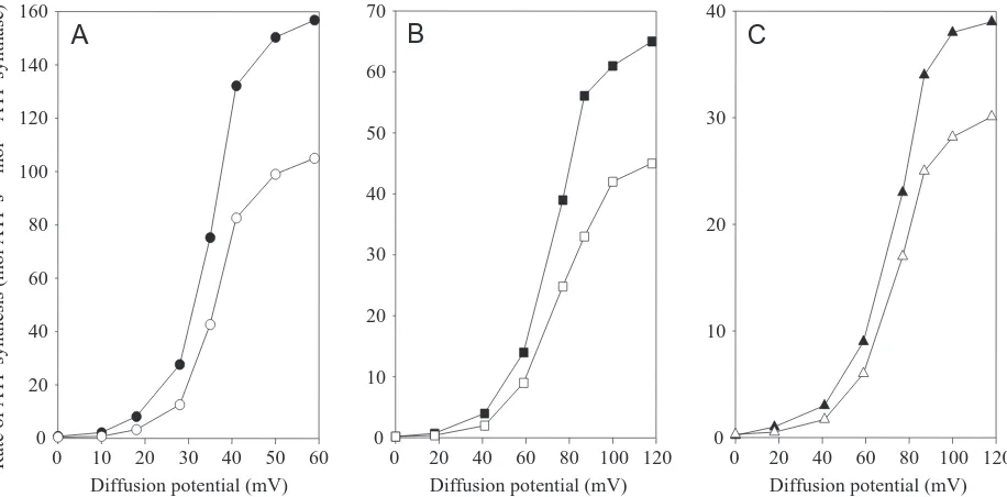
The obligatory role of the electrical potential (∆ψ) for torque generation in the Fo motor of the ATP synthase strongly indicated that this force was also needed to produce ATP. This supposition has been verified using reconstituted ATP synthases from P. modestum, E. coli and spinach chloroplasts (Kaim and Dimroth, 1998a,b, 1999). In neither case was ATP synthesis observed if only an ion gradient (∆pNa+or ∆pH) was applied (Fig. 4). The rate of ATP synthesis increased exponentially with increasing membrane potential, approaching saturation at
∆ψ>60 mV for the chloroplast ATP synthase and at
∆ψ>120 mV for the P. modestum and E. coli ATP synthases, respectively. The midpoint potential was approximately 30 mV for the ATP synthase from chloroplasts and approximately 70 mV for those from P. modestum and E. coli. Despite these differences, it is clear that the membrane potential is an indispensable driving force for all three ATP synthases. This is a surprising result, especially for the chloroplast enzyme, which has been reported to operate on ∆pH alone (Jagendorf and Uribe, 1966). Furthermore, numerous studies seemed to indicate that ∆pH and ∆ψprovided not only thermodynamically but also kinetically equivalent driving forces for the synthesis of ATP (Junesch and Gräber, 1991; Turina et al., 1991). We noted in our recent studies, however, that the chemical concentration gradient (∆pH or ∆pNa) contributed to the rate of
ATP synthesis in all cases investigated, but far less than expected if ∆ψ and ∆pH (∆pNa) were kinetically equivalent (Fig. 4) (Kaim and Dimroth, 1999).
These discrepancies could be traced back to the introduction of the ‘succinate-bath method’ for ∆pH generation (Jagendorf and Uribe, 1966), which has been the method of choice ever since. First, chloroplasts were incubated with 10 mmol l−1 succinate, pH 4–5, to acidify the interior reservoir, and the ∆pH was generated by subsequent 1:1 dilution into Tris buffer, pH 8.5. Unexpectedly, however, this method not only establishes the desired ∆pH but also a ∆ψof appreciable size because of diffusion of the succinate monoanion (Kaim and Dimroth, 1998a, 1999). Immediately after the acid–base transition, the concentrations of the succinate monoanion in the acidic interior or the basic exterior are 7.2 mmol l−1 and 0.006 mmol l−1, respectively. Diffusion of the charged succinate species across the membrane could therefore generate a Nernst potential of 180 mV. This is in reasonable agreement with the
∆ψof 140 mV determined experimentally (Kaim and Dimroth, 1999). The membrane permeability for the anionic succinate species probably increases significantly upon its folding into a ring in which the negative charge is delocalised to both carboxylic groups. Succinate, maleinate, malate or malonate, which can all adopt an intramolecular ring structure, develop a membrane potential in the ‘acid-bath procedure’, whereas fumarate with a trans double bond does not.
Model for torque generation in the Fomotor Fig. 5 shows a model of the Fomotor of P. modestum that
A
Diffusion potential (mV) 0 10 20 30 40 50 60 0
20 40 60 80 100 120 140 160
Diffusion potential (mV) 0 20 40 60 80 100 120 0
10 20 30 40 50 60 70
B
Diffusion potential (mV) 0 20 40 60 80 100 120 0
10 20 30 40
C
Rate of ATP synthesis (mol ATP s
−
1 mol
−
[image:6.609.75.533.72.298.2]1 ATP synthase)
Fig. 4. Relative contribution of the electrical potential (∆ψ) and the H+or Na+concentration gradient (∆pH and ∆pNa respectively) to the rate
of ATP formation by three different ATP synthases. (A) Reconstituted spinach chloroplast ATP synthase. K+/valinomycin diffusion potentials
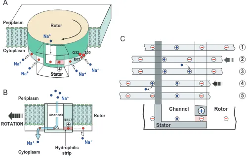
takes into account the experimental work mentioned in this article (Dimroth et al., 1999). The counter-rotating assemblies of the Fomotor make up the rotor, consisting of a ring of 9–12 copies of subunits c, and the stator, comprising subunit a. The rotor contains the Na+-binding sites, consisting of the triad Q32, E65, S66 near the cytoplasmic surface of the membrane. To prevent ion leakage between the reservoirs, the entire rotor–stator interface is hydrophobic except for two regions: (i) an ion-selective blind channel within the stator that leads from the periplasm to the level of the rotor sites; and (ii) a hydrophobic strip that connects the channel laterally with the cytoplasmic reservoir. This permits charged (unoccupied) rotor sites to enter the rotor–stator interface from the right and to pass as far to the left as the edge of the stator channel. The universally conserved positive stator charge (R227) has been
placed close to this strip to prevent ions from leaking through this route to the cytoplasm. Having bound an ion from the channel, a rotor site can pass through the hydrophobic barrier and exit the stator, whereupon the site can discharge its ion to the cytoplasm. Hence, the flux of ions is from the periplasm through the stator channel onto an empty rotor site, and from there to the cytoplasm after the rotor has turned.
Now let us consider how this ion flux is coupled to the generation of rotary torque and what may be the fundamental role of the membrane potential in the torque-generating mechanism. Owing to random Brownian motion, the progression of the rotor is stochastic but, as a result of several electrostatic forces, its diffusion is biased to the left (Fig. 5). Let us start with a rotor site on the right edge of the stator interface. Occupation of this site is in dissociation equilibrium
Channel
Stator
Rotor 1
2
3
5 4
Periplasm
Cytoplasm
Rotor
Stator
Na+
Na+
Na+
Na+
S66 Q32
E65
Stator
A
B
C
Periplasm
Cytoplasm ROTATION
Hydrophilic strip
Na+
Na+ Na+
Channel Rotor
R227
Na+
+
+
Fig. 5. (A) Diagrams of the rotor–stator assembly of the Propionigenium modestum ATP synthase. The rotor contains 9–12 Na+-binding sites
near the cytoplasmic membrane surface. The stator contains an aqueous channel that conducts ions from the periplasmic reservoir to the level of the rotor sites. The channel is laterally connected with the cytoplasm by a hydrophilic strip. The positive stator charge (R227) prevents leakage of ions along this strip to the cytoplasm. (B) Face-on view of the rotor–stator assembly. Rotation during ATP synthesis is to the left. The stator channel admits Na+from the periplasm, but these ions can only exit to the cytoplasm by boarding a rotor site and passing through the
dielectric barrier forming the left wall of the channel. (C) Typical sequence of events that advances the rotor by one step. In position 1, the third rotor site from the left has been captured by the stator charge. 1→2: the rotor site can escape from the potential well by thermal fluctuations, and the membrane potential biases its thermal escape to the left. However, it cannot pass the hydrophobic barrier that forms the left edge of the channel. 2→3: once the site has moved out of the potential well towards the channel, it picks up one Na+, which prevents its backwards
[image:7.609.60.562.75.398.2]with the cytoplasmic Na+concentration. If the site is empty, it is electrostatically attracted with its negatively charged E65 by the positive stator charge R227. The captured site can escape by thermal fluctuations but, without the membrane potential, it is as likely to escape to the right as to the left. If the channel is aqueous, the bulk of the potential drop will be across the hydrophilic strip. This biases the thermal escape of the rotor site from its potential well to the left. Once in contact with the channel, the site will quickly pick up an ion, which reduces its electrostatic field to that of a dipole. This prevents it from being attracted backwards by the stator charge and enables it to pass easily through the hydrophobic barrier, when the next site diffuses into the stator and is captured by the stator charge. When the site emerges from the stator, it quickly loses its ion to the cytoplasm. With its negative charge, the site now encounters the dielectric barrier of the stator and cannot diffuse back to the right. Thus, the membrane potential plays the pivotal role in biased diffusion of the rotor to the left, which is essential for the generation of rotary torque. A low Na+concentration on the cytoplasmic side accelerates rotation, because most rotor sites are empty and therefore attracted more frequently to the stator charge. A high Na+concentration on the periplasmic side also accelerates rotation because an empty rotor site, having escaped from the stator charge to the channel, picks up a Na+ more quickly, and this favours its progression to the left. It is important to note that Oster and colleagues have made quantitative simulations of the motor’s performance using the experimental data obtained from the P. modestum ATP synthase (Dimroth et al., 1999). These calculations show that the Na+Fomotor generates sufficient torque for ATP synthesis and that almost all the torque is due to the membrane potential.
Future trends
Rapid progress has been made in the last few years in understanding the operating principles of the Fomotor of the ATP synthase. However, high-resolution structural information is lacking. The validity of our present working models needs to be checked with the structure to verify or falsify details of Fo function. A structure for Fo would undoubtedly also be extremely valuable in designing new experiments to answer specific questions about the motor’s operation. For instance, it would be very rewarding to see the channel in the a subunit, the exact position of the stator charge R227, and the location of the rotor sites with respect to the membrane surface. Another unresolved problem is the connection of the c subunit ring to the other rotor subunits, ε and γ. This will be important in understanding how rotational torque generated by the Fomotor is transmitted to the rotating parts within the F1headpiece. Will the Fo-driven rotation of the γsubunit be continuous or will it proceed in three steps of 120 ° with an elastic spring in between? In what respects does the rotary Fomotor resemble the bacterial flagellar motor, and what are the differences?
The mechanochemistry and osmochemistry of the V-ATPases are further challenging problems for the future. The c subunit ring of this enzyme contains only half the number of binding
sites of the F-type ATPases. If F- and V-ATPases work on the same principle, twice as much ATP has to be consumed in the case of the V-ATPases to pump the same number of protons across the membrane; this obviously allows a steeper pH gradient to be generated. In the opposite direction, the Vomotor can use only every second rotor site for the torque-generating mechanism, so that, for ATP synthesis, the proton-motive force has to be substantially higher than for the F-ATPases. Many unresolved questions still have to be answered to understand in molecular detail how biological mechano-osmotic and mechanochemical energy-converting engines work.
References
Birkenhäger, R., Hoppert, M., Deckers-Hebestreit, G., Mayer, F. and Altendorf, K. (1995). The Focomplex of the Escherichia coli
ATP synthase. Investigation by electron spectroscopic imaging and immunoelectron microscopy. Eur. J. Biochem. 230, 58–67.
Cain, B. D. and Simoni, R. D. (1988). Interaction between Glu-219
and His-245 within the a subunit of F1Fo-ATPase in Escherichia
coli. J. Biol. Chem. 263, 6066–6612.
Dimroth, P. (1997). Primary sodium ion translocating enzymes.
Biochim. Biophys. Acta 1318, 11–51.
Dimroth, P., Kaim, G. and Matthey, U. (1998). The motor of the
ATP synthase. Biochim. Biophys. Acta 1365, 87–92.
Dimroth, P., Wang, H., Grabe, M. and Oster, G. (1999). Energy
transduction in the sodium F-ATPase of Propionigenium
modestum. Proc. Natl. Acad. Sci. USA 96, 4924–4929.
Girvin, M. E. and Fillingame, R. H. (1994). Hairpin folding of
subunit c of F1Fo ATP synthase: 1H distance measurements to
nitroxide-derivatized aspartyl-61. Biochemistry 33, 665–674.
Girvin, M. E., Rastogi, V. K., Abilgaard, F., Markley, J. L. and Fillingame, R. H. (1998). Solution structure of the transmembrane
H+-transporting subunit c of the F1FoATP synthase. Biochemistry
37, 8817–8824.
Gräber, P. and Witt, H. T. (1976). Relations between the electric
potential, pH gradient, proton flux and phosphorylation in the photosynthetic membrane. Biochim. Biophys. Acta 423, 141–163.
Hoffmann, A. and Dimroth, P. (1991). The electrochemical proton
potential of Bacillus alcalophilus. Eur. J. Biochem. 201, 467–473.
Jagendorf, A. T. and Uribe, E. (1966). ATP formation caused by
acid–base transition of spinach chloroplasts. Proc. Natl. Acad. Sci. USA 55, 170–177.
Jäger, H. F., Birkenhäger, R., Stalz, W.-D., Altendorf, K. and Deckers-Hebestestreit, G. (1998). Topology of subunit a of the
Escherichia coli ATP synthase. Eur. J. Biochem. 251, 122–132.
Junesch, U. and Gräber, P. (1991). The rate of ATP synthesis as a
function of ∆ψcatalyzed by the active, reduced H+-ATPase from
chloroplasts. FEBS Lett. 294, 275–278.
Junge, W., Lill, H. and Engelbrecht, S. (1997). ATP synthase: an
electrochemical transducer with rotary mechanics. Trends Biochem. Sci. 22, 420–423.
Kaim, G. and Dimroth, P. (1993). Formation of a functionally active
sodium-translocating F1Fo ATPase in Escherichia coli by
homologous recombination. Eur. J. Biochem. 218, 937–944.
Kaim, G. and Dimroth, P. (1998a). ATP synthesis by the F1FoATP
synthase of Escherichia coli is obligatorily dependent on the electric potential. FEBS Lett. 434, 57–60.
Kaim, G. and Dimroth, P. (1998b). Voltage-generated torque drives
Kaim, G. and Dimroth, P. (1998c). A triple mutation in the a subunit
of the Escherichia coli/Propionigenium modestum F1Fo ATPase
hybrid causes a switch from Na+stimulation to Na+ inhibition.
Biochemistry 37, 4626–4634.
Kaim, G. and Dimroth, P. (1999). ATP synthesis by F-type ATP
synthase is obligatorily dependent on the transmembrane voltage. EMBO J. 18, 4118–4127.
Kaim, G., Matthey, U. and Dimroth, P. (1998). Mode of interaction
of the single a subunit with the multimeric c subunits during the translocation of the coupling ions by F1FoATPases. EMBO J. 17,
688–695.
Kaim, G., Wehrle, F., Gerike, U. and Dimroth, P. (1997).
Molecular basis for the coupling ion selectivity of F1Fo ATP
synthases: Probing the liganding groups for Na+and Li+in the c
subunit of the ATP synthase from Propionigenium modestum. Biochemistry 36, 9185–9194.
Kluge, C. and Dimroth, P. (1992). Studies on Na+ and H+
translocation through the Fo part of the Na+-translocating F1Fo
ATPase from Propionigenium modestum: discovery of a membrane potential dependent step. Biochemistry 31, 12665–12672.
Kluge, C. and Dimroth, P. (1993a). Kinetics of inactivation of the
F1Fo ATPase of Propionigenium modestum by
dicyclohexylcarbodiimide in relationship to H+ and Na+
concentration: probing the binding site for the coupling ions. Biochemistry 32, 10378–10386.
Kluge, C. and Dimroth, P. (1993b). Specific protection by Na+or
Li+of the F1Fo ATPase of Propionigenium modestum from the
reaction with dicyclohexylcarbodiimide. J. Biol. Chem. 268, 14557–14560.
Krulwich, T. A. (1995). Alkaliphiles: basic molecular problems of
pH tolerance and bioenergetics. Mol. Microbiol. 15, 403–410.
Laubinger, W. and Dimroth, P. (1988). Characterization of the ATP
synthase of Propionigenium modestum as a primary sodium pump. Biochemistry 27, 7531–7537.
Lightowlers, R. N., Howitt, S., Hatch, L., Gibson, F. and Cox, G. B. (1987). The proton pore in the Escherichia coli FoF1-ATPase: a
requirement for arginine at position 210 of the a subunit. Biochim. Biophys. Acta 894, 399–406.
Long, J. C., Wang, S. and Vik, S. B. (1998). Membrane topology
of subunit a of the F1FoATP synthase as determined by labeling
of unique cysteine residues. J. Biol. Chem. 273, 16235–16240.
Matthey, U., Kaim, G., Braun, D., Wüthrich, K. and Dimroth, P.
(1999). NMR studies of subunit c of the ATP synthase from Propionigenium modestum in dodecylsulfate micells. Eur. J. Biochem. 261, 459–467.
Matthey, U., Kaim, G. and Dimroth, P. (1997). Subunit c from the
sodium-ion-translocating F1Fo ATPase of Propionigenium
modestum. Production, purification and properties of the protein in dodecylsulfate solution. Eur. J. Biochem. 247, 820–825.
Neumann, S., Matthey, U., Kaim, G. and Dimroth, P. (1998).
Purification and properties of the F1Fo ATPase of Ilyobacter
tartaricus, a sodium ion pump. J. Bacteriol. 180, 3312–3316.
Singh, S., Turina, P., Bustamante, C. J., Keller, D. J. and Capaldi, R. (1996). Topographical structure of membrane-bound Escherichia
coli F1FoATP synthase in aqueous buffer. FEBS Lett. 397, 30–34.
Takeyasu, K., Omote, H., Nettikadan, S., Tokumasu, F., Iwamotu-Kihara, A. and Futai, M. (1996). Molecular imaging of
Escherichia coli F1Fo-ATPase in reconstituted membranes using
atomic force microscopy. FEBS Lett. 392, 110–113.
Thayer, W. S. and Hinkle, P. C. (1975). Synthesis of adenosine
triphosphate by an artificially imposed electrochemical proton gradient in bovine heart submitochondrial particles. J. Biol. Chem.
250, 5330–5335.
Turina, P., Melandri, B. A. and Gräber, P. (1991). ATP synthesis
in chromatophores driven by artificially induced ion gradients. Eur. J. Biochem. 196, 225–229.
Uribe, E. (1973). ATP synthesis by a K+-valinomycin-induced charge
imbalance across chloroplast grana membranes. FEBS Lett. 36, 143–147.
Valiyaveetil, F. I. and Fillingame, R. H. (1998). Transmembrane
topology of subunit a in the Escherichia coli F1FoATP synthase.
J. Biol. Chem. 273, 16241–16247.
Yamada, H., Moriyama, Y., Maeda, M. and Futai, M. (1996).
Transmembrane topology of Escherichia coli H+-ATPase subunit
a. FEBS Lett. 390, 34–38.
Zhang, Y. and Fillingame, R. H. (1995). Changing the ion binding
specificity of the Escherichia coli H+-transporting ATP synthase by