Brain Infarction
Blake A. Johnson, Joseph E. Heiserman, Burton P. Drayer, and Paul J. Keller
PURPOSE: To determine the contribution of cranial MR angiography (MRA) for the evaluation of patients with acute and subacute brain infarction. METHODS: MR and MRA studies performed on
78 adult patients with acute and subacute stroke were retrospectively reviewed and correlated with the clinical records. RESULTS: There were 50 acute and 28 subacute infarctions in our series. Five of 78 MRA exams (6%) were nondiagnostic. Sixty examinations (80%) were positive for stenosis or occlusion. The distribution of stenotic or occlusive vascular lesions correlated with the location of infarction in 56 of the 60 positive cases (93%). MRA provided information not obtained from the MR images in 40 cases (55%). One hundred four individual vessels in 8 patients who underwent conventional cerebral angiography were compared with the MRA appearance. The MRA interpretations correlated with the conventional angiographic evaluations for 90 vessels
(87%). CONCLUSIONS: Vascular lesions demonstrated on intracranial MRA show a high correla-tion with infarct distribucorrela-tion. MRA provides information adjunctive to conventional MR in a majority of cases. We conclude that MRA is an important component of the complete evaluation of brain infarction.
Index terms: Magnetic resonance angiography (MRA); Brain, infarction; Brain, magnetic resonance
AJNR Am J Neuroradio/15:901-908, May 1994
The cost of brain infarction to society in terms of morbidity, mortality, and economics is
pro-found. Stroke is the third most common cause of death in the United States (1, 2) and has devastating sequelae for survivors and their
fam-ilies. In certain countries in the Far East, it is the most common cause of death (3). Heightened
interest in the early diagnosis and treatment of
acute stroke challenges neuroimagers to optimize
available modalities and to develop new tech-niques for the evaluation of cerebrovascular
dis-ease. Magnetic resonance (MR) provides accurate anatomic information regarding the distribution and characteristics of early infarction. MR
angiog-raphy (MRA) complements the cross-sectional
Received August 5, 1993; accepted pending revision October 27;
revision received December 17.
Presented at the Annual Meeting of the American Society of
Neuro-radiology, Vancouver, Canada, May 16-21, 1993. From the Barrow Neurological Institute, Phoenix.
Address reprint requests to Burton P. Drayer, MD,
Radiologist-in-Chief, Barrow Neurological Institute, 350 W Thomas Rd, Phoenix, AZ 85031.
AJNR 15:901-908, May 1994 0195-6108/94/1505-0901 © American Society of Neuroradiology
901
parenchymal imaging modalities, providing a noninvasive means for assessing cerebrovascular structures. Our goal in this investigation was to evaluate the role of MRA in the diagnostic eval-uation of patients who present with acute and subacute stroke. We correlated the distribution of infarction on conventional MR with the distri-bution of cerebrovascular disease depicted on MRA and sought additional information provided by MRA that could not be derived from conven-tional MR alone.
Methods
Patients
We retrospectively reviewed the clinical records and imaging studies of 78 consecutive patients who presented to our institution between August 30, 1991, and January 14, 1993, with acute or subacute stroke with positive MR findings, and underwent MRA. Clinical records were inter-rogated for information regarding symptoms and time of onset.
Imaging
902 JOHNSON
20/1 (repetition time/echo time/excitations), and long-repetition-time dual-echo axial sequences (2500/30,90/1) were performed.
Various forms of three-dimensional time-of-flight MRA were used during this investigation. Improved protocols were implemented as they became available. Twenty-nine of 78 examinations were conducted with the unmodified pulse sequence supplied by the vendor, or with the local addition of magnetization transfer binomial radio-frequency pulses (4). The MRA parameters included: 50/4.9/1, 25° flip angle, 19-cm field of view, 64 partitions 0.7 mm thick, 256 X 256 matrix, first moment flow compensation on the section- and frequency-encoding axes, radio-frequency phase-spoiled acquisition, and scan time of 13.6 minutes. When magnetization transfer was used, parameters were the same with the exception of the repetition time (30 msec) and acquisition time (8.3 minutes).
The remaining 49 patients were evaluated using multiple overlapped thin-slab acquisition (MOTSA) (5), a technique in which multiple thin 3-D slabs are sequentially acquired. The slabs in this case consisted of 16 0. 75-mm-thick sections, and the slab locations overlapped each other by 50%. Other relevant scan parameters were: 31/5.3/1 (early versions required longer echo times, up to 7 msec), 30° flip angle, 11 slabs, 16- to 14-cm field of view, 256 X 128 matrix, first moment flow compensation, and scan time of 11.9 minutes.
Axial source images were printed, and a maximum intensity pixel algorithm was used to generate subvolume projections of the anterior and posterior circulations. The entire maximum intensity pixel volume set was also re-viewed in a single base-view projection. The anterior and posterior circulation projection images were printed at 18° increments, rotated in a counterclockwise fashion over 180°.
Image Analysis
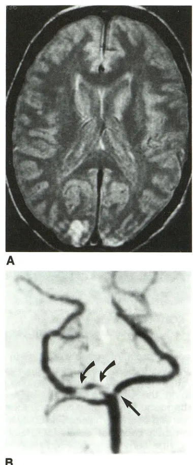
All imaging studies were reviewed by two neuroradiol-ogists (J.E.H. and B.A.J.) who were blinded to the clinical information, MR results, and conventional angiographic findings. The source images and projection MRA images were reviewed first. The intracranial vessels were individ-ually graded as normal, stenotic, or occluded on the MRA examinations. A stenotic lesion consisted of focal signal attenuation of 50% diameter or greater or a signal void with distal reconstitution of intravascular signal (Fig 1). An occlusion was interpreted when there was no distal recon-stitution of signal. The readers then documented the loca-tion, size, and signal characteristics of the infarction based on the conventional MR study. A consensus opinion was formed at the time of image interpretation. A computerized spreadsheet was generated during the evaluation of the films for recording the results of the MR and MRA interpre-tations.
The vascular distribution of infarction as depicted on MR was then compared with the MRA findings to assess for correlations. MRA findings were considered to correlate with the MR if a stenotic or occlusive lesion occurred in
AJNR: 15, May 1994
A
8
Fig. 1. A, Intermediate-weighted image demonstrates small bilateral occipital cortical infarcts.
B, MRA projection image of the posterior circulation shows tandem stenotic lesions in the proximal right posterior cerebral artery (curved arrows) and a single focus of stenosis at the origin of the left posterior cerebral artery (straight arrow). The superior cerebellar artery is not visualized, although there was no infarction in the superior cerebellar distribution.
[image:2.612.341.531.76.534.2]Data for the MOTSA and 3-D time-of-flight MRA were
compared by computing the normal deviate z derived from
the
x
2 statistic assuming independent samples. Acorrec-tion was applied for continuity (6).
Conventional cerebral angiography was performed on 8
of 78 patients (11 %). All but one angiogram was obtained within 1 week of the MR study. Individual vessels were
assessed by both readers for stenosis or occlusion. On each
study, the internal carotid, anterior cerebral, middle
cere-bral, posterior cerecere-bral, vertecere-bral, basilar, and superior
cerebellar arteries were evaluated. We read the
conven-tional angiograms while blinded to the MRA results and subsequently compared them with the results of the MRA
for correspondence of the interpretation for each of 104
vessels.
Results
The cohort comprised 40 men and 38 women, 26 to 87 years of age (mean, 67 years). There were 50 acute (<48-hour) and 28 subacute (3- to
14-day) infarctions in our series. The interval
between ictus and imaging ranged from 0 to 14
days (mean, 3.2 days). Five (6%) of the MRAs were considered nondiagnostic because of patient motion and were excluded from further statistical
analysis. Clinical data were available for all but 7
patients; imaging criteria were used to categorize
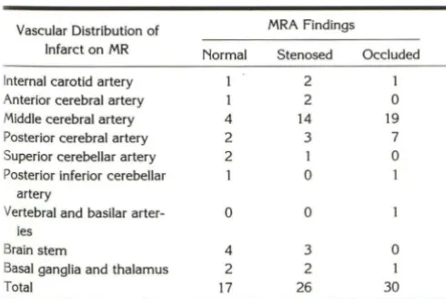
the lesions as acute or subacute infarction in these individuals. Only 13 of the 73 diagnostic MRA findings (18%) were normal. The remaining 60 patients (82%) demonstrated a stenotic or
occlusive vascular lesion on MRA. The vascular
occlusion or stenosis correlated with the
distri-bution of infarction in 56 of the 60 positive cases
(93%). Twenty-six patients with stenoses and 30
patients with occlusive lesions were in this
cate-gory. Table 1 shows the distribution of infarction
in each patient and the status of the correspond-ing vessels as evaluated by MRA.
TABLE 1: MR distribution of infarctions versus MRA findings
Vascular Distribution of MRA Findings
Infarct on MR Normal Stenosed Occluded
Internal carotid artery 2 1
Anterior cerebral artery 2 0
Middle cerebral artery 4 14 19
Posterior cerebral artery 2 3 7 Superior cerebellar artery 2 I 0
Posterior inferior cerebellar 0
artery
Vertebral and basilar arter- 0 0
ies
Brain stem 4 3 0
Basal ganglia and thalamus 2 2 I
Total 17 26 30
The MOTSA MRA technique was used in 48 of 73 cases (66%). Forty of the MOTSA studies
(83%) had findings that correlated with the
dis-tribution of infarction, versus 15 of 25 (60%) of
the studies that antedated our use of MOTSA. This difference in results was marginally signifi -cant (P= .054). The mean age of patients studied with the MOTSA technique was 67 years, with a mean of 2 days between ictus and imaging. The mean age for the other group was 66 years, with a mean of 2 days between ictus and imaging. The clinical presentations for these groups were comparable as well.
There was no significant difference in results between patients with acute infarction and those with subacute infarction. The MRA findings cor-related with the distribution of infarction in 34 of 46 (7 4%) patients with acute infarcts and in 21 of 27 (78%) patients with subacute infarcts. How-ever, of the patients with clinical records docu-menting an event occurring 24 hours or less before scanning, 14 of 26 (54%) had occlusions in the vascular distribution of the infarction, ver-sus 14 of 40 (35%) of those who presented after 24 hours.
The size of the infarct correlated with the MRA
findings. Of 14 patients with infarcts smaller than
2 em, 9 had MRA findings that correlated with the vascular distribution (64% ). Forty-six of 59 patients (78%) with infarcts 2 em or larger had correlative MRA findings.
Using the criteria described in the previous section, we determined that MRA provided infor-mation that could not be derived directly from the MR images in 40 patients (55%). This in-cluded cases in which collateral circulation was demonstrated on MRA (Fig 2). Other examples include cases in which conventional MR sug-gested compromise or patency of a major vessel or a branch, and MRA demonstrated otherwise. Such cases met this criterion only if the infor-mation obtained from MRA could not be gleaned from the conventional MR alone.
One hundred four individual vessels in 8 pa-tients who underwent conventional angiography
were evaluated. The same grading scheme used
for analyzing the MRA exams was used to eval-uate the contrast angiograms; individual vessels were graded as normal, stenotic, or occluded.
The MRA readings correlated with the
conven-tional angiographic findings in 90 vessels (87%)
(Table 2). Two that appeared normal on
conven-tional angiography were read as occluded on
[image:3.615.52.297.577.741.2]904 JOHNSON AJNR: 15, May 1994
('
A
Bc
Fig. 2. A, T2-weighted image shows a large infarction in the left middle cerebral artery distribution with lenticulostriate territory involvement, which suggests proximal MCA occlusion.
8, Coronal projection and C, base-view MRA images show bilateral internal carotid artery occlusions. A potential collateral pathway is suggested by the prominent right external carotid and ophthalmic arteries (arrow).
TABLE 2: Conventional angiography versus MRA findings
MRA Normal
Stenosed
Occluded
Conventional Angiography
Normal
73 7 2
Stenosed
5 10 0
Occluded
0 0 7
one case, the angiogram was performed 6 days after the MRA, and recanalization was apparent in the distribution of infarction. In the second case, more than 1 week elapsed (after the MRA) before the angiogram was performed. Both pa-tients had posterior cerebral artery distribution
infarcts. Vessels that were read as normal on MRA and stenotic on contrast angiography (or vice versa) were as follows: superior cerebellar artery (5 vessels), anterior cerebral artery (3 ves-sels), vertebral artery (2 vesves-sels), internal carotid artery (1 vessel), and posterior cerebral artery (1 vessel).
None of the discordant interpretations of sten-otic vessels was in the distribution of infarction.
Discussion
Stroke Evaluation
Existing and potential therapeutic regimens for cerebrovascular disease provide motivation for
early and accurate assessment of cerebrovascular disease (7). MR is an excellent tool for evaluating cerebral infarction, and it affords the ability to diagnose stroke early in the course of the disease
(8, 9). However, MR and other cross-sectional imaging modalities alone are insufficient for the comprehensive evaluation of cerebrovascular dis-ease because they fail to delineate the complex cerebrovascular anatomy adequately. This infor-mation is significant because prognosis correlates with the location and magnitude of intravascular disease ( 1 0) and with the presence of collateral channels or early recanalization (11). Information regarding the severity, location, and number of cerebrovascular lesions may help clinicians select the appropriate therapy for cerebrovascular dis-ease (12-14 ). The lack of correlation between initial clinical presentation and angiographic find-ings ( 1 0) further justifies obtaining appropriate vascular imaging studies to exclude clinically un-suspected abnormalities.
Contrast Angiography
[image:4.612.57.561.78.252.2] [image:4.612.54.298.331.410.2]asso-ciation may provide important prognostic infor-mation and may significantly affect the decision to anticoagulate or institute thrombolytic therapy. Evaluation of the cerebral vasculature should be performed in a timely fashion, because throm-boembolic disease is a dynamic process, and vascular imaging findings are time dependent (12, 16, 18-20).
Risks of Angiography
Tempering the valuable information provided by conventional cerebral arteriography is the small but significant risk associated with this invasive procedure (21, 22). The risk is further
increased in patients with cerebrovascular disease
(23). The risk/benefit considerations are espe-cially important in patients with transient is-chemic attacks. Because as many as 50% of such patients will subsequently suffer a stroke
(24), vascular evaluation is important. The yield of angiography is low in these patients (16), yet
the risk of the procedure is still present. Thus
performing an angiogram may be more difficult to justify in this setting, whereas a noninvasive
screening modality is certainly warranted.
MRA
MRA is effective for the evaluation of carotid bifurcation disease (25-28) and intracranial
aneu-rysms (29, 30). Limited clinical trials evaluating
the clinical efficacy of intracranial MRA in acute
or subacute stroke are available (7), although
excellent correlation with cerebral angiography as already been demonstrated for the diagnosis
of intracerebral vascular occlusion (31 ). The
util-ity of this important technology has also been
demonstrated in large-vessel cerebral occlusive
disease in the pediatric and adult populations (32). Warach et at (33) correlated MR, dynamic
con-trast-enhanced MR, and time-of-flight MRA in the
evaluation of patients presenting with cerebral
ischemia. In their series, there was a high
corre-lation between stenosis and occlusion as
dem-onstrated by MRA and reduction in blood volume
and delayed transit time assessed by dynamic
enhanced MR.
In our series, there was a high correlation
between the vascular distribution of infarction
and abnormalities on MRA. In addition, patterns
of flow and the presence or absence of vascular
disease that was not suspected or inapparent on
conventional MR was demonstrated on MRA in
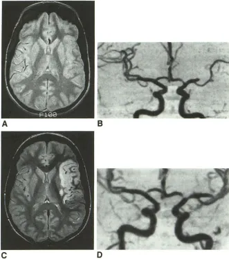
55% of cases. Although vascular stenosis or occlusion may often be inferred based on the MR exam alone (Fig 3A), vascular imaging provides
the location and degree of vascular compromise
and may reveal collateral pathways or unsus
-pected vascular disease at other locations. Stroke neurologists previously sought this information with an invasive procedure, so MRA appeals to those with this prior standard of practice. The
MRA and cross-sectional brain images complete
the diagnostic imaging evaluation of patients who have had strokes, directing the clinician to the specific location (or absence) of vascular abnor-malities. As thrombolytic regimens and other early intervention protocols mature, such timely information will be even more important. In ad-dition, serial examinations provide a noninvasive means for following the evolution of vascular compromise (Fig 3).
Correlation with Conventional Angiography
We compared our MRA interpretations against the standard of conventional angiography to ver-ify the accuracy of the MRA readings. The high correlation between MRA and conventional an
-giography (87%) in our series (Table 2) is in agreement with previous results of Heiserman et at (31 ). A stronger correlation was demonstrated with normal and occluded vessels than with sten-otic arteries, which is also in agreement with prior series. Although intracranial MRA may not cor
-relate with angiography as well for stenotic lesions as for complete vascular occlusions, this short-coming is mitigated by the fact that distal vessel disease in patients studied angiographically for cerebrovascular disease tends to be occlusive
rather than stenotic ( 16).
Two vessels in our series were read as occluded on MRA and as patent with conventional angiog-raphy. In the first case, a patient with a left posterior cerebral artery distribution infarct had a nonvisualized posterior cerebral artery on MRA.
The conventional angiogram performed 6 days later showed a patent vessel. It is presumed that this vessel recanalized in the interim, given the large caliber of this vessel on the subsequent study. The second case in which MRA suggested vascular occlusion and the conventional angio-gram revealed a patent vessel was also a left posterior cerebral artery occlusion on the MRA. In this case the conventional angiogram was per -formed more than 1 week after the MRA. This
-906 JOHNSON
Fig. 3. A, Intermediate-weighted image
obtained 6 hours after the onset of speech difficulties and right-sided weakness shows
subtle swelling of the left insular cortex.
There is also absence of flow void within the
opercular branches of the middle cerebral artery, implying slow or no flow.
8, Coronal projection MRA image
ob-tained at the same time demonstrates
occlu-sion of a proximal middle cerebral artery
branch with a paucity of distal vessel filling.
C, Intermediate-weighted image 3 days later shows a large high-signal-intensity in-farct in the left middle cerebral artery terri-tory.
D, MRA demonstrates partial recanaliza-tion of the occluded vessel with increased perfusion of multiple distal branches. The A distribution of infarction is similar to that
seen in Figure 2, despite significant
differ-ences in the distribution of vascular
compro-mise.
c
zation, although technical limitations of MRA must be considered as well. Turbulent dephasing or very slow flow within a patent vessel can cause signal loss. Ideally, MRA/conventional angiogram comparisons should be made in studies per-formed within 24 hours of each other to limit discrepancies caused by the natural history of thrombosis.
Umitations of MRA
Some MRA pitfalls occur because this method is not simply an anatomic exam. Problems with
focal signal loss caused by complex flow
phenom-enon initially jaded reader confidence (34); how-ever, limiting the echo delay time to minimize phase shifts and turbulent dephasing, which con-tribute to intravascular signal loss, has produced significant improvements (31 ).
A second problem encountered with 3-D
time-of-flight MRA occurs as a result of saturation
effects at the margins of the volume or slab (35).
Using thin overlapping slabs, the degree of spin
AJNR: 15, May 1994
8
D
saturation is significantly reduced (36). Overlap-ping the slabs allows the peripheral partitions within the slab to be discarded because the cor-responding anatomic information is available from the adjacent slab. In the current study, the MRA exams obtained with overlapping slabs showed a higher correlation with the distribution of infarction (83%) than did the other exams (60%).
A third problem encountered in MR is limitation of spatial resolution. Warach et al showed occlu -sive or severe stenotic lesions of major vessels in the distribution of cerebral infarction in 16 of 24 patients but found MR unreliable in infarcts smaller than 2 em (33). In our series infarct~
smaller than 2 em also showed a lower incidencE of correlative MRA findings (64%) than did in -farcts 2 em and larger (78%). The lower correla -tion with smaller infarcts may result from limita -tions in spatial resolution, preventing visualizatior of occluded distal branch vessels with small di
[image:6.612.227.558.81.455.2]A fourth potential problem with MRA results from the projection algorithm used to reconstruct
vascular anatomic images from the source data.
Low-intensity features of vessels may be lost on
maximum intensity pixel images, contributing to
artifactual narrowing of vessel caliber (37). MRA pulse sequences that maximize flow signal and minimize background signal provide the best
compensation for such artifacts. Further
refine-ments in MRA techniques will address the limited
spatial resolution, saturation effects from
recir-culation, and signal loss from higher-order motion
(38, 39). Such improvements will further improve
the utility of MRA in the evaluation of cerebro
-vascular disease.
Limitations of Study
A variable was introduced into this trial as a
result of evolving MRA techniques. A marginally
significant difference between results with
MOTSA studies and those that antedated them was noted. Because the MOTSA studies showed a higher correlation between MRA abnormalities
and the distribution of infarction, inclusion of the
older studies likely results in an underestimation of the utility of MRA for the evaluation of stroke. This trial was not randomized to determine
whether the information obtained from the MRA
studies contributed to or affected patient man-agement. Our goal was not outcome analysis, although such studies are warranted to evaluate
further the clinical utility of this important
tech-nology.
It is also important to be aware that although we compared MRA with conventional
angiogra-phy when the latter was available, a limited
num-ber of these comparative cases were available.
The purpose of this study was not to compare
the two modalities, but rather to determine the incidence of positive MRA studies in individuals with acute and subacute infarction.
Summary
Vascular lesions demonstrated on intracranial
MRA show a high correlation with infarct
distri-bution. In a majority of cases, this noninvasive
modality provides information that cannot be
acquired from cross-sectional MR alone. As early
intervention for stroke therapy continues to
evolve, the expeditious and complete evaluation
of such patients becomes even more critical.
Combining MR and MRA provides a more
com-prehensive means of evaluating intravascular dis-ease. MRA could be used to screen potential candidates for thrombolytic therapy or surgical
intervention, and to provide information that
helps prescribe and monitor less invasive therapy.
Although it may not currently serve as a
replace-ment for conventional angiography, we conclude
that MRA is useful for selecting patients who
warrant the more invasive examination and for
screening those who do not.
References
1. Bryan RN. Imaging of acute stroke. Radiology 1990;177:615-616 2. Wilson M. Angiography in cerebrovascular occlusive disease. Am J
Med Sci 1965;5:554-576
3. Hachinski V, Norris JW. In Plum F, Baringer JR, Gilman S, eds. The acute stroke. Philadelphia: Davis, 1985:286
4. Pike GB, Hu BS, Glover GH, Enzmann Dr. Magnetization transfer time-of-flight magnetic resonance angiography. Magn Reson Med
1992;25:372-379
5. Parker DL, Yuan C, Blatter DD. MR angiography by multiple thin slab
3D acquisition. Magn Reson Med 1991;17:434-451
6. Snedcor GW, Cochran WG. Statistical methods. 8th ed. Ames: Iowa State University Press, 1989:124-127
7. Masaryk T J. Ischemic brain disease, vascular brain disease, and
magnetic resonance angiography. Curr Opin Radioll992;4:79-88
8. Elster AD, Moody DM. Early cerebral infarction: gadopentetate di-meglumine enhancement. Radiology 1990;177:627-632
9. Yuh WTC, Crain MR, Loes DJ, et al. MR imaging of cerebral ischemia:
Findings in the first 24 hours. AJNR Am J Neuroradioll991; 12:621-629
10. Accession J, Boyd WN, Hugh AE, Hutchinson EC. Cerebral angi
og-raphy in ischemic cerebrovascular disease. Arch Neurol1969;
20:527-532
11. Kobayashi N, Saito Y. Infarction and circulation in cerebrum: effect
of recanalization and/or collateral circulation on the lesion and prog-nosis. Neuroradiology 1978; 16:108-112
12. Wolpert SM, Caplan LR. Current role of cerebral angiography in the diagnosis of cerebrovascular diseases. AJR Am J Roentgenol
1992;159:191-197
13. Caplan LR, Rosenbaum AE. Role of cerebral angiography in
vertebra-basilar occlusive disease. J Neural Neurosurg Psychiatry
1975;38:601-612
14. Pessin MS, Hinton RC, Davis KR, et al. Mechanisms of acute carotid stroke. Ann Neural 1979;6:245-252
15. Kohlmeyer K, Graser C. Comparative study of computed tomography (CT) and carotid angiography (CAG) in stroke patients. Neuroradiol -ogy 1978;16:162-163
16. Goldenberg G, Reisner TH. Angiographic findings in relation to clinical
course and results of computed tomography in cerebrovascular
disease. Eur Neural 1983;22: 124-130
17. Bozzao L, Angeloni U, Bastianello S, et al. Early angiographic and CT
findings in patients with hemorrhagic infarction in the distribution of
the middle cerebral artery. AJNR Am J Neuroradiol1991;
12:1115-1121
18. Fisher M, Sotak CH, Minematsu K, Li L. New magnetic resonance
techniques for evaluating cerebrovascular disease. Ann Neural
1992;32: 1 15-122
908 JOHNSON
20. Alter M, Kieffer S, Resch J, Ansari K. Cerebral infarction: clinical and angiographic correlations. Neurology 1972;22:590-602
21. Dion JE, Gates PC, Fox AJ, et al. Clinical events following neuroan-giography: a prospective study. Stroke 1987;18:997-1004
22. Grzyska U, Freitag J, Zeumer H. Selective cerebral intraarterial DSA. Complication rate and control of risk factors. Neuroradiology
1990;32:296-299
23. Hankey GJ, Warlow CP, Sellar RJ. Cerebral angiographic risk in mild cerebrovascular disease. Stroke 1990;21 :209-222
24. Davis DO, Pressman BD. Angiography of cerebrovascular disease. In Wilkins RH, ed. Clinical neurosurgery. Baltimore: Williams & Wilkins, 1975:163-184
25. Masaryk T J, Modic MT, Ruggieri PM, et al. Three-dimensional (vol-ume) gradient-echo imaging of the carotid bifurcation: preliminary clinical experience. Radiology 1989;171:801-806
26. Litt AW, Eidelman EM, Pinto RS, et al. Diagnosis of carotid artery stenosis: comparison of 20FT time-of-flight MR angiography with contrast angiography in 50 patients. AJNR Am J Neuroradio/
1991 ;12:149-154
27. Masaryk AM, Ross JS, DiCello MC et al. 30FT MR angiography of the carotid bifurcation: potential and limitations as a screening ex-amination. Radiology 1991; 179:797-804
28. Heiserman JE, Drayer BP, Fram EK, et al. Carotid artery stenosis:
clinical efficacy of two dimensional time of flight MR angiography. Radiology 1992;182:761-768
29. Sevick RJ, Tsuruda JS, Schmalbrock P. Three-dimensional time of flight MR angiography in the evaluation of cerebral aneurysms. J
Comput Assist Tomogr 1990;14:874-881
AJNR: 15, May 1994
30. Ross JS, Masaryk T J, Modic MT, Ruggieri PM, Haacke EM, Selman WR. Intracranial aneurysms: evaluation with MR angiography. AJNR Am J Neuroradio/ 1990; 11 :449-456
31. Heiserman JE, Drayer BP, Keller PJ, Fram EK. Intracranial vascular stenosis and occlusion: evaluation with three-dimensional time-of-flight MR angiography. Radiology 1992; 185:667-673
32. Ruggieri PM, Masaryk T J, Ross JS, Modic MT. Magnetic resonance angiography of the intracranial vasculature. Top Magn Reson Imaging
1991 ;3(3):23-33
33. Warach S, Wei L, Ronthal M, Edelman RR. Acute cerebral ischemia: evaluation with dynamic contrast-enhanced MR imaging and MR angiography. Radiology 1992;182:41-47
34. Masaryk T J, Modic MT, Ross JS, et al. Intracranial circulation: preliminary clinical results with three-dimensional (volume) MR an-giography. Radiology 1989;171:793-799
35. Keller PJ. Time of flight magnetic resonance angiography. Radio/ C/in North Am 1992;2:639-656
36. Blatter DD, Parker DL, Robison RO. Cerebral MR angiography with multiple overlapping thin slab acquisition: part I. Quantitative analysis of vessel visibility. Radiology 1991 ; 179:805-811
37. Anderson CM, Saloner D, Tsuruda JS, Shapeero LG, Lee RE. Artifacts in maximum-intensity-projection display of MR angiograms. AJR Am
J Roentgeno/1990; 154:623-629
38. Edelman RR. Basic principles of magnetic resonance angiography. Cardiovasc lntervent Radio/ 1992; 15:3-13