Progressive Posttraumatic Myelomalacic Myelopathy: Imaging and
Clinical Features
Steven Falcone, Robert M. Quencer, Barth A. Green, Sherri J. Patchen, and M. Judith Donovan Post
PURPOSE: To describe the imaging features, surgical management, and clinical outcome of progressive posttraumatic myelomalacic myelopathy (PPMM), a relatively unrecognized but im-portant cause of progressive myelopathy in patients with previous spinal cord injuries. METHODS: The clinical records, imaging studies, and postoperative outcome of 10 patients with PPMM were reviewed. Fifteen preoperative and five postoperative MRs were analyzed for intramedullary signal abnormalities, the nature of these signal abnormalities, and cord tethering. All patients had intraoperative sonography. RESULTS: Neurologic signs and symptoms found in our patients included 1) progressive loss of motor function (6/1 0), 2) sensory level changes (4/1 0), 3) increased spasticity (4/10), 4) autonomic dysreflexia (4/10), 5) loss of bowel or bladder control (4/10), and 6) local and/or radicular pain (4/1 0). Preoperative MR in nine patients revealed intramedullary T1 / T2 lengthening (9/9), extramedullary tethering/adhesions (9/9), ill-defined lesional borders (6/9),
cord expansion (5/9), and increased signal intensity of the lesion on T1-weighted images compared with CSF (7 /9). Proton density images in five patients demonstrated a relative increase in signal intensity over CSF. In all five postoperative MRs, there was evidence of untethering of the spinal cord and a decrease in cord size in two patients. Intraoperative sonography revealed cord tethering and abnormal cord echotexture in all cases. Postoperative clinical evaluation revealed neurologic improvement in nine patients. CONCLUSIONS: PPMM may clinically and radiographically mimic progressive posttraumatic cystic myelopathy (PPCM). MR provides clues to the diagnosis of myelomalacia preoperatively. Intraoperative sonography confirms the absence of a confluent cyst. These points are crucial in the surgical procedures in PPMM vs PPCM. In PPMM, lysis of intradural adhesions results in an improvement in symptoms in a manner similar to the shunting of PPCM.
Index terms: Spinal cord, myelopathy; Spinal cord, injuries; Spinal cord, magnetic resonance; Spinal cord, ultrasound
AJNR Am J Neuroradio/15:747-754, Apr 1994
Traditional views state that all posttraumatic myelopathies are related to cord cysts (1-3). This idea is reflected in the nomenclature ( posttrau-matic syringomyelia, ascending cystic degenera-tion of the cord, progressive posttraumatic cystic myelopathy; and posttraumatic cystic myelopa-thy) which has attempted to describe the etiology
Received January 12, 1993; accepted pending revision March 23; revision received June 29.
From the Departments of Radiology (S.F., R.M.Q., M.J.D.P.) and Neurosurgery (B.A.G., S.J.P.), University of Miami/Jackson Memorial Medical Center.
Address reprint requests to Dr Steven Falcone, Department of Radiol-ogy (R109), University of Miami, 1611 NW 12th Ave, Miami, FL 33136.
Presented at the 30th Annual Meeting of the ASNR, May 31-June 5, 1992, St. Louis, Missouri.
AJNR 15:747-754, Apr 1994 0195-6108/94/0004-0747 © American Society of Neuroradiology
747
of the problem. However, there are other causes
of posttraumatic myelopathies, including arach-noiditis (4-6), cord compression, secondary to spinal instability (7) or bone fragment narrowing the canal (4), cord tethering (4, 8), loculated subarachnoid cysts with resultant cord compres-sion (4), cord atrophy (4), and microcystic spinal cord degeneration/gliosis (9, 10).
The syndrome of posttraumatic myelomalacic myelopathy (PPMM) encompasses a continuum
of interrelated disease processes and may precede the formation of a confluent cyst (11). The purpose therefore of this study is to describe the imaging features and clinical outcome of PPMM not only because this entity must be separated diagnostic -ally from progressive posttraumatic cystic
748 FALCONE
Materials and Methods
The clinical records, imaging studies, and postoperative outcome of 10 patients with PPMM were reviewed. In this retrospective study, patients were included based on the following criteria: 1) there was previous severe spinal cord injury (ie, permanent neurologic deficit referrable to the cord), 2) there was clinical evidence of a progressive my-elopathy, which adversely affected the patients' quality of life, 3) there was operative intervention for this progressive myelopathy, 4) there was absence of a confluent, shuntable intramedullary spinal cord cyst as demonstrated by intra-operative sonography, and 5) there was no coexistent process that could explain the patients' symptoms (ie, cord compression).
There were 1 0 patients who had a posttraumatic pro-gressive myelopathy who met all the above criteria. Six men and four women between the ages of 21 and 63 were studied. All patients were incomplete quadraplegics or paraplegics with an initial level of injury as shown in Table 1.
Nine patients had preoperative magnetic resonance (MR). Five of these patients also had a postoperative MR. Noncontrast MR scans were obtained using either a 1.0-T or 1.5-T superconducting magnet. Spin-echo images were acquired in the following manner: T1-weighted sagittal
AJNR: 15, April1994
(600-700/20/2 [repetition time/echo time/excitations]); T1-weighted axial (700-850/20/2); and dual-echo sagittal (1500-3000/20-80). T2*-weighted sequences were per-formed in sagittal (600/18°/4) or axial (1100/18°/2) plane.
In one patient, a computed tomographic (CT) myelogram was obtained before surgery. All patients had intraoperative sonography (a prerequisite for inclusion in this study).
The imaging features evaluated on preoperative MR include the presence of hypointense or hyperintense intra-medullary signal abnormalities, the well-defined or poorly defined margins of these intramedullary lesions, the pres-ence of cord expansion at the site of the lesion, and the presence or absence of cord tethering.
Intraoperative sonography was performed after a lami-nectomy with a 7 .5-MHz transducer. Axial and sagittal scanning was performed. We determined whether the in-tramedullary signal abnormalities as seen on MR were microcystic, hyperechoic, or hypoechoic relative to normal cord tissue. We also evaluated for the presence of cord tethering and the presence of extramedullary cysts. As mentioned previously, at the time of operation, there was absence of a confluent intramedullary cyst as demonstrated by intraoperative sonography in all 10 patients.
With intraoperative sonography more definitely exclud-ing the presence of a shuntable intramedullary cyst, the
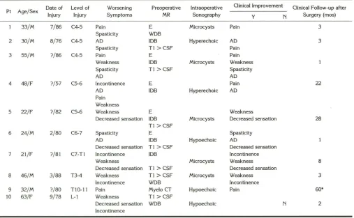
TABLE 1: Features of progressive posttraumatic myelomalacic myelopathy
Age/Sex Date of Level of Worsening Preoperative Intraoperative
Clinical Improvement Clinical Follow-up after Pt
Injury Injury Symptoms MR Sonography y N Surgery (mos)
33/M 7/86 C4-5 Pain E Microcysts Pain 3
Spasticity WDB
2 30/M 8/76 C4-5 AD IDB Hyperechoic AD 3
Spasticity T1 > CSF Pain
3 55/M ?/86 C4-5 Pain E Pain
Weakness IDB Microcysts Weakness
Spasticity T1 > CSF Spasticity
AD AD
4 48/F ?/57 C5-6 Incontinence E Pain 22
AD IDB Hyperechoic AD
Pain Weakness
5 22/F ?/82 C5-6 Weakness E Weakness
Decreased sensation IDB Microcysts Decreased sensation 28
T1 > CSF
6 24/M 2/80 C6-7 Spasticity E Spasticity
AD IDB Hypoechoic AD
Decreased sensation T1 > CSF Decreased sensation
7 21/F ?/81 C7-T1 Incontinence IDB Incontinence
Weakness Microcysts Weakness 8
Decreased sensation T1 > CSF Decreased sensation
8 46/M 3/88 T3-4 Weakness T1 > CSF Microcysts Weakness 3
Incontinence WDB Incontinence
9 32/M ?/80 T10-11 Pain Myelo CT Hypoechoic Pain 60• 10 63/F 9/78 L-1 Weakness T1 > CSF
Decreased sensation WDB Hypoechoic N 2
Incontinence
Note.- E indicates expansion of cord; microcysts, cysts < 5 mm; AD, autonomic dysreflexia; WDB, well-defined borders; IDB, ill-defined borders;
and T1 > CSF, signal intensity of lesion greater than cerebrospinal fluid on T1-weighted image.
[image:2.612.53.560.393.706.2]AJNR: 15,Ap~ 1994 MYELOPATHY 749
A
8
c
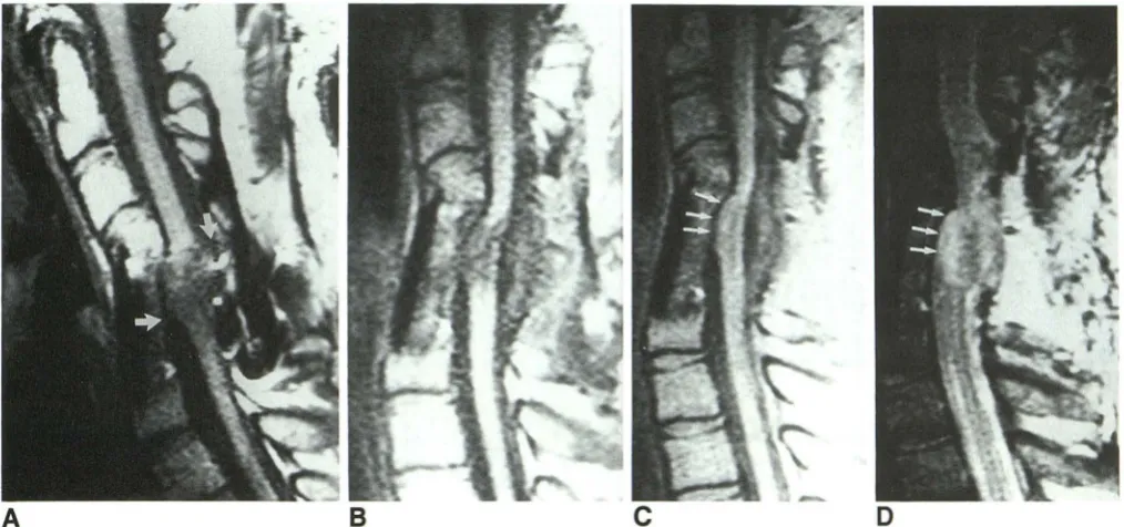
DFig. 1. Case 3. A, Preoperative T1-weighted (600/20) midline sagittal images demonstrates abnormal signal and expansion of cervical
cord at C4-5. Although there is decreased signal intensity of the cord it is not as low as cerebrospinal fluid and the lesional borders are
ill defined. Note evidence of previous anterior and posterior fusion. At surgery a confluent cyst was not demonstrated. There is anterior and posterior tethering with adhesions (arrows). B, Postoperative T1-weighted (600/20) sagittal; C, proton density-weighted (1783/20) sagittal; D, T2-weighted (1783/80) sagittal images. After posterior untethering of the cord the cord approaches a more normal size at C4-5 with residual anterior tethering and reestablishment of a dorsal subarachnoid space. Majority of residual signal abnormality of cord does not follow CSF signal intensity as best demonstrated on proton density image (arrows).
cord was untethered with meticulous microdissection of intradural adhesions. An expansile duraplasty was then performed in eight of 1 0 cases using a dural allograft allowing for a widened subarachnoid space and lessening the opportunity for retethering of the cord to occur.
Results
All patients presented with the clinical signs and symptoms of a progressive posttraumatic myelopathy. Their presentation was similar to those patients who have the classic cystic my-elopathy and included: increasing motor loss or weakness in six patients, sensory changes in four patients, increased spasticity in four patients, autonomic dysreflexia in four cases, worsening bowel and/ or bladder function in four cases, and local of radicular pain in four patients (Table 1).
After surgery (described above) nine of ten patients showed improvement in one or more of their presenting symptoms (Table 1). One patient did not improve after surgery.
In our cases, preoperative MR showed intra-medullary spinal cord lesions which demonstrated T1, T2, and T2* lengthening relative to the nor-mal spinal cord signal. Intramedullary T1
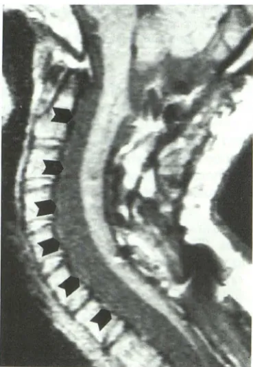
abnor-malities however, were not as low as cerebrospi-nal fluid (CSF) in seven cases (Figs 1 and 2). Proton density images were obtained in five cases, and those images revealed intramedullary signal abnormalities which did not follow CSF (Fig 1 ). Characterization of lesion margins re-vealed irregular /ill-defined margins in five cases (Fig 1 ). The cord was focally expanded in the region of intramedullary signal abnormality in four cases (Figs 1 and 2). Evidence of cord tethering was seen in all cases (Figs 1, 2, and 3). In the one patient who had a CT myelogram before surgery, some uptake of the contrast was seen centrally in the cord on delayed CT.
Intraoperative sonography showed three basic patterns of abnormal cord echogenicity. In three patients, the intramedullary spinal cord lesions were hypoechoic to the normal spinal cord. Two patients showed intramedullary abnormalities which were hyperechoic to normal cord (Fig 4).
In four patients, a heterogenous pattern was seen with small (<5 mm) microcysts occupying the
cord (Fig 2). In all cases, the normal central echo
was interrupted at the site of the cord lesion.
At surgery, severe intradural scar formation
[image:3.613.54.561.78.316.2]750 FALCONE AJNR: 15, April 1994
Fig. 2. Case 5. A, Tl-weighted (600/20) midline sagittal image. Focal cord expansion with intramedullary abnormalities which have greater signal intensity than CSF and ill-defined and irregular borders typical of PPMM. Note evidence of prior anterior cervical fusion. B, Intraoperative spinal sonography via laminectomy defect. Transverse image demonstrates focal cord expansion with dorsal tethering and microcysts (arrows). C, Tl-weighted (500/20) midline sagittal image. Eleven months after cord untethering, recreation of the dorsal subarachnoid space is seen. Arrows point to dural allograft. C
Fig. 3. Case 7. Tl-weighted (600/20) midline sagittal. An example of dorsal tethering of the spinal cord with lack of a posterior subarachnoid space and large expanded ventral su b-arachnoid space (arrows). Note evidence of previous lamin ec-tomy.
scar had a white fibrous appearance. The dura
was "stuck" to the cord with obliteration of the
subarachnoid space.
Discussion
The syndrome of progressive posttraumatic myelopathy has been reported to occur in 0.3%
to 3.2% of chronically injured spinal cord patients from 2 months to 36 years after injury (12). It was once believed that this progressive posttrau-matic myelopathy was by definition related to the formation of an intramedullary spinal cord cyst (1, 2). However, we have found that patients may have a progressive posttraumatic myelopathy in the absence of a definable confluent intramedul-lary spinal cord cyst.
[image:4.615.50.561.76.282.2] [image:4.615.82.267.355.623.2]sonog-AJNR: 15, April1994
A
B
c
Draphy may be the only means whereby this dis-tinction is definitive.
Fox et al (10) had one patient with progressive posttraumatic myelopathy in whom only intense gliosis was found at surgery. This patient's MR revealed an area of T 1 and T2 prolongation within the cord which did not follow CSF signal intensity on proton density images. Surgical findings re-ported by MacDonald et al (9) revealed what they called a "microcystic myelopathy" with arach-noiditis in two patients with posttraumatic my-elopathy. Both of these patients had preoperative MR scans which were interpreted as being
com-patible with a syrinx. Stevens et al (14) had five
cases of posttraumatic myelopathy which showed an atrophic cord in four of five patients, a gelatinous cord in two cases, and axonal frag-mentation and demyelination in one at the time of surgical exploration. MR was not performed in
these patients. Quencer et al (11, 16) in two
separate reports noted nonshuntable intramedul-lary cysts because the cysts were either too small
(<5 mm; ie, microcysts) (two patients),
nondefin-able (two patients) by intraoperative sonography,
or collapsed after myelotomy (one patient).
Al-though preoperative MR was not performed in
these cases, the preoperative metrazamide CT
was erroneously diagnosed as showing a shunta-ble intramedullary cyst in all patients. Similarly, Gebarski et al (15) in their series of nine patients
with progressive posttraumatic myelopathy had
MYELOPATHY 751
Fig. 4. Case 2. A, Sagittal. B, Axial. In-traoperative spinal sonography via laminec-tomy defect. The cord is tethered and
"'stuck" against a thickened dura (arrow -heads). There is abnormal increased echo
-genicity of the cord with lack of a normal central echo. The ventral subarachnoid
space is enlarged (short arrows). C, Sagittal. D, Axial (after untethering). After careful
untethering of the cord and removal of thick-ened dura the cord assumes a more normal
position in the spinal canal with reduction in size of ventral subarachnoid space (long a
r-rows). Signal abnormality in cord persists.
Note dural edge (short arrows).
one patient with no confluent shuntable cyst but small (<5 mm) intramedullary microcysts. In this case MR correctly classified the intramedullary signal abnormality as being of the small cystic type; metrazimide CT suggested the presence of
a large confluent cyst. In the largest series of
cases, Osborne et al (4) reported on 14 patients who had a progressive posttraumatic myelopathy but no intramedullary cyst as seen at surgery. Tethering and/or arachnoiditis was described in 13 of these patients, cord compression from bone/metal or subarachnoid cysts in three cases, and a small noncystic "cord cavity" in one patient.
MR was not obtained in these patients. These
cases, along with the cases in this series, lend credence to the observations that posttraumatic progressive myelopathy need not be the result of an expanding intramedullary spinal cord cyst.
In order to describe more accurately patients
with the clinical syndrome of a progressive post
-traumatic myelopathy in whom there is absence of a confluent shuntable intramedullary spinal cord cyst, or a compressive lesion of the spinal
cord, the term progressive posttraumatic
mye-lomalacic myelopathy is most apt. The term
myelomalacia is derived from the Greek myelo
and malakia and simply means "softening of the
spinal cord" (17); however, we feel that pathologic
correlates may exist for this term. Preliminary
observations at our institution have shown that
[image:5.617.56.384.72.327.2]post-752 FALCONE
traumatic myelomalacic cord show a reactive
astrocytosis, microcysts, and thickening of the
pia-arachnoid (Fig 5).
As mentioned by many authors (2, 9, 10, 12, 15, 16, 18, 19), a myelomalacic or microcystic cord may be the precursor to a confluent intra-medullary spinal cord cyst; the concept that these
pathologic changes may be associated with a
progressive myelopathy should be emphasized. The fact that gliosis is commonly associated with syringomyelia (20), in particular about the periph-ery of the cyst, may be evidence that myelomala-cia precedes the development of a cyst. If the myelomalacia/ gliosis were initially present, one could then imagine how a residual peripheral rim of gliosis may exist as a central confluent cyst develops. Sherman et at (20) found by MR in one
case several small "cysts" which eventually
coa-lesced to form a confluent cyst. This case gives
us indirect evidence by imaging that microcystic
changes evolve into confluent cysts.
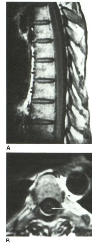
As suggested in the literature, if an area of intramedullary signal abnormality is small, a cyst may be mistaken for a small area of myelomala-cia exhibiting a well-circumscribed low-signal area on T1-weighted images within a normal sized
or focally expanded cord (21). Well-
defined/well-circumscribed margins were seen in three of our cases, a feature usually associated with a cyst (Fig 6). The presence of ill-defined margins, how-ever, would favor myelomalacia over a confluent cyst. Perhaps the best distinguishing feature of a cyst versus myelomalacia on MR imaging is
in-creased intramedullary signal intensity on proton
Fig. 5. Histopathologic transverse
sec-tion through a posttraumatic myelomalacic
cord at the C-4 level in a cadaver. This trichrome strain demonstrates an area of abnormal red staining which represents an astrocytosis (long arrow). In the center of
the cord there is a loose matrix of blood
vessels and astrocytes. Dorsally is a small
microcyst (arrowhead). The pia-arachnoid is
abnormally thickened by blue staining con-nective tissue (short arrow).
AJNR: 15, April 1994
density images (21). This characteristic was seen
in all five of our patients in whom a "dual echo"
sequence was obtained. However, one can antic-ipate some pitfalls in using this criterion. If a posttraumatic intramedullary spinal cord cyst had a high protein content, it might not show a signal
equivalent to CSF on the first echo of a dual
-echo sequence. In this situation, myelomalacia would be falsely diagnosed. Similarly, the signal
intensity on the T1-weighted image would likely
be greater than CSF. In general, however, if in these patients an intramedullary lesion demon-strating irregular ill-defined margins is seen and does not follow CSF signal intensity, it is most likely an area of myelomalacia rather than a cyst. CT myelopathy with a water-soluble contrast agent has been used in the past to distinguish myelomalacia from an intramedullary cyst. In particular, delayed CT approximately 4 hours after intrathecal instillation of contrast is recom-mended (11, 19). This time interval allows for the diffusion of contrast into the cyst and therefore demonstration of the cyst by CT. However, as seen in one of our patients who had a delayed CT myelogram, false-positive results can occur. False-positive results for the detection of an in-tramedullary spinal cord cyst by CT myelography
have been reported ( 11, 16).
Short of myelotomy, intraoperative
sonogra-phy is the standard of reference in the
documen-tation of a spinal cord cyst. As in other anatomic
locations, a lesion that is anechoic and
preopera-AJNR: 15, Apri11994
tive MR or CT myelography may not be able to distinguish between an intramedullary cyst and myelomalacia in some cases, intraoperative so
-nography becomes a crucial tool in the diagnosis and management of these patients.
In our experience, a predominant feature which is often seen in both posttraumatic cystic and noncystic myelopathy is associated tethering of the spinal cord. We feel that this tethering and scar formation of the spinal cord plays an impor-tant role in the pathophysiology of a myeloma-lacic cord. Although purely speculative, this scar-ring and tethescar-ring along with local chemical changes, recumbent position of the patient, chronic ischemia, and changes in local CSF dy-namics are likely interrelated factors contributing to changes in the cord substance. Our series of cases demonstrates that in patients with PPMM,
surgical lysis of adhesions and untethering of the spinal cord will lead to clinical improvement in the majority of patients, as in nine of our 10
cases.
In fact, at our institution, if symptoms warrant,
surgery may be performed on the basis of the imaging studies, even if a cyst is not suspected. In the presence of myelomalacia, untethering of spinal cord and nerve roots can be performed which may lead to clinical improvement.
We did realize, however, that performing sur-gery on these patients simply for the lysis of adhesions is controversial and not universally accepted. The main criticism for this type of surgery is that after surgery there may even be worse arachnoid scarring. The problem of post-operative scarring is addressed by the placement of a dural allograft after lysis of adhesions to try to recreate a subarachnoid space and by encour-aging early postoperative mobilization of the pa-tient to try to prevent adhesion formation which may occur because of the dorsal location of the
cord in the recumbent position. Although no definite conclusion can be made regarding the
success of surgery because of the small patient population and short follow-up periods, initial results are very encouraging.
At surgery, careful and meticulous microdis-section of scar tissue from the cord and nerve
roots is performed. If there is residual scar tissue
which is stuck to the cord, overzealous dissection is not advised because mechanical damage to the
spinal cord and ischemic changes secondary to
stripping of normal vessels can occur. If a plane
cannot be achieved between the cord and the
pia-arachnoid, further removal of scar tissue is
MYELOPATHY 753
A
B
Fig. 6. Case 8. Initial injury at T3-4 level. A, Tl-weighted (700/
20) midline sagittal of lower thoracic cord. There is abnormal
decreased signal intensity of the cord which is slightly greater in
intensity than surrounding CSF. Note well-defined tapering inferior
border. B, Tl-weighted (700/20) axial section. A hypointensity of
the cord is again seen which is fairly well marginated. There is a septation (arrow). Although a confluent cyst was not found with intraoperative sonography, the MR findings could be interpreted as being compatible with a cyst.
not performed. After the cord is untethered and released freely into the subarachnoid space, a dural allograft is sewn in place. This expansile
[image:7.615.334.526.71.574.2]754 FALCONE
space and helps prevent retetethering of the spinal cord.
Although this initial report appears encourag-ing for the successful management of patients with PPMM, this is a small series of patients. In addition, further study is needed to determine whether there are long-term benefits to surgery. The importance of this investigation, however, is that it serves to emphasize a new concept in the management of a certain subset of chronic spinal
cord injury patients. Radiographically, we can
assist not only in differentiating cystic from non -cystic myelopathy, but in showing the extent and level of scarring and cord tethering.
In conclusion, PPMM is a relatively unrecog-nized syndrome which clinically simulates post-traumatic cystic myelopathy. Although there are certain MR characteristics that may suggest the presence of myelomalacia, it may be difficult to distinguish a confluent intramedullary cyst from myelomalacia. Therefore, intraoperative sonog -raphy becomes a crucial imaging tool in distin -guishing between myelomalacia and a confluent intramedullary cyst in patients with posttraumatic progressive myelopathy. This is now of great importance because the type of surgery to be performed is different in the presence of myelo -malacia. In patients with PPMM, lysis of
surround-ing spinal cord adhesions and untethering of the spinal cord resulted in clinical improvement in the majority of our patients.
References
I. Barnett HJM, Jousse AT. Syringomyelia as a late sequel to traumatic paraplegia and quadraplegia. In: Walton JN, ed. Major problems in neurology. London: Saunders, 1973: 129-153
2. Barnett HJM, Jousse AT, Ball MJ. Pathology and pathogenesis of progressive cystic myelopathy as a late sequel to spinal cord injury. In: Walton JN, ed. Major problems in neurology. London: Saunders, 1973:179-219
3. Barnett HJM, Jousse AT. Nature, prognosis and management of post-traumatic syringomyelia. In: Walton JN, ed. Major problems in ne u-rology. London: Saunders, 1973:154-164
AJNR: 15, April 1994
4. Osborne DRS, Vavoulis G, Nashold BS, Dubois PJ, Drayer BP, Heinz RE. Late sequelae of spinal cord trauma: myelographic and surgical correlation. J Neurosurg 1982;57:18-23
5. Donaldson I, Gibson R. Spinal cord atrophy associated with arach-noiditis as demonstrated by computed tomography. Neuroradiology 1982;24: 101-105
6. Shaw MDM, Russell JA, Grossert KW. The changing pattern of spinal arachnoiditis. J Neural Neurosurg Psychiatry 1978;41 :97 -I 07 7. Schneider RC, Knighton R. Chronic neurologic sequelae of acute
trauma to the spine and spinal cord. Part Ill. The syndrome of chronic injury to the cervical spinal cord in the region of the central canal. J Bone Joint Surg (Am) 1959;905-919
8. Ragnarsson TS, Durward QJ, Nordgren RE. Spinal cord tethering after traumatic paraplegia with late neurological deterioration. J Neurosurg 1986;64:397-401
9. MacDonald RL, Findlay JM, Tator CH. Microcystic spinal cord degen-eration causing post-traumatic myelopathy. Report of two cases. J Neurosurg 1988;68:466-471
10. Fox JL, Wener L, Drennan DC, Manz HJ, Won DJ, AI-Mefty 0. Central spinal cord injury: magnetic resonance imaging confirmation and operative considerations. Neurosurgery 1988;22:340-347 11. Quencer RM, Green BA, Eismont FJ. Post-traumatic spinal cord
cysts: Clinical features and characterization with metrizamide com-puted tomography. Radiology 1983; 146:415-423
12. Umbach I, Heilporn A. Review article: Post-spinal cord injury syrin-gomyelia. Paraplegia 1991;29:219-221
13. Rossier AB, Foo D, Shillito J, Dyro FM. Post-traumatic cervical syringomyelia. Brain 1985; I 08:439-461
14. Stevens JM, Olney JS, Kendall BE. Post-traumatic cystic and non-cystic myelopathy. Neuroradiology 1985;27:48-56
15. Gebarski JS, Maynard FW, Gabrielsen TO, Knake JE, Latack JT, Hoff JT. Post-traumatic progressive myelopathy. Radiology 1985; 1157:379-385
16. Quencer RM, Morse BM, Green BA, Eismont FJ, Brost P. Intraoper-ative spinal sonography: Adjunct to metrizamide CT in the assessment and surgical decompression of post-traumatic spinal cord cysts. AJNR Am J Neuroradiol1984;5:71-79
17. Stedmans Medical Dictionary 24th ed. Baltimore: Williams and Wil-kins, 1982:917
18. Mclean DR, Miller JDR, Allen PBR, Ezzedin SA. Posttraumatic syrin-gomyelia. J Neurosurg 1973;39:485-492
19. Seibert CE, Dreisbach JN, Swanson WB, Edgar RE, Williams P, Hahn H. Progressive post-traumatic cystic myelopathy. AJNR Am J Neu-roradiol1981 ;2:115-119
20. Sherman JL, Barkovich AJ, Citrin CM. The MR appearance of syringomyelia: New observations. AJNR Am J Neuroradiol 1986; 7:985-995