1378
MS
Dear Editor-in-Chief:I wish to congratulate Eric Russell for his con-scientious and thought-provoking commentary on the status of carotid stenting. The technique of ca-rotid stenting is about to begin a new phase of ad-ditional rigorous scrutiny and investigation. The National Institute of Neurologic Disorders and Stroke (NINDS) recently has approved and funded a large multicenter, multinational, randomized, con-trolled trial comparing carotid stenting with carotid endarterectomy. The Carotid Revascularization Endarterectomy vs. Stent Trial (CREST) plans to begin training and credentialing interventionalists this summer, and recruitment is expected to begin late this year or in early 2000. The trial will address the relevant efficacy of carotid stenting and carotid endarterectomy in a North American Symptomatic Carotid Endarterectomy Trial (NASCET)-like pop-ulation of patients with symptomatic high-grade stenoses. We anticipate that 2500 patients will need to be recruited to satisfy the statistical requirements of the study. It is essential that this landmark trial have the full involvement of and commitment from the neuroradiologic community.
This landmark trial is the initiative of a neuro-radiologist, Robert Ferguson, Chairman of the De-partment of Radiology at Queens University Hos-pital in Kingston, Ontario. After his pivotal work in organizing the North American Cerebral Percu-taneous Transluminal Angioplasty Registry (NACPTAR), Dr. Ferguson collaborated with Rob-ert Hobson, Director of Vascular Surgery at the New Jersey Medical School, Newark, New Jersey and the principal investigator of the VA Coopera-tive Trial of Endarterectomy, as well as with me and others, to form the CREST Trial group. Indeed, neuroradiologists have been instrumental in the rapid development and current success of this tech-nique. Beginning with the pivotal and ongoing work of Jacques Theron in Caen, France and the pioneering contributions of Jiri J. Vitek at the Uni-versity of Alabama at Birmingham, the technique has now gained wide acceptance. Eric Russell di-minishes the rigor of the well-planned IRB-ap-proved clinical trials that were begun at the Uni-versity of Alabama at Birmingham in 1994 (1). The results of these well-conducted and critically au-dited prospective registries provided the clinical findings that gained the acceptance of physicians throughout the world and, more importantly, in-dustry support, without which, further device and technique development would have been impossi-ble. Most importantly, study participants were sub-jected to independent neurologic evaluation 24 hours postprocedurally, a standard to which the NASCET has never been held. The statement that no long-term outcome information is available is incorrect. Late results have been reported, and mul-tiple groups now have confirmed the rarity of neu-rologic events after stent intervention (2).
Similar-ly, our experience has been analyzed carefully in terms of risk stratification on the basis of clinical symptoms and morphologic characteristics of le-sions (3). Similarly, a great deal of information has been promulgated concerning the use of coronary wires and balloons to cross and predilate high-grade lesions. Furthermore, cerebral protection dur-ing stent placement is an active area of investiga-tion, and current carotid stent symposia have focussed on a variety of new devices designed to provide cerebral protection. The political, regula-tory, and reimbursement ramifications of this tech-nique remain beyond our control. We consider the definition of carotid stenting as a significant-risk procedure to be quite arbitrary. This definition ig-nores the 6% incidence of death and stroke that occurs with carotid endarterectomy (4). In our most recent experience, we have completed 136 proce-dures with no deaths, no major strokes, and a 1.6% incidence of minor nondisabling strokes. In our in-stitution, the neurologist would like to consider ca-rotid stenting as the standard of care for their pa-tients, but this technique remains incorrectly labeled as high-risk, federally unapproved, and nonreimbursable.
At least one prospective, randomized, controlled trial has been completed comparing carotid angio-plasty (stenting in 1 of 3 of cases), to carotid end-arterectomy. This trial compared the learning curve of the radiologists involved against the mature sults of experienced vascular surgeons in major re-gional centers. The results from CAVATAS have been reported and will be published soon. There was no difference between the incidence of minor or ma-jor neurologic events in this randomized trial. I am confident that the CREST study will validate the results that have emerged from numerous registries throughout the world. The results of CREST, how-ever, will not be available for at least 6 years. In the meantime, the CREST group will be conducting, with the NINDS and the FDA, industry-supported, rigorous, prospective registry studies in the very large population of patients who will never and can never be studied in prospective, randomized trials. Neuroradiologic involvement in carotid stenting has been pivotal in its development, and will continue to be essential if the procedure is to emerge as it should as a safer, more comfortable, and less inva-sive alternative to carotid endarterectomy.
Sincerely, Gary S. Roubin, M.D., Ph.D.
Co-Principal Investigator Intervention CREST Group
References
1. Yadav JS, Roubin GS, Iyer S, Vitek JJ, et al. Elective stenting of extracranial carotid arteries. Circulation 1997;95:376–381 2. Yadav SS, Roubin GS, Iyer SS, King P, Parks JM, Jain SP, Goods
3. Mathur A, Roubin GS, Iyer SS, Piamsonboon C, Liu MW, Gomez CR, Vitek JJ, et al. Predictors of stroke complicating carotid artery stenting. Circulation 1998;97:1239–1245
4. Cebul RD, Snow RJ, Pine R, Hertzer NR, Norris DG. Indications, outcomes and provider volumes for carotid endarterectomy. JAMA 1998;279:1282–1287
Reply
Dr. Roubin’s comments concerning the AJNR commentary on carotid artery balloon angioplasty and stenting (CABAS) are welcome (1). Dr. Roubin is known as a thoughtful and innovative investi-gator, and he is commended for his approach to this evolving technology. He has treated a high-risk pa-tient population, and yet has shown results com-petitive with surgical carotid endarterectomy (CEA). He understands the risks involved. Recently he has been quoted as saying, ‘‘It is important that a realistic and comprehensive classification of the neurologic complications be used when a newly evolving technique such as carotid stenting is eval-uated.’’ He has also asserted that ‘‘independent oversight when the incidence of neurologic com-plications is assessed cannot be overemphasized (2).’’ Unfortunately, these rules are not uniformly adhered to by all performing either CABAS or sur-gical endarterectomy.
His summary of events since the formulation of the previous commentary (1) provides an update of this fast-moving field. Dr. Roubin anticipates that the European CAVATAS trial, comparing angio-plasty (with or without stenting) and CEA, will show no difference between either treatment ap-proach or minor and major neurologic events. One hopes that publication of those results will include sufficient case material regarding stenting and an ad-equate categorization of degree of stenosis to pro-vide guidance. There also will be a need to look carefully at patient data to consider whether this study will be applicable to practice in North America.
Dr. Roubin also suggests that the rigor of the well-planned, IRB-approved clinical trials begun at his former institution, the University of Alabama at Birmingham, was demeaned in the commentary (1). This is not what was written. In fact, the data from the referenced study were outlined in the commen-tary in a factual and favorable manner. That study, which included pre- and postprocedural neurologic evaluation, complete cerebral angiography, and ap-propriate use of neuroimaging follow-up, serves as a model for similar efforts. It is clear that Dr. Roub-in’s multidisciplinary group, which included an in-terventional neuroradiologist, deserves much credit for having the vision and drive to show that CABAS is a competitive technique.
Controversy in the initial phases of development of a new therapy is common and should not dis-suade active pursuit of a potentially valuable tech-nique. Nevertheless, the practice that prompted the commentary is the increasingly widespread pro-motion and performance of CABAS by those less
experienced, less thoughtful, and less competent than Dr. Roubin and his colleagues, before there is scientific proof of its safety and efficacy. It is nat-ural that groups of physicians with appropriate in-dividual or team expertise will continue to inves-tigate this procedure, considering encouraging short- and intermediate-term outcome data already available. We must realize, however, that the fa-vorable data to date comes from a few highly skilled teams. Others who also perform CABAS may not have adequate training and experience, and may not have comparable results. One recent report in the Journal of Vascular Surgery suggests that the learning curve for CABAS may be quite steep in some circumstances, despite internal institutional control of a carefully designed study (3). This re-cent European single-re-center study, meant to be a randomized comparison of CABAS and CEA, was halted when five of seven patients who underwent CABAS had a stroke.
Dr. Roubin also writes that the ‘‘statement that no long-term outcome information is available is incorrect,’’ and he goes on to say that ‘‘late results have been reported and multiple groups now have confirmed the rarity of neurologic events after stent intervention.’’ In support, he cites one reference to his own published abstract presented in 1996 (4). Despite that study’s title, the abstract refers to only 22 cases maximally followed, and these were only followed for 6 months. For stroke disease preven-tion, this is characterized best as little more than short-term and certainly far from long-term. That this patient group was followed longer term is not indicated in the abstract; if that was done subse-quently, the accurate peer-reviewed publication ref-erence is anticipated. It is wondered, in fact, wheth-er the patients wwheth-ere incorporated into a latwheth-er (1997) report by the same group, discussed in the com-mentary (5), dealing with Palmaz stent collapse in 11 (16%) of 70 cases, requiring repeat angioplasty in five and stenting with an alternative device in three. The carotid is an artery that is apt to be sub-jected to a degree of mobility and torsion greater than others for which stents are approved for use, and caution about enthusiastic promotion before completion of randomized trials is appropriate.
The general call by many for long-term follow-up also relates to published data regarding the real incidence of restenosis after stenting in the carotid and elsewhere; an incidence perhaps higher than that observed with CEA (6–13). Also, we should remember that today’s ‘‘best medical therapy’’ has advanced beyond that available in the past and that neither CEA nor CABAS have been compared with this advancing target.
in-novator (14), has presented results showing that an-gioplasty and stenting shower particulate material with the potential to damage the brain (Annual meeting of the Radiological Society of North Amer-ica [abstract], December 1998). From personal ob-servation, intraprocedural and postprocedural neu-rologic testing is employed variably in the community during the performance of CABAS. Be-cause avoiding, detecting, and treating any compli-cation is a fundamental aspect of the practice of any medical procedure, direct intracranial endovascular rescue, which requires special expertise, should be an available option at any institution performing CABAS. In recognizing the essential role that the neuroradiologist plays in CABAS, Dr. Roubin also clearly appreciates the training and expertise that a neuroradiologist possesses. This is important for performing and interpreting the diagnostic neuroan-giogram, and for performing intracranial vascular navigation and intra-arterial stroke rescue.
Dr. Roubin provides a welcome announcement of the recent approval of funding for the Carotid Revascularization Endarterectomy versus Stenting Trial (CREST) in North America. This landmark event, important and welcome, is in no small part because of the efforts of Dr. Roubin.
One hopes that within fewer than the 10 years required for NASCET, CREST will produce solid, randomized, clinical outcome data comparing ap-propriate parameters of nondisabling stroke, disa-bling stroke, and death for CABAS and CEA. The CREST trialists have an important responsibility to assure that their case material is at least as rigorous as the landmark CEA trials, with patient popula-tions clearly defined.
With the CREST approval, there is the opportu-nity to caution that protocol methods be very precise in the definition of carotid stenosis measurement and other clinical parameters, striving to go beyond some limitations inherent in previous studies. For example, despite a well-organized and detailed method for verifying duplex sonographic assessment for carotid stenosis in centers participating in the ACAS trial, the ACAS data indicate that 8% of pa-tients were declared ineligible for CEA after angi-ographic verification of the degree of stenosis (15). Most of this discrepancy was likely because of in-adequate correlations between sonography and an-giography, despite detailed planning.
For those patients who are excluded from con-ventional angiographic study in the CREST, it is hoped that an improved correlation with angio-graphic stenosis assessment beyond that of ACAS is being developed by CREST organizers for two reasons. First, improved correlation would enable the sufficient recruitment of CREST collaborators. Second, this improvement would help clarify the ambiguities others have highlighted in the multiple methods available for stenosis measurement. Al-though CREST investigators expect a ‘‘NASCET-like’’ patient population, how these patients will be categorized in CREST for various parameters,
in-cluding stenosis measurement, needs to be clear. Many authors during the last 5 years have referred to the so-called ‘‘NASCET method’’ of stenosis as-sessment from angiography without attention to the details of how the measurement was actually car-ried out in NASCET (6–20).
A commonly overlooked detail in NASCET methods is the assessment criteria of ‘‘near occlu-sion.’’ Accurate assessment of ‘‘near occlusion’’ is important. This group of patients is at the highest risk for intervention, whether by CEA or CABAS. Skewing the patient population in either arm can alter the results of any trial. Measurement of the distal ‘‘normal’’ internal carotid, of course, will be misleading if the diameter has begun to decrease because of a critical proximal stenosis. When deemed appropriate to measure, the distal normal carotid was measured in NASCET ‘‘where the walls [were] parallel,’’ another detail often not ad-hered to in publications claiming to use the ‘‘NAS-CET protocol.’’
Although there has been some lack of attention to detail in the literature regarding stenosis mea-surement, one hopes that CREST trialists will be very rigorous in detailing stenosis categorization. The collaboration of physicians from multiple dis-ciplines, including some without the tradition of detailed evaluation of cerebral and cervical vascu-lature, demands a precise method of stenosis mea-surement. This precision should ensure that ‘‘NAS-CET protocol’’ stenosis measurement begins with a thorough assessment of the intracranial circula-tion from all potential routes in order to uncover subtle signs of near occlusion that are frequently encountered. Without this, the future results of CREST cannot be considered a true comparison and extrapolation from NASCET. Now that the NINDS has decided to support CREST, all those who have an interest in CABAS and CEA need to support the trial, and join to maximize its validity. Dr. Roubin provides details concerning the pre-liminary registry studies for CABAS that have sup-ported the planning and acceptance of the random-ized CREST. He points out the importance of the basic data provided by the North American Cere-bral Percutaneous Transluminal Angioplasty Reg-istry (NACPTAR), but provides no references in-dicating where readers may review the data. Such studies are important but do not replace the need for the CREST, as Dr. Roubin is well aware.
indi-cation yet of safety and efficacy without the results of controlled trials. That CREST will perform a randomized trial is an important step forward. Ex-perienced interventional neuroradiologic teams need to gear up to support this trial. At the same time, the patients for study need to undergo rigor-ous assessment and categorization within the trial so that its results will have broad-reaching appli-cation, as a natural extension of the recent, exem-plary endarterectomy studies.
Sincerely yours,
Eric J. Russell
Member, Editorial Board
References
1. Russell EJ. Carotid artery balloon angioplasty and stenting (CABAS): a neuroradiologic perspective. AJNR Am J Neuro-radiol 1998;19:1535–1539
2. American Heart Association press release, April 6, 1998 3. Naylor AR, Bolia A, Abbott RJ, et al. Randomized study of
carotid angioplasty and stenting versus carotid endarterecto-my: a stopped trial. J Vasc Surg 1998;28:326–334
4. Yadav SS, Roubin GS, Iyer SS, et al. Immediate and late out-come after carotid angioplasty (PTA) and stenting. J Am Coll Card 1996;27:277A
5. Mathur A, et al. Palmaz stint compression in patients following carotid artery stenting. Catheterization and Cardiovascular Di-agnosis 1997;41:137–140
6. Block PC. Restenosis after percutaneous transluminal coro-nary angioplasty—anatomic and pathophysiological mecha-nisms. Strategies for prevention. (Review) Circulation 1990; 81(3 Suppl):IV2–4
7. Foley JB, Brown RIG, Penn IM. Thrombosis and restenosis af-ter stenting in failed angioplasty: comparison with elective stenting. Am Heart J 1994;128:12–20
8. Bray AE, Liu WG, Lewis WA, Harrison C, Maullin A. Strecker stents in the femoropopliteal arteries: value of duplex ultra-sonography in restenosis assessment. J Endovasc Surg 1995;2: 150–160
9. Bergeron P, Pinot JJ, Poyen V et al. Long term results with the Palmaz stent in the superficial femoral artery. J Endovasc Surg 1995;2:161–167
10. Rogers C, Edelman ER. Endovascular stent design dictates ex-perimental restenosis and thrombosis. Circulation 1995;91: 2995–3001
11. Frericks H, Kievit J, van Baalen JM, van Bockel JH. Carotid recurrent stenosis and risk of ipsilateral stroke: a systematic review of the literature. Stroke 1998;29:244–250
12. Lattimer CR, Burnand KG. Recurrent carotid stenosis after ca-rotid endarterectomy. Br J Surg 1997;84:1206–1219
13. Ricotta JJ, O’Brien-Irr MS. Conservative management of resid-ual and recurrent lesions after carotid endarterectomy: long term results. J Vasc Surg 1997;26:963–970
14. Theron JG, Payelle GG, Coskun O, et al. Carotid artery stenosis: treatment with protected balloon angioplasty and stent place-ment. Radiology 1996;201:627–636
15. Executive Committee for the Asymptomatic Carotid Atheroscle-rosis Study (ACAS). Endarterectomy for asymptomatic carotid artery stenosis. JAMA 1995;273:1421–1428
16. North American Symptomatic Carotid Endarterectomy Trial (NASCET) Steering Committee. North American Symptomatic Carotid Endarterectomy Trial: methods, patient characteris-tics and progress. Stroke 1991; 22:711–720
17. North American Symptomatic Carotid Endarterectomy Trial Col-laborators. Beneficial effect of carotid endarterectomy in symp-tomatic patients with high grade carotid stenosis. N Engl J Med 1991;325:445–453
18. Fox AJ. How to measure carotid stenosis. Radiology 1993;186: 316–318
19. Barnett HJM, Taylor DW, Eliasziw M, et al, for the North Amer-ican Symptomatic Carotid Endarterectomy Trial Collaborators. Benefit of carotid endarterectomy in patients with
sympto-matic moderate or severe stenosis. N Engl J Med 1998;339: 1415–1425
20. Morgenstern LB, Fox AJ, Sharpe BL, et al, for the North Amer-ican Symptomatic Carotid Endarterectomy Trial (NASCET) Group. The risks and benefits of carotid endarterectomy in patients with near occlusion of the carotid artery. Neurology 1997;48:911–915
21. Bettman MA, Katzen BT, Whisnant J, et al. Carotid stenting and angioplasty: a statement for healthcare professionals from the Councils on Cardiovascular Radiology, Stroke, Cardiovascu-lar Surgery, Epidemiology and Prevention, and Clinical Car-diology, American Heart Association. JVIR 1998;9:3–5
Gradient- and Spin-Echo MR Imaging of the Brain
In their study comparing gradient- and spin-echo (GRASE) and T2-weighted fast spin-echo imaging of the brain, Rockwell et al (1) conclude that T2-weight-ed GRASE images are better at depicting lesions with paramagnetic susceptibility effects. Although this is likely true, as fast-spin-echo images have diminished susceptibility artifact, the authors fail to note previous reporting of hemosiderin-containing lesions from hemorrhage that were not visible on GRASE images but were seen on conventional spin-echo images (2). Therefore, neither GRASE nor fast spin-echo images should supplant gradient-echo images for the detec-tion of hemosiderin.
Furthermore, we wish to caution readers that the contrast properties and sensitivity to detection of T2 hyperintense lesions on rapid, hybrid imaging sequences depend on the specific implementation of the pulse sequence, and particularly the k-space trajectory used for data acquisition (3–5). Two pri-or studies have shown that GRASE images have diminished sensitivity to T2 hyperintense lesions when compared with conventional spin-echo im-ages (2, 6), although, admittedly, conventional spin-echo images may be more sensitive to small lesions than fast spin-echo images. Consequently, before reliance is placed on using rapid hybrid se-quences in clinical practice, each site should de-velop its own experience and familiarity with these sequences.
Mahesh R. Patel
Neuroradiology Service Beth Israel Deaconess Medical Center
Roman A. Klufas
Neuroradiology Service Brigham and Women’s Hospital Boston, MA
References
1. Rockwell DT, Melhem ER, Bhatia RG. GRASE (gradient- and spin-echo) MR of the brain. AJNR Am J Neuroradiol 1997;18: 1923–1928
2. Patel MR, Klufas RA, Shapiro AW. MR imaging of diseases of the brain: comparison of GRASE and conventional spin-echo T2-weighted pulse sequences. AJR 1995;165:963–966
compar-ison with fast spin-echo NM in diseases of the brain. AJNR Am J Neuroradiol 1997;18:1635–1640
4. Feinberg DA, Kiefer B, Litt AW. Dual contrast GRASE (gradi-ent-spin echo) imaging using mixed bandwidth. Magn Reson Med 1994;31:461–464
5. Feinberg DA, Kiefer B, Litt AW. High resolution GRASE MRI of the brain and spine: 512 and 1024 matrix imaging. J Comput Assist Tomogr 1995;19:1–7
6. Fellner F, Schmitt R, Trenkler J, Fellner C, Bohm-Jurkovic H. Tur-bo gradient-spin-echo (GRASE): first clinical experiences with a fast T2-weighted sequence in MRI of the brain. Eur J Radiol 1995;19:171–176
Reply
I would like to thank Drs. Patel and Klufas for their interest and comments regarding our recent publication in the AJNR (1).
We agree with Drs. Patel and Klufas regarding the issue of paramagnetic brain lesion demonstration as a function of the MR imaging technique used. We have demonstrated that GRASE imaging performs better than fast spin-echo imaing in this respect. This can be explained by two factors. First, despite the implementation of multiple 180 refocusing RF puls-es, the effects of static magnetic field inhomogeneity in GRASE imaging are not minimized to the same degree as in fast spin-echo imaging. This is related to the off-resonance echoes used to fill the periphery of k-space (2). The greater the number of these off-resonance echoes, the greater is their representation in the center of k-space and, hence, the greater is their contribution to the overall image signal. Second, the effects of time-varying magnetic field variability are more pronounced in GRASE compared to fast spin-echo techniques. This is related to differences be-tween the two techniques regarding the shortest achievable time interval between the Hahn spin-echo. In GRASE imaging, this interval has to be longer in order to accommodate the multiple gradient-echo re-versals (3–5).
Formal comparison between GRASE imaging and conventional spin-echo and gradient-recall echo im-aging, as related to the demonstration of paramag-netic brain lesions, is lacking. We believe, however, that both conventional spin-echo and optimized gra-dient-recall echo readouts are superior for the dem-onstration of paramagnetic brain lesions to the GRA-SE technique currently in question. As a result of much longer echo-times (echo-spacing), selective T2 relaxation enhancement from molecular diffusion through regions of variable magnetic field is much greater in conventional spin-echo imaging than in GRASE imaging. The effects of accumulated phase and chemical shifts in the few non-Hahn echoes ac-quired in our GRASE technique are probably not enough to outweigh the differences in echo-times (echo-spacing). This, however, may not be true for GRASE techniques implementing a greater number of gradient echoes per 180–180 interval. Also, in gra-dient-echo recall imaging, with relatively long echo-times optimized for paramagnetic brain lesion detec-tion, both static and time-varying magnetic field inhomogeneities contribute to selective T2 relaxation
enhancement, resulting in exaggeration of signal loss compared to GRASE imaging (6).
Hyperintense brain lesion demonstration on MR images depends on contrast resolution, spatial res-olution, signal-to-noise, and artifacts related to hu-man and technical factors. When contrast resolu-tion, spatial resoluresolu-tion, and signal-to-noise are identical across the different MR imaging tech-niques, and the human factor is eliminated, con-ventional spin-echo techniques probably will be su-perior to the different fast hybrid spin echo–based techniques. This is because of the inherent tech-nique-related artifacts. In both fast spin-echo and GRASE imaging, there is further modulation in signal along the phase-encoding direction related to T2-decay resulting from variable echo times. This modulation affects the spatial-encoding process in the phase direction and results in ghosting and blur-ring artifacts. These artifacts are proportional to the number of echoes per TR interval, the echo-spac-ing, and the scheme used to fill k-space. Shorter echo-spacing (between the echoes within a 180– 180 interval) and nonsequential (interleaved) phase-encode ordering through the echo train in GRASE imaging actually may reduce signal mod-ulation between segments of k-space compared to fast spin-echo techniques (7).
It is important to emphasize that, with fast hybrid MR imaging techniques, high temporal resolution can be exchanged for better spatial resolution, sig-nal-to-noise, and contrast resolution (longer TR). In our study, a longer TR was used with the faster GRASE technique whereas the scan time per se-quence similar remained the same. This may ex-plain partially the better demonstration of hyper-intense brain lesions on GRASE images. Also, fast imaging reduces the chance of human-related im-age artifacts. The effect of these artifacts common-ly outweigh differences in inherent technique-spe-cific artifacts.
We agree with Drs. Patel and Klufas that the ad-vantages and disadad-vantages of fast MR imaging techniques compared to conventional spin-echo have to be tested in the clinical setting. In addition to differences in MR imaging hardware and soft-ware available at various clinical sites, practicing radiologists have to factor in the type of patients being imaged.
Continual improvements in MR hardware per-formance and pulse sequence design has and will continue to reduce the need for T2-weighted con-ventional spin-echo techniques. I also would like to point out that optimized high resolution (2563 512 matrix), T2-weighted GRASE imaging already has replaced both conventional and fast spin-echo techniques for routine brain imaging at several clin-ical sites in Europe and the United States.
Elias R. Melhem, M.D.
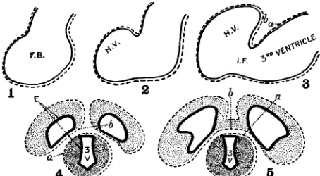
FIG. Diagram demonstrating the formation of the double-layered tela choroidea (velum interpositum) of the third ventricle in the human.
1) The developing neural tube includes the forebrain (FB) which is surrounded by the pia mater (broken line).
2) and 3) The expanding vesicle of the cerebral hemisphere (HV) carries its own pia mater (b) which overlaps the pia mater (a) of the third ventricle. IF signifies intraventricular foramen.
4) A double layer of pia mater (b, a) is interposed between the two cerebral vesicles and the third ventricle (3V). E signifies ependyma.
5) The two layers of pia mater (b, a) over the third ventricle persist after connection of the hemispheres by the commissures.
References
1. Rockwell D, Melhem ER, Bhatia R. GRASE (gradient- and spin-echo) MR imaging of the brain. AJNR Am J Neuroradiol 1997;18:1923–1928
2. Mugler JP III, Brookeman JR. Off-resonance image artifacts in interleaved-EPI and GRASE pulse sequences. Magn Reson Med 1996;36:306–313
3. Feinberg DA, Oshio K. GRASE (gradient- and spin-echo) MR imaging: a new fast clinical imaging technique. Radiology 1991;181:597–602
4. Oshio K, Feinberg DA. GRASE (gradient- and spin-echo) MR imaging: A novel fast MRI technique. Magn Reson Med 1991; 20:344–349
5. Feinberg DA, Berthold K, Litt AW. Dual contrast GRASE (gra-dient-spin echo) imaging using mixed bandwidth. Magn Reson Med 1994;31:461–464
6. Luedeke KM, Roeschmann P, Tischler R. Susceptibility artifacts in NMR imaging. Magn Reson Imaging 1985;3:329–343 7. Melhem ER, Patel R, Rockwell D, Whitehead RE, Bhatia R, Jara
H. Utility of dual-echo gradient- and spin-echo (GRASE) MR imaging of the brain: a comparison to fast spin-echo. AJR Am J Roentgenol 1998;171:797–802
The Evolutionary and Embryologic Basis for the Development and Anatomy of the Cavum
Veli Interpositi
I read with interest the recent article by Chen et al entitled, ‘‘Sonographic Characteristic of the Ca-vum Velum Interpositum’’ (1). The regional anat-omy of the cavum veli interpositi is complex and difficult to understand. I would like to point out the following anatomic information that is in variance with some of the statements made in the above-mentioned publication.
The ependymal roof of the third ventricle is cov-ered by a double layer of pia mater, the tela choroidea (2). The embryologic basis for this double-layered tela choroidea was outlined diagrammatically in 1932 By Frazer (3), modified by Brash (4), and reproduced in the neuroradiologic literature (5). It results from the overlapping of the third ventricle by the enlarging forebrain (Fig). During an early fetal stage, the pros-encephalon and dipros-encephalon are covered by a con-tinuous layer of pia mater. With further brain devel-opment, the expanding cerebral vesicles of the forebrain, covered by its own pia mater, overlaps the pia mater of the third ventricle, resulting in the dou-ble-layered tela choroidea (velum interpositum) of the roof of the third ventricle. The anterior aspect of the tela choiroidea, which is closed, is at the inter-ventricular foramen where the pia mater folds on it-self. When the posterior end remains open, the po-tential space between the double layers of the tela choroidea forms the cavum veli interpositi that com-municates with the quadrigeminal cistern. The inter-nal cerebral veins are located between the two layers of the cavum veli interpositi (2).
Certain human embryologic changes are a ‘‘re-capitulation’’ of evolutionary modifications. There-fore, examining the changing anatomy of various adult vertebrate brains facilitates understanding the overlapping of the third ventricle by the expanding human fetal cerebrum (5). In the shark’s linear brain, the cerebrum is anterior to the diencephalon.
The entire roof of the diencephalon (third ventricle) is visible, and consists of a single layer of pia mater covered by a prominent venous plexus. In reptiles and primates, the expanding cerebrum overlaps the diencephalon, resulting in the double-layered tela choroidea of the third ventricle.
In summary:
•The velum interpositum is the double-layered tela choroidea of the third ventricle.
•The cavum veli interpositi is within the double-layered tela choroidea of the third ventricle, not superior to it.
•The internal cerebral veins are within the ca-vum veli interpositi, not inferior to it.
•The correct nomenclature is velum interpositum and cavum veli interpositi.
•Finally, a discussion of fluid-filled structures in the pineal region should include an enlarged supra-pineal recess of the third ventricle. This recess may be quite large, and may extend posteriorly below the splenium (5, 6).
References
1. Chen CY, Chen FH, Lee CC, Lee KW, Hsiao HS. Sonographic characteristic of the Cavum Velum Interpositum. AJNR Am J Neuroradiol 1998;19:1631–1635
2. Williams PL, Warwick R, Dyson M, Bannister LH, ed. Gray’s Anatomy. 37th ed. London: Churchill Livingstone; 1989:1019,1081 3. Frazer JE. A Manual of Embryology. New York: William Wood;
1932
4. Brash JC. Cunningham’s Textbook of Anatomy. 9th ed. New York: Oxford University Press; 1951
5. Kier EL. The cerebral ventricles: a phylogentic and ontogentic study. In: Newton TH, Potts DG, eds. Radiology of the Skull and Brain. St. Louis: C V Mosby; 1977;2787–2914
Reply
We would like to thank Dr. Kier for pointing out issues regarding the evolutionary and embryologic basis for the development and anatomy of cavum veli interpositi (CVI) in our recent article. First, we want to emphasize that we did use the term ‘‘Ca-vum veli interpositi’’ in our original submission. During the peer review process, however, a referee opined that ‘‘Cavum veli interpositi’’ is not an ap-propriate term. We agree with Dr. Kier that ‘‘Ca-vum veli interpositi’’ is probably more appropriate than ‘‘Cavum velum interpositum’’ (1, 2), which is rarely used in the literature. Relative to the issues of embryogenesis and formation of CVI and its re-lated radiologic anatomy there are, to my knowl-edge, only a few published papers in the English-language literature (3, 4). The main point is whether the tela choroidea, by embryologic or an-atomic definitions, includes the cisternal space of CVI at or near the term age of human brain devel-opment. According to Zellweger and van Epps (4), the tela choroidea of the third ventricle originates from the roof plate of the diencephalic region by a protrusion of a fold of pia mater into the primitive neural tube at about the third fetal month. With further development, the pia mater fold is pushed backward, forming the final tela choroidea of the third ventricle. It encloses a horizontal sac or fis-sure under the fornix, which opens behind and un-der the splenium of the corpus callosum where its pia mater is connected to the pia mater covering the median fissure of the cerebrum. The sac-like pia fold carries the name ‘‘transverse or choroidal fissure.’’ In the majority of cases, the choroidal fis-sure closes. When it persists, the choroidal fisfis-sure is called ‘‘CVI or cisterna interventricularis.’’ CVI is a true cisternal structure communicating with the quadrigeminal cistern, as has been shown by com-paring pneumoencephalography and autopsy spec-imens (3). Some investigators suggest that it is a part of the anterior extension of the quadrigeminal cistern. There is no doubt that the tela choroidea forms the roof of the third ventricle; the tela cho-roidea itself is, by definition, the structure where the pia and ependyma approximate. It contains, however, a pair of internal cerebral veins, so it is debatable whether the CVI and its roof (the hip-picampal commissure) still can be included as parts of tela choroidea or part of the third ventricle as it relates to radiologic anatomy. In the published lit-erature, the anatomic location of this cavum has
been described frequently as above or superior to the third ventricle. The internal cerebral veins course within the tela choroidea on the roof of the third ventricle. Thus, anatomically, they are inferior or lateral to the CVI CSF space. Although our study did not aim to provide the embryologic evi-dence of the origin of the CVI, we reported that the color-coded internal cerebral veins on sono-graphic studies are anatomic landmarks to CVI. They are inferior or inferolateral to the CVI, but do not enter it. This raises a similar question as to whether the mega cisterna magna is a part of the fourth ventricle because it is now widely accepted that the mega cisterna magna is formed by the evagination of the tela choroidea of the fourth ventricle.
In summary, we believe:
•The velum interpositum originates from a fold of pia mater protrusion, which forms the final tela choroidea of the third ventricle.
•The cavum veli interpositi is a true cistern sit-uated above (but not communicating with) the third ventricle.
•The internal cerebral veins form parts of the inferolateral or lateral boundaries of the CVI but are not anatomically within it.
•Both terms ‘‘Cavum veli interpositi’’ and ‘‘Ca-vum velum interpositum’’ have been used inter-changeably in the English-language literature, and the former is more appropriate.
•An enlarged suprapineal recess of the third ven-tricle is a frequent finding in patients with obstruc-tive hydrocephalus, but is extremely rare if ever seen as a normal variant in neonates and infants.
Cheng-Yu Chen, M.D.
Department of Radiology National Defense Medical Center and Tri-Service
General Hospital, Taipei, Taiwan, Republic of China
References
1. Meller W, Tsai LY, Chiu LC. Cavum velum interpositum in a boy with infantile autism. J Autism Dev Disord 1985 Mar;15: 109–111
2. Larroche JC, Baudey J. Cavum septi lucidi, cavum vergae, ca-vum veli interpositi: cavites de la ligne mediane. Biol Neonat 1961;3:193–236
3. Picard L, Leymarie F, et al. Cavum veli interpositi. Roentgen anatomy: pathology and physiology. Neuroradiology 1976;10: 215–220