Received 9 January 2017 Accepted 15 January 2017
Edited by W. T. A. Harrison, University of Aberdeen, Scotland
‡ Additional correspondence author, e-mail: [email protected].
Keywords:crystal structure; rhenium; dithio-carbamate; carbonyl; Hirshfeld surface analysis.
CCDC reference:1527565
Supporting information:this article has supporting information at journals.iucr.org/e
fac
-Acetonitriletricarbonyl(dimethylcarbamodithio-ato-
j
2S
,
S
000)rhenium(I): crystal structure and
Hirshfeld surface analysis
Sang Loon Tan,aSee Mun Lee,aPeter J. Heard,b‡ Nathan R. Halcovitchcand Edward R. T. Tiekinka*
aResearch Centre for Crystalline Materials, School of Science and Technology, Sunway University, 47500 Bandar Sunway, Selangor Darul Ehsan, Malaysia,bOffice of the Provost, Sunway University, 47500 Bandar Sunway, Selangor Darul Ehsan, Malaysia, andcDepartment of Chemistry, Lancaster University, Lancaster LA1 4YB, UK. *Correspondence e-mail: [email protected]
The title compound, [Re(C3H6NS2)(C2H3N)(CO)3], features an octahedrally
coordinated ReI atom within a C3NS2 donor set defined by three carbonyl
ligands in afacialarrangement, an acetonitrile N atom and two S atoms derived from a symmetrically coordinating dithiocarbamate ligand. In the crystal, dithiocarbamate-methyl-H O(carbonyl) interactions lead to supramolecular chains along [361]; both dithiocarbamate S atoms participate in intramolecular methyl-H S interactions. Further but weaker acetonitrile-C—H O(carbon-yl) interactions assemble molecules in the ab plane. The nature of the supramolecular assembly was also probed by a Hirshfeld surface analysis. Despite their weak nature, the C—H O contacts are predominant on the Hirshfeld surface and, indeed, on those of related [Re(CO)3(C3H6NS2)L]
structures.
1. Chemical context
The reaction between a secondary amine and carbon disulfide in the presence of an alkali metal hydroxide yields a class of ligands, the dithiocarbamates,()S2CNRR0. These ligands have
long attracted the attention of coordination chemists owing to their high affinity for heavy-atom centres drawn from trans-ition metals, main group elements as well as lanthanides and actinides. The motivation for their study ranges across various disciplines and in the present time focuses upon their devel-opment as drugs (Hogarth, 2012; Bertrand & Casini, 2014), as chelating agents for the removal of toxic levels of metals in bio-remediation, etc. (Gallagher & Vo, 2015), as imaging/ radio-pharmaceutical agents (Berry et al., 2012) and as synthetic precursors for metal sulfide nanoparticles (Lewiset al., 2015; Knapp & Carmalt, 2016). In terms of crystal engin-eering endeavours, dithiocarbamates are not nearly as well studied as carboxylates. This partly arises as a result of the greater chelating ability of dithiocarbamate by virtue of the significant contribution of the (2)S2 CN(+)RR0 canonical
organometallic derivatives, metal-bound substituents (Tiekink, 2006; Tiekink & Zukerman-Schpector, 2010). Another consequence of the tight chelating mode of the di-thiocarbamate ligands is the formation of aromatic MS2C
chelate rings that can function as acceptors for C—H
interactions, i.e. C—H (chelate) interactions (Tiekink & Zukerman-Schpector, 2011; Jotaniet al., 2016). As a result of the above, a very large number of crystal structure determinations have been reported in the literature, with the last systematic reviews published over a decade ago (Heard, 2005; Hogarth, 2005).
Reflecting the wealth of structural information on metal dithiocarbamates, a search of the Cambridge Crystallographic Database (Groom et al., 2016) for rhenium dithiocarbamate structures reveals over 70 ‘hits’. One structure that attracted the attention of the authors was that of twofold symmetric, binuclear [(CO)3Re(S2CNEt2)]2, whereby each
dithio-carbamate ligand is 2-tridentate, simultaneously chelating
one ReI atom while bridging a second (Flo¨rke, 2014). The unusual feature of the structure is that the dithiocarbamate ligands lie to one side of the molecule and might be described as being syn. This arrangement is the same as that found in analogous, isoelectronic PtIVcomplexes (Heardet al., 2000),
but contradicts the observations seen in the overwhelming majority of the binary dithiocarbamates of the zinc triad elements, a focus of present research, whereby binuclear molecules with equal numbers of chelating and2-tridentate
ligands lead to binuclear molecules of the general formula, {M(S2CNRR’)2}2(Cox & Tiekink, 2009; Tiekink, 2003; Tanet
al., 2016; Jotani et al., 2016). This disparity lead to the attempted synthesis of the dimethyldithiocarbamate analogue of [(CO)3Re(S2CNEt2)]2, which when recrystallized from
acetonitrile resulted in the isolation of mononuclear (CO)3Re(S2CNMe2)(N CMe), (I). Herein, the molecular
and crystal structures of (I) are described along with a detailed analysis of the self-assemblyviaa Hirshfeld surface analysis.
2. Structural commentary
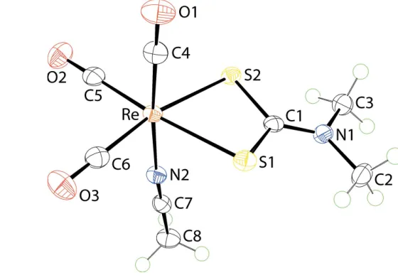
The molecular structure of (I) is shown in Fig. 1 and selected geometric parameters are collected in Table 1. The ReIatom is
coordinated by threefacially-orientated carbonyl ligands, two dithiocarbamate-S atoms and an acetonitrile-N atom. The dithiocarbamate ligand is chelating in a symmetric mode with the difference between the long and short Re—S bond lengths being less than 0.01 A˚ . This mode of coordination is reflected in the equivalence of the associated C—S bond lengths and a relatively short C1—N1 bond length, Table 1, all pointing to a significant contribution of the (2)S2C N
(+)
Me2 canonical
form to the overall electronic structure of the dithiocarbamate ligand. From the geometric data collected in Table 1, there is evidence that the shortest Re—CO bond length is formed by the carbonyltrans to the acetonitrile-N atom as opposed to those trans to the dithiocarbamate-S atoms. However, the experimental errors do not allow definitive conclusions to be made. This point is discussed further inDatabase surveybelow.
3. Supramolecular features
[image:2.610.313.564.93.171.2]Based on the standard criteria inPLATON(Spek, 2009), the most specific directional interaction between molecules in (I) is a dithiocarbamate-methyl-H O(carbonyl) interaction, Table 2. These lead to linear supramolecular chains along [361], Fig. 2a. Further searching for intermolecular inter-actions reveals that the two remaining carbonyl-O atoms participate in weak C—H O interactions just below the sum Table 1
Selected geometric parameters (A˚ ,).
Re—S1 2.4956 (6) Re—C5 1.924 (2)
Re—S2 2.5034 (6) C1—S1 1.722 (2)
Re—N2 2.153 (2) C1—S2 1.727 (2)
Re—C4 1.909 (3) C1—N1 1.320 (3)
Re—C6 1.921 (3)
S1—Re—C5 169.42 (7) N2—Re—C4 175.53 (9)
[image:2.610.88.271.518.687.2] [image:2.610.281.561.520.717.2]S2—Re—C6 168.98 (7)
Figure 1
of the van der Waals radii, each with an acetonitrile-C—H atom, Table 2. The combination of these weak interactions leads to supramolecular layers in theabplane, Fig. 2b. The two other potentially basic sites, namely the dithiocarbamate-S atoms, form intramolecular interactions with dithiocarbamate-methyl-H atoms, Table 2. The layers stack along thecaxis as shown in Fig. 2c,i.e. without directional interactions between them.
4. Hirshfeld surface analysis
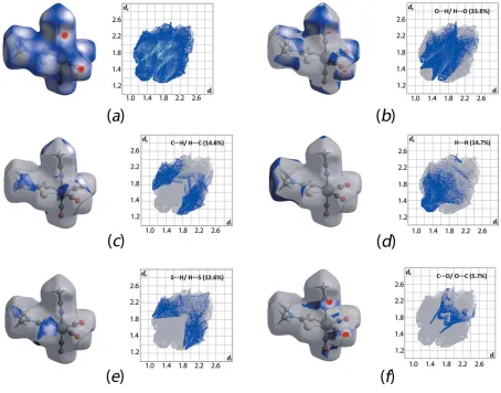
The protocols for the Hirshfeld surface analysis were as described recently (Yeoet al., 2016). In general, the Hirshfeld surface of (I) features some close interaction contacts as evidenced from the intense-red spots, Fig. 3a, being indicative of dnorm contact distances shorter than the sum of van der
Waals radii (McKinnonet al., 2007). The combination of thedi
andde, in intervals of 0.01 A˚ , resulted in the sparrow-like
two-dimensional fingerprint plot. This has been decomposed into several close contacts as shown in Fig. 3b–f. Specifically, the intense-red spots resulting from O H/H O as well as C O/O C contacts give bat- and scarab-shaped fingerprint profiles with correspondingde+dicontact distances tipped at
ca 2.5 and 3.0 A˚ , respectively; Fig. 3b and f. These contact distances are approximately 0.25 A˚ shorter than the sum of the respective van der Waals radii (Batsanov, 2001) and constitute about 33.8 and 5.7%, respectively, of the overall Hirshfeld surface contacts for the molecule. Other major contacts include C H/H C (14.8%), H H (14.7%) and S H/H S (12.6%) which result in the pincer, bust sculpture and pincer forms of the respective decomposed fingerprint plots, despite the fact their contact distance are very close or equivalent to the sum of van der Waals radii withde+divalues
of 2.8, 2.4 and 2.9 A˚ , respectively; see Fig. 3c–e.
5. Database survey
A series of eight closely related structural analogues with the formula [Re(CO)3(S2CNMe2)L], where L= ammonia (NH3)
(1), pyridine (py) (2), imidazole (Im) (3), pyrazole (pz) (4), triphenylphosphine (PPh3) (5),
1,3,5-triaza-7-phosphaada-mantane (PTA) (6), t-butyl isocyanide (tBuNC) (7) and cyclohexyl isocyanide (CyNC) (8) have been reported previously (Herricket al., 2009). The bond lengths about the ReIatom in 1–8 and (I) are collated in Table 3; the numbering schemes correspond to that shown in Fig. 1. There are a few
general observations that can be noted. Firstly, neitherd(Re— S1) nord(Re—S2) show major deviations in their respective bond lengths as evidenced from the mean difference of 0.005 A˚ for each. Despite the small differences, a trend is observed in thatd(Re—S2) is generally longer thand(Re— S1). A consistent pattern is observed in the relatedd(Re— C5), i.e. trans to S1, and d(Re—C6), i.e. trans to S2, bond lengths for which the latter registers an average elongation of 0.005 A˚ . Secondly, thed(Re—L) bond lengths are found to consistently increase from C-donor ligands to N-donors, with a Figure 2
[image:3.610.306.566.72.544.2]The molecular packing in (I): (a) supramolecular chain sustained by methyl-C—H O(carbonyl) interactions shown as orange dashed lines, (b) view of the supramolecular layers in the ab plane with non-participating H atoms removed and (c) a view of the unit-cell contents in projection down theaaxis.
Table 2
Hydrogen-bond geometry (A˚ ,).
D—H A D—H H A D A D—H A
C2—H2B O1i 0.98 2.59 3.260 (3) 126
C8—H8C O2ii 0.98 2.69 3.332 (3) 123
C8—H8B O3iii 0.98 2.69 3.244 (3) 116
C2—H2C S1 0.98 2.49 3.030 (2) 114
C3—H3A S2 0.98 2.64 3.035 (2) 105
ca0.10 A˚ or 5% increment, followed by P-donors with about a 0.26 A˚ or 12% increase,cf. the N-donor ligands. However, the observed trend deviates from expectation in that thed(M—L) bond length is anticipated to increase in the order N < C < P-donor type ligand by approximately 2.6 and 27.4%, respec-tively, based on their calculated covalent bond radii. Further, it is observed that d(Re—C4), i.e. with C4 trans to L, is
marginally longer thand(Re—C5) andd(Re—C6) byca0.01– 0.02 A˚ . Finally, d(C4 O1) is generally shorter, by about 0.01 A˚ , cf. d(C5 O2) andd(C6 O3), i.e. with C5 and C6
[image:4.610.45.565.117.229.2]transto the S1 and S2 atoms, respectively. These observations show the presence of strong-backbonding prevailing in the C-donor type ligands that result in shorter Re—Land longer Re—C4 bonds as well as shorter C4 O1 bond lengths when Table 3
Selected bonding parameters (A˚ ) for (I) and literature analogues [Re(CO)3(S2CNMe2)L]..
L= ammonia (NH3) (1), pyridine (py) (2), imidazole (Im) (3), pyrazole (pz) (4), triphenylphosphine (PPh3) (5), 1,3,5-triaza-7-phosphaadamantane (PTA) (6),t -butyl isocyanide (tBuNC) (7) and cyclohexyl isocyanide (CyNC) (8) (Herricket al., 2009).
L Re—S1 Re—S2 Re—C C O Re—C C O Re—C C O Re—L
(transto S1) (transto S2) (transtoL)
(1) 2.497 (2) 2.506 (2) 1.915 (7) 1.164 (8) 1.912 (6) 1.161 (7) 1.916 (7) 1.153 (9) 2.228 (5)
(2) 2.505 (2) 2.498 (1) 1.925 (6) 1.147 (7) 1.929 (5) 1.137 (7) 1.926 (5) 1.141 (7) 2.219 (4)
(3) 2.501 (2) 2.518 (3) 1.937 (7) 1.135 (8) 1.914 (7) 1.157 (9) 1.918 (7) 1.166 (8) 2.189 (6)
(4) 2.489 (4) 2.501 (4) 1.906 (14) 1.147 (17) 1.900 (14) 1.153 (17) 1.912 (13) 1.133 (16) 2.173 (10) (5) 2.513 (3) 2.506 (3) 1.910 (10) 1.169 (13) 1.895 (10) 1.179 (12) 1.931 (10) 1.152 (12) 2.474 (3) (6) 2.527 (5) 2.529 (4) 1.925 (15) 1.147 (19) 1.898 (16) 1.160 (20) 1.983 (18) 1.110 (20) 2.437 (5)
(7) 2.512 (3) 2.521 (2) 1.906 (7) 1.176 (9) 1.941 (8) 1.137 (9) 1.955 (8) 1.152 (9) 2.102 (7)
(8) 2.502 (2) 2.512 (2) 1.914 (9) 1.142 (12) 1.908 (10) 1.168 (12) 1.953 (9) 1.125 (11) 2.082 (9)
(I) 2.496 (1) 2.503 (1) 1.924 (2) 1.150 (3) 1.921 (3) 1.145 (3) 1.909 (3) 1.155 (3) 2.153 (2)
Figure 3
[image:4.610.74.528.366.722.2]compared to the other structural analogues. Further, these trends are clearly reflected in the blue shift of the CO vibrational band forL= C-type donor ligands, with an average
= 180 cm1, compared with those for N- and P-type donors (Herricket al., 2009). In the present study,(CO) for (I) was observed at 1883 cm1.
The molecular packing in each of 1–8 was also studied through Hirshfeld surface analysis by calculating the relative composition of each intermolecular close contact present in the structure usingCrystal Explorer(Wolffet al., 2012); Fig. 4. Generally, the intermolecular close contacts are dominated by O H/H O, H H, followed by either C H/H C or S H/H S contacts, with the exceptional cases being for 5 and 6, with hydrogen-rich P-donor ligands, for which the dominance is in the order H H > O H/H O > C H/ H C > S H/S H. These results highlight the relative importance of the C—H O contacts in these structures despite their relatively weak nature.
6. Synthesis and crystallization
All chemicals and solvents were used as purchased without purification, and all reactions were carried out under ambient conditions. The melting point was determined using an Elec-trothermal digital melting point apparatus and was uncor-rected. The IR spectra were obtained on a Perkin Elmer Spectrum 400 FT Mid-IR/Far-IR spectrophotometer from 4000 to 400 cm1 (abbreviations: vs, very strong; s, strong).
1
H NMR spectra were recorded at room temperature in
DMSO-d6 solution on a Bruker AVANCE-400 MHz
instru-ment.
Bromopentacarbonylrhenium(I) (0.25 mmol, 0.102 g) in acetone (10 ml) was added to sodium dimethyldithio-carbamate hydrate (0.25 mmol, 0.036 g) in acetone (10 ml). The resulting mixture was stirred and refluxed for 2 h. The filtrate was evaporated until a precipitate was obtained. The precipitate was recrystallized from its acetonitrile solution. Colourless blocks were obtained from the slow evaporation of the filtrate. Yield: 0.064 g, 60%; M.p. 478–479 K. IR (cm1): 2009 (s), 1883 (vs).1H NMR (in DMSO-d6):3.21 (s, 6H, N–
CH3), 2.07 (s, 3H, C–CH3).
7. Refinement
Crystal data, data collection and structure refinement details are summarized in Table 4. Carbon-bound H atoms were placed in calculated positions (C—H = 0.98 A˚ ) and were included in the refinement in the riding-model approximation, with Uiso(H) set to 1.2Ueq(C). The maximum and minimum
residual electron density peaks of 0.80 and 1.21 e A˚3 were
located 0.87 and 0.91 A˚ , respectively, from the Re atom.
Acknowledgements
[image:5.610.94.506.73.373.2]We thank Sunway University for support of biological and crystal engineering studies of metal dithiocarbamates. Figure 4
References
Batsanov, S. S. (2001).Inorg. Mater.,37, 1031–1046.
Berry, D. J., Torres Martin de Rosales, R., Charoenphun, P. & Blower, P. J. (2012).Mini Rev. Med. Chem.12, 1174–1183.
Bertrand, B. & Casini, A. (2014).Dalton Trans.43, 4209–4219. Brandenburg, K. (2006).DIAMOND. Crystal Impact GbR, Bonn,
Germany.
Cox, M. J. & Tiekink, E. R. T. (2009).Z. Kristallogr.214, 184–190. Farrugia, L. J. (2012).J. Appl. Cryst.45, 849–854.
Flo¨rke, U. (2014). Private communication (refcode KOGRIJ). CCDC, Cambridge, England.
Gallagher, W. P. & Vo, A. (2015).Org. Process Res. Dev. 19, 1369– 1373.
Groom, C. R., Bruno, I. J., Lightfoot, M. P. & Ward, S. C. (2016).Acta Cryst.B72, 171–179.
Heard, P. J. (2005).Prog. Inorg. Chem.53, 1–69.
Heard, P. J., Kite, K., Nielsen, J. S. & Tocher, D. A. (2000).J. Chem. Soc. Dalton Trans.pp. 1349–1356.
Herrick, R. S., Ziegler, C. J., Sripothongnak, S., Barone, N., Costa, R., Cupelo, W. & Gambella, A. (2009).J. Organomet. Chem.694, 3929– 3934.
Hogarth, G. (2005).Prog. Inorg. Chem.53, 71–561. Hogarth, G. (2012).Mini Rev. Med. Chem.12, 1202–1215.
Jotani, M. M., Tan, Y. S. & Tiekink, E. R. T. (2016).Z. Kristallogr.231, 403–413.
Knapp, C. E. & Carmalt, C. J. (2016).Chem. Soc. Rev.45, 1036–1064. Lewis, E. A., McNaughter, P. D., Yin, Z. J., Chen, Y. Q., Brent, J. R., Saah, S. A., Raftery, J., Awudza, J. A. M., Malik, M. A., O’Brien, P. & Haigh, S. J. (2015).Chem. Mater.27, 2127–2136.
McKinnon, J. J., Jayatilaka, D. & Spackman, M. A. (2007).Chem. Commun.pp. 3814–3816.
Rigaku Oxford Diffraction (2015).CrysAlis PRO. Agilent Technol-ogies Inc., Santa Clara, CA, USA.
Sheldrick, G. M. (2008).Acta Cryst.A64, 112–122. Sheldrick, G. M. (2015).Acta Cryst.C71, 3–8. Spek, A. L. (2009).Acta Cryst.D65, 148–155.
Tan, Y. S., Halim, S. N. A. & Tiekink, E. R. T. (2016).Z. Kristallogr.
231, 113–126.
Tiekink, E. R. T. (2003).CrystEngComm,5, 101–113. Tiekink, E. R. T. (2006).CrystEngComm,8, 104–118.
Tiekink, E. R. T. & Zukerman-Schpector, J. (2010).Coord. Chem. Rev.254, 46–76.
Tiekink, E. R. T. & Zukerman-Schpector, J. (2011).Chem. Commun.
47, 6623–6625.
Westrip, S. P. (2010).J. Appl. Cryst.43, 920–925.
Wolff, S. K., Grimwood, D. J., McKinnon, J. J., Turner, M. J., Jayatilaka, D. & Spackman, M. A. (2012).Crystal Explorer. The University of Western Australia.
[image:6.610.43.294.90.376.2]Yeo, C. I., Tan, S. L. & Tiekink, E. R. T. (2016).Acta Cryst.E72, 1446– 1452.
Table 4
Experimental details.
Crystal data
Chemical formula [Re(C3H6NS2)(C2H3N)(CO)3]
Mr 431.49
Crystal system, space group Triclinic,P1
Temperature (K) 100
a,b,c(A˚ ) 5.7442 (1), 7.5022 (1), 14.6644 (2) ,,() 91.496 (1), 95.517 (1), 102.371 (1)
V(A˚3) 613.71 (2)
Z 2
Radiation type MoK
(mm1) 10.23
Crystal size (mm) 0.150.110.11
Data collection
Diffractometer Agilent SuperNova Dual Source
diffractometer with an AtlasS2 detector
Absorption correction Gaussian (CrysAlis PRO; Rigaku Oxford Diffraction, 2015)
Tmin,Tmax 0.371, 0.503
No. of measured, independent and observed [I> 2 (I)] reflections
32146, 3244, 3153
Rint 0.033
(sin/)max(A˚
1) 0.698
Refinement
R[F2> 2 (F2)],wR(F2),S 0.016, 0.035, 1.09
No. of reflections 3244
No. of parameters 148
H-atom treatment H-atom parameters constrained
max,min(e A˚
3) 0.80,1.21
Computer programs:CrysAlis PRO (Rigaku Oxford Diffraction, 2015),SHELXS97
sup-1
Acta Cryst. (2017). E73, 213-218
supporting information
Acta Cryst. (2017). E73, 213-218 [https://doi.org/10.1107/S2056989017000755]
fac
-Acetonitriletricarbonyl(dimethylcarbamodithioato-
κ
2S
,
S
′
)rhenium(I): crystal
structure and Hirshfeld surface analysis
Sang Loon Tan, See Mun Lee, Peter J. Heard, Nathan R. Halcovitch and Edward R. T. Tiekink
Computing details
Data collection: CrysAlis PRO (Rigaku Oxford Diffraction, 2015); cell refinement: CrysAlis PRO (Rigaku Oxford
Diffraction, 2015); data reduction: CrysAlis PRO (Rigaku Oxford Diffraction, 2015); program(s) used to solve structure:
SHELXS97 (Sheldrick, 2008); program(s) used to refine structure: SHELXL2014 (Sheldrick, 2015); molecular graphics:
ORTEP-3 for Windows (Farrugia, 2012) and DIAMOND (Brandenburg, 2006); software used to prepare material for
publication: publCIF (Westrip, 2010).
fac-Acetonitriletricarbonyl(dimethylcarbamodithioato-κ2S,S′)rhenium(I)
Crystal data
[Re(C3H6NS2)(C2H3N)(CO)3]
Mr = 431.49
Triclinic, P1
a = 5.7442 (1) Å
b = 7.5022 (1) Å
c = 14.6644 (2) Å
α = 91.496 (1)°
β = 95.517 (1)°
γ = 102.371 (1)°
V = 613.71 (2) Å3
Z = 2
F(000) = 404
Dx = 2.335 Mg m−3
Mo Kα radiation, λ = 0.71073 Å Cell parameters from 22533 reflections
θ = 3.0–29.4°
µ = 10.23 mm−1
T = 100 K Block, colourless 0.15 × 0.11 × 0.11 mm
Data collection
Agilent SuperNova Dual Source
diffractometer with an AtlasS2 detector Radiation source: micro-focus sealed X-ray
tube, SuperNova (Mo) X-ray Source Mirror monochromator
ω scans
Absorption correction: gaussian
(CrysAlis PRO; Rigaku Oxford Diffraction, 2015)
Tmin = 0.371, Tmax = 0.503 32146 measured reflections 3244 independent reflections 3153 reflections with I > 2σ(I)
Rint = 0.033
θmax = 29.7°, θmin = 2.8°
h = −7→7
k = −10→10
l = −20→20
Refinement
Refinement on F2 Least-squares matrix: full
R[F2 > 2σ(F2)] = 0.016
wR(F2) = 0.035
S = 1.09 3244 reflections
148 parameters 0 restraints
Hydrogen site location: inferred from neighbouring sites
sup-2
Acta Cryst. (2017). E73, 213-218
w = 1/[σ2(F
o2) + (0.0185P)2 + 0.3859P] where P = (Fo2 + 2Fc2)/3
(Δ/σ)max = 0.002
Δρmax = 0.80 e Å−3 Δρmin = −1.21 e Å−3
Special details
Geometry. All esds (except the esd in the dihedral angle between two l.s. planes) are estimated using the full covariance
matrix. The cell esds are taken into account individually in the estimation of esds in distances, angles and torsion angles; correlations between esds in cell parameters are only used when they are defined by crystal symmetry. An approximate (isotropic) treatment of cell esds is used for estimating esds involving l.s. planes.
Fractional atomic coordinates and isotropic or equivalent isotropic displacement parameters (Å2)
x y z Uiso*/Ueq
Re 0.53372 (2) 0.29366 (2) 0.78801 (2) 0.01195 (4) S1 0.40857 (10) 0.56377 (8) 0.71937 (4) 0.01596 (12) S2 0.74400 (10) 0.37175 (8) 0.64780 (4) 0.01389 (11) O1 0.1152 (3) 0.0231 (3) 0.68068 (14) 0.0259 (4) O2 0.7673 (3) −0.0198 (3) 0.85255 (14) 0.0258 (4) O3 0.2514 (3) 0.2703 (3) 0.95601 (13) 0.0267 (4) N1 0.6870 (4) 0.6973 (3) 0.58960 (14) 0.0156 (4) N2 0.8304 (4) 0.4965 (3) 0.85327 (14) 0.0151 (4) C1 0.6222 (4) 0.5632 (3) 0.64470 (16) 0.0132 (4) C2 0.5732 (5) 0.8540 (3) 0.58562 (18) 0.0203 (5) H2A 0.4691 0.8457 0.5280 0.031* H2B 0.6968 0.9672 0.5888 0.031* H2C 0.4773 0.8542 0.6374 0.031* C3 0.8671 (4) 0.6924 (3) 0.52607 (17) 0.0190 (5) H3A 0.9870 0.6289 0.5534 0.028* H3B 0.9456 0.8176 0.5137 0.028* H3C 0.7895 0.6277 0.4685 0.028* C4 0.2721 (4) 0.1247 (3) 0.72181 (17) 0.0174 (5) C5 0.6800 (4) 0.0972 (3) 0.82800 (17) 0.0169 (5) C6 0.3585 (4) 0.2794 (3) 0.89373 (17) 0.0176 (5) C7 0.9880 (4) 0.6097 (3) 0.88229 (16) 0.0155 (5) C8 1.1894 (5) 0.7539 (4) 0.91890 (18) 0.0211 (5) H8A 1.3395 0.7194 0.9075 0.032* H8B 1.1849 0.7720 0.9851 0.032* H8C 1.1795 0.8676 0.8888 0.032*
Atomic displacement parameters (Å2)
U11 U22 U33 U12 U13 U23
sup-3
Acta Cryst. (2017). E73, 213-218
N2 0.0146 (10) 0.0153 (10) 0.0152 (10) 0.0027 (8) 0.0023 (8) −0.0009 (8) C1 0.0119 (10) 0.0130 (10) 0.0133 (11) 0.0020 (9) −0.0022 (8) −0.0030 (8) C2 0.0227 (12) 0.0163 (11) 0.0232 (13) 0.0073 (10) 0.0004 (10) 0.0038 (10) C3 0.0194 (12) 0.0175 (12) 0.0190 (12) 0.0008 (10) 0.0037 (10) 0.0020 (10) C4 0.0142 (11) 0.0192 (12) 0.0186 (12) 0.0025 (10) 0.0037 (9) −0.0006 (10) C5 0.0143 (11) 0.0154 (11) 0.0176 (12) −0.0034 (9) 0.0010 (9) −0.0027 (9) C6 0.0164 (11) 0.0170 (11) 0.0189 (12) 0.0043 (10) −0.0008 (9) −0.0014 (9) C7 0.0154 (11) 0.0179 (11) 0.0139 (11) 0.0050 (10) 0.0018 (9) 0.0002 (9) C8 0.0171 (12) 0.0202 (12) 0.0228 (13) −0.0012 (10) −0.0004 (10) −0.0049 (10)
Geometric parameters (Å, º)
Re—S1 2.4956 (6) N1—C3 1.463 (3) Re—S2 2.5034 (6) N2—C7 1.140 (3) Re—N2 2.153 (2) C2—H2A 0.9800 Re—C4 1.909 (3) C2—H2B 0.9800 Re—C6 1.921 (3) C2—H2C 0.9800 Re—C5 1.924 (2) C3—H3A 0.9800 C1—S1 1.722 (2) C3—H3B 0.9800 C1—S2 1.727 (2) C3—H3C 0.9800 O1—C4 1.155 (3) C7—C8 1.453 (3) O2—C5 1.150 (3) C8—H8A 0.9800 O3—C6 1.145 (3) C8—H8B 0.9800 C1—N1 1.320 (3) C8—H8C 0.9800 N1—C2 1.462 (3)
sup-4
Acta Cryst. (2017). E73, 213-218
N1—C1—S2 122.84 (18) H8B—C8—H8C 109.5
C2—N1—C1—S1 −2.5 (3) Re—S1—C1—N1 −170.0 (2) C3—N1—C1—S1 −179.31 (18) Re—S1—C1—S2 10.24 (11) C2—N1—C1—S2 177.28 (18) Re—S2—C1—N1 170.0 (2) C3—N1—C1—S2 0.5 (3) Re—S2—C1—S1 −10.21 (11)
Hydrogen-bond geometry (Å, º)
D—H···A D—H H···A D···A D—H···A
C2—H2B···O1i 0.98 2.59 3.260 (3) 126 C8—H8C···O2ii 0.98 2.69 3.332 (3) 123 C8—H8B···O3iii 0.98 2.69 3.244 (3) 116 C2—H2C···S1 0.98 2.49 3.030 (2) 114 C3—H3A···S2 0.98 2.64 3.035 (2) 105