Supported by the Sichuan Agricultural University (Shuangzhi Project).
Effect of ZnO Nanoparticle on Cell Viability, Zinc
Uptake Efficiency, and Zinc Transporters Gene
Expression: a Comparison with ZnO and ZnSO
4
Xiangfei Zhang, Zhisheng Wang*, Lei Mao, Xianwen Dong,
Quanhui Peng, Juncai Chen, Cui Tan, Rui Hu
Animal Nutrition Institute, Sichuan Agricultural University, Ya’an, P.R. China *Corresponding author: [email protected]
ABSTRACT
Zhang X., Wang Z., Mao L., Dong X., Peng Q., Chen J., Tan C., Hu R. (2017): Effect of ZnO nanoparticle
on cell viability, zinc uptake efficiency, and zinc transporters gene expression: a comparison with ZnO and ZnSO4. Czech J. Anim. Sci., 62, 32–41.
Zinc plays an important role in functional and structural integrity of cells. The aim of the current study was to compare cell viability, zinc uptake efficiency, and gene expression of metallothionein (MT), divalent metal transporter (DMT-1), and other important zinc transporters (ZnTs) under experimental treatment of TPEN (N, N, N', N'-Tetrakis (2-pyridylmethyl) ethylenediamine) (2 µM), and three zinc sources (zinc oxide
nanopar-ticle (nano-ZnO), bulk zinc oxide (ZnO), and zinc sulfate (ZnSO4)) at different levels (25, 50, and 100 µM) in
rat intestinal epithelial cell line IEC-6. Cells were classified into TPEN group and TPEN + zinc sources groups. In the present study, significantly decreased cell viability was observed in TPEN group, while
supplementa-tions with nano-ZnO at all levels and ZnO (50 and 100 µM) significantly increased the cell viability. ZnSO4
ata high concentration (100 µM) inhibited cell viability. Furthermore, cells of nano-ZnO group showed the
highest viability at a 25 µM concentration. The uptake efficiency of nano-ZnO is higher than that of ZnSO4 and
ZnO. Additionally, a significant down-regulation for ZnT-1, ZnT-4, MT, DMT-1 mRNA with TPEN treatment was detected. Compared with the unchanged ZnT-4, all zinc treatments up-regulated the gene expressions of ZnT-1, ZnT-5, ZnT-7, MT, and DMT-1. Our results indicate that nano-ZnO is more effective than ZnO and
ZnSO4 in enhancing cell viability, and its lower cytotoxicity, higher uptake efficiency, and comparative
trans-portation at low concentration also favour its potential use as a new zinc source in feed additives.
Keywords: nano-ZnO; bulk ZnO; zinc transportation; TPEN; IEC-6 cells
The intestine is the central organ for zinc ab-sorption and transportation. Intestinal cells take up zinc from the diet, and then distribute it to different organs. Zinc is an important co-factor for many enzymes, transcription factor as well as hormones and, hence, is a vital component and intracellular mediator of the cell. In recent years, the importance of some zinc compounds supple-mentation in animal diets has gained recognition,
Two of the most commonly used inorganic zinc sources in feed industry up to now are zinc oxide
(ZnO) and zinc sulfate (ZnSO4). Several previous
studies compared the absorption characteristics of these two chemical forms using the animal test as a measure of bioavailability, however, the results were controversial. Some of these animal studies showed much lower bioavailability of zinc oxide in comparison with zinc sulfate (Sandoval et al. 1997), but some more recent studies showed that zinc oxide was effective regardless of its poor Zn bioavailability (Mavromichalis et al. 2000; Case and Carlson 2002). The studies also indicated that zinc may have a local effect in intestine, rather than a systemic effect.
Nanomaterials comprised of ZnO have gener-ated considerable concern in recent years due to their potential use in biological applications aris-ing from their special physiochemical properties (Stark 2011). A new form of zinc – nano-ZnO – should have an enhancing effect on zinc uptake and bioavailability. It is generally accepted now that nano-ZnO, compared to traditional Zn forms, could efficiently penetrate into cell, internalize and disrupt membrane architecture. But as a conse-quence of its rapid absorption characteristics, most of the studies showed cytotoxicity of nano-ZnO because of particle dissolution and zinc release in the medium or uptake in the cells leading to cell injury (Xia et al. 2008; De Berardis et al. 2010). In this regard, many recent reports have confirmed that nano-ZnO has adverse biological outcomes on the prokaryotic and eukaryotic system, while in an appropriate concentration it is nontoxic to natural biomolecules (Hanley et al. 2008; Shen et al. 2008). A majority of those studies have focused on some low tolerance cells, including immune cells, or even on some immortalized cells lines, which showed changed sensitivities to some chemicals by altering metabolic processes of cells. Thus, cyto-toxicity by susceptibility to nano-ZnO appears to be affected by the cell type and is also related to other physiologically relevant parameters, including the concentration used and inherent differences in cellular uptake processes. These variable toxic ef-fects of nano-ZnO on various types of cells indicate that a more detailed investigation of its effects on intestinal epithelial cells is needed.
The intestine is the first tissue confronted with zinc, and an abundant uptake of zinc from the small intestine is indispensable for cellular processes
in the body. However, a high level of intracellu-lar zinc is deleterious and poisonous to the cell. Therefore, maintaining zinc level in an appropriate variation range in tissue is of great importance. As a result, zinc metabolism is tightly regulated. Zinc homeostasis is controlled by a large group of zinc-regulatory proteins involved in zinc me-tabolism (zinc absorption, storage, transporta-tion, and excretion). These key proteins include metallothionein (MT), divalent metal transporter (DMT-1), and transmembrane zinc transporters (ZnT). In mammalian cells, ZnT family consists of ten homologous SLC30 proteins (ZnT-1–ZnT-10) (Cousins et al. 2006) and most of the zinc transport-ers have been confirmed as directly or indirectly involved in absorption, storage or utilization of Zn in cells (Kambe et al. 2002; Palmiter and Huang 2004; Qin et al. 2009). ZnT-1 was the first identi-fied mammalian zinc transporter located in the plasma membrane and responsible for transport-ing cytoplasmic zinc across the membrane to the extracellular space (Qin et al. 2009). ZnT-4 is widely expressed, but more abundantly in brain and epi-thelial cells (Cousins and McMahon 2000). It is very important for maintaining the body Zn homeostasis (Huang and Tepaamorndech 2013). ZnT-5 is also highly expressed in a large number of cells, and a higher level was detected in pancreatic beta cells (Kambe et al. 2002). ZnT-7, which is more abundant in duodenum and jejunum, is mainly expressed in lung and small intestine. A previous study described ZnT-7 as the Golgi apparatus protein involved in the accumulation of Zn from cytoplasm, because in the Golgi apparatus ZnT-7 occurs at a subcel-lular level (Kirschke and Huang 2003). In addition, metallothionein (MT) is a kind of low-molecular-weight proteins, which can maintain homeostasis of some required elements (Hoadley and Leinart 1987; Cherian et al. 1994). DMT-1 is located in the apical membrane of intestinal cells and can transport a wide range of divalent metal ions, but studies concerning Zn transportation are limited.
Little attention has been given to the uptake and regulation of ZnTs with ZnO nanoparticles in previous studies. Therefore, in the present study, we investigated the effects of nano-ZnO, bulk ZnO,
and ZnSO4 on cell viability and uptake efficiency
MATERIAL AND METHODS
Epithelial cell culture. The rat intestinal IEC-6 cells were obtained from the American Type Cul-ture Collection (Manassas, USA). Cells were
rou-tinely grown in plastic tissue culture flasks (25 cm2
growth area) in Dulbecco’s Modified Eagle Medium (DMEM) containing 10% (v/v) fetal bovine serum
(FBS), 2 mM glutamine, and 1 × 10⁵ U/l penicillin/
streptomycin, and maintained at 37°C in an
atmos-phere of 5% CO2: 95% air at 90% relative humidity.
In all experiments, IEC-6 cells were seeded on tissue culture plates and monitored using phase-contrast microscopy. Cell passages were maintained between 10 and 15 for all experiments, and medium was changed every 2 days.
Cell viability assay. Cellular viability with or without experimental agents was determined using the water-soluble tetrazolium salt WST-8 (2-(2-methoxy-4-nitrophenyl)-3-(4-nitrophenyl)-5-(2,4-disulphophenyl)-2H-tetrazolium, mono-sodium salt) assay, which is based on the ability of viable cells to convert dissolved WST-8 to an insoluble orange-coloured formazan product. Briefly, IEC-6 cells propagated in 96-well plates
upon reaching confluency at 37°C and 5% CO2
atmosphere. The culture medium was aspirated and cells were incubated with N, N, N', N'-Tetrakis (2-pyridylmethyl) ethylenediamine (TPEN) (2 µM)
and nano-ZnO, ZnO, and ZnSO4 with the
con-centration of 25, 50, and 100 µM for 24 and 48 h, respectively. After that, CCK-8 (Beyotime Institute of Biotechonology, China) solution (10 µl/100 µl culture medium) was added and incubation for
2 h at 37°C in a humidified incubator (5% CO2)
followed. Colour development at a wavelength of 450 nm was measured using an automatic plate reader Thermo Multiskan MK (Thermo Fisher Scientific Inc., USA). Relative cell viability (%) was calculated as follows:
Cell viability (%) = (OD450nm(sample)/OD450nm(control)) ×
× 100
where:
OD450nm(sample) = absorbance of treated cell sample OD450nm(control) = absorbance of control cell sample
Determination of zinc uptake efficiency. IEC-6 cells propagated in 6-well plates upon reaching confluency, and the media in the wells were then replaced with IEC-6 zinc-free medium (0.82 µM
TPEN was used to chelate zinc in the IEC-6 medi-um) with three zinc sources (nano-ZnO, bulk ZnO,
and ZnSO4) at different levels (25, 50, and 100 µM).
After incubation for 24 h at 37°C, the media were removed, and the cells were then washed once with 5% acetic acid and 10 mmol/l ethylenediaminetet-raacetic acid (EDTA), respectively, then twice by phosphate buffered saline (PBS). After that, cells were incubated with 1 ml of ultrapure nitric acid overnight until the cell pellets were completely digested. The solutions were finally diluted to 3 ml with ultrapure water and used to analyze Zn absorption. Zn concentrations in both control and supplemented media were determined by a flame atomic absorption spectrometer NovAA 400 (Analytik Jena AG, Germany). The calculation formula of uptake efficiency is as follows:
Uptake efficiency (%) = (Zn retained in cells/Zn added to the cell medium) × 100
Effects of incubation time. IEC-6 cells were cultured as described above. The medium in the wells was then replaced with zinc-free medium
with 50 µM nano-ZnO, ZnO, and ZnSO4, all groups
were incubated at 37°C for 1, 3, 6, 12, and 24 h, respectively. Zinc concentrations in the cell sample were then determined by flame atomic absorption spectroscopy.
RNA isolation and real-time PCR analysis. Total RNA was isolated from both treated and untreated IEC-6 cells with Trizol reagent (Invitrogen Inc., USA) as described by the manufacturer’s protocol. Reverse transcription of RNA into cDNA was per-formed using random hexamers, oligo (dT) primer,
and PrimeScript™ RT reagent Kit (TaKaRa, China),
according to the manufacturer’s instructions. Real-time PCR analysis of cDNA was performed using
the iCycler iQ™ Real-Time PCR Detection System
(Bio-Rad Laboratories Inc., USA) and SYBR® Premix
Ex Taq™ II (TaKaRa). Relative gene expression was
achieved by the Δ-Ct method. One housekeeping gene (β-actin) was used for normalization purpose. The reactions of PCR amplification were heated to 95°C for 10 s and immediately cycled 40 times through the 5 s denaturing step at 95°C, 20 s an-nealing step, and 15 s elongation step at 72°C. All primers used are listed in Table 1.
particle diameter (purity > 99%) was bestowed by the College of Chemistry and Chemical Engineer-ing, Xiamen University (China). Zinc oxide and zinc sulfate (purity > 99% both) were purchased from Sigma Aldrich (USA). Cell counting kit solution (CCK-8) was purchased from Beyotime Institute of Biotechonology (China). RNAiso Regent,
Prime-Script™ RT Reagent Kit, SYBR® Premix Ex Taq™ II
were purchased from TaKaRa (China).
Statistical analysis. All data were expressed as means ± SD. One-way analysis of variance (ANOVA) and Dunnett’s post-hoc test were used to perform comparisons by the SAS software (Statistical Analy-sis System, Version 9.2, 2008). Group differences resulting in P-values of less than 0.05 were consid-ered to be statistically significant.
RESULTS
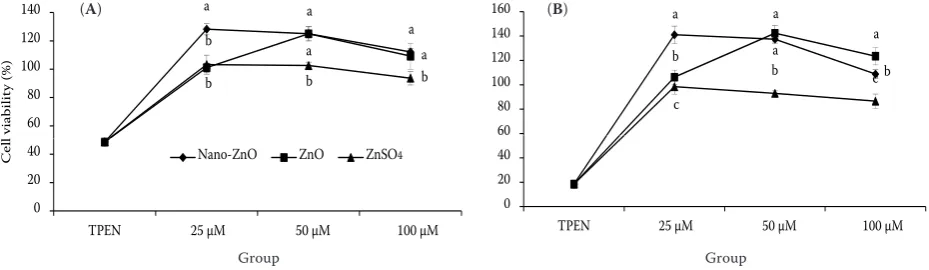
Effects of TPEN and three zinc sources on IEC-6 cell viability. Viability of cells cultured in the presence or absence of the three zinc sources is shown in Figure 3. The results showed that viability of cells which were exposed to TPEN decreased significantly (P < 0.05). Compared with untreated control group, viability of cells treated with 25, 50, 100 µM nano-ZnO or 50 and 100 µM ZnO increased significantly (P < 0.05), but had a decreasing tendency in 100 µM concentration.
Furthermore, 25 and 50 µM ZnSO4 for 24 h and
25 µM for 48 h did not significantly affect cell viability (P > 0.05), but it induced a dramatical decrease with 100 µM concentrations similarly (P < 0.05). At the level of 25 µM, cell viability with nano-ZnO was significantly higher than with ZnO
and ZnSO4 (P < 0.05). When nano-ZnO and ZnO
were added with the concentrations of 50 and 100 µM, cell viability was significantly higher than
if ZnSO4 was added (P < 0.05).
Effects of three zinc sources on uptake efficiency in IEC-6 cells. The uptake efficiencies of the three zinc sources in IEC-6 were the next objective of the current study. Therefore, differences in the
uptake efficiency of nano-ZnO, ZnO, and ZnSO4,
which were used for the performance of dose- and time-response kinetics, were confirmed by atomic absorption spectroscopy. As shown in Figure 1, cells incubated with nano-ZnO showed the highest
uptake efficiency with a following order of ZnSO4
and ZnO at all levels (P < 0.05). As for the uptake efficiencies of different levels of nano-ZnO, the highest value was observed in cells treated with the concentration of 25 µM compared to 50 and 100 µM, and this variation was similar as in the ZnO treatment. The uptake efficiency of 50 µM
ZnSO4 concentration was greater than that of
25 µM and 100 µM by the end of 24 h (P < 0.05). To further investigate the effects of incuba-tion time on uptake efficiency, cells were treated
with 50 µM nano-ZnO, ZnO, and ZnSO4 for 1, 3,
[image:4.595.61.534.112.327.2]6, 12, and 24 h, respectively. As seen in Figure 2,
Table 1. Primers used in the study
Gene Primer sequences(5'-3') Annealing temperature (°C) Product size (bp)
ZnT-1 F: GAACATGCGAGGAGTGTTTCTG R: CGTCTTCAGTACAACCCTTCCAG 64.5 110
ZnT-4 F: GCAGAGGAAGGTGAAGACCAG R: GCCACAAAGCAAGAAGAGTGAG 55.0 170
ZnT-5 F: GGAGGAGTGGTAGTGAGTGCTGT R: TGAAGTGATAGAGCGGTGTTCC 64.5 127
ZnT-7 F: GCCACCCAAGTTCAATCTGTTC R: GCCGTAGAGTAGTTCCACAAAAGC 61.4 133
DMT-1 F: GCCCTTCACCACCTACTTTGAT R: ATCCAGCCACTGCTCCAGACT 64.5 170
MT F: GGCTGCAAGAACTGCAAATG R: CACTTGTCCGAGGCACCTTT 61.4 113
the uptake efficiency of cells in nano-ZnO group
was significantly higher than that of ZnSO4 and
ZnO from 1 h to 24 h. Our results indicated that nano-ZnO reached maximum uptake efficiency at 3 h (P < 0.05), and then showed a significant and time-related decrease, suggesting the possible uptake saturation of nano-ZnO in the cells. In ad-dition, there was no significant difference in uptake efficiency of ZnO from 1 h to 24 h (P > 0.05). The
cellular uptake of ZnSO4 reached the highest level
at 12 h (P < 0.05) and decreased with increasing incubation time.
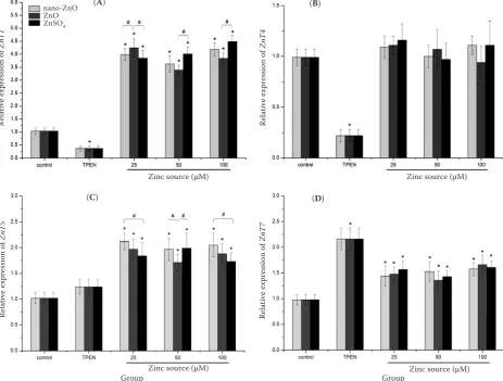
Effects of three zinc sources on Zn transport-ers mRNA expressions in IEC-6 cells. To test
the effects of nano-ZnO, ZnO, and ZnSO4 on
the transcriptional level of different related zinc transporters, cells with the three Zn sources were incubated for 24 h and total RNAs were prepared. Reverse transcription PCR results showed that all of the genes were expressed in IEC-6 cells (Fig-ures 4 and 5). Significant (P < 0.05) decreases in
a 14 a 10 12 (% )
ZnSO4 Nano-ZnO ZnO
a a 8 10 ficiency a 6 ption ef f
b b b b
2 4
Abso
r
b b b b
c c c c c
0
1 4 7 10 13 16 19 22 25
1 4 7 10 13 16 19 22 25
[image:5.595.66.288.90.264.2]Time (h)
Figure 1. Uptake efficiency of nano-ZnO, feed-grade ZnO, and ZnSO4 in IEC-6 cells
cells propagated on transwell filters were cultured with various concentrations of three zinc forms for 24 h; medium treated with 2 µM TPEN (Zn-deficient medium)
*significant difference when compared with 25 µM Zn level (P < 0.05); #significant difference when comparing between
[image:5.595.303.534.98.256.2]Zn sources (P < 0.05) data are means ± SD (n = 8)
Figure 2. Absorption efficiency of nano-ZnO, feed-grade ZnO, and ZnSO4 in IEC-6 cells
cells propagated on transwell filters were cultured with 50 µM Zn for different times as indicated; medium treated with 2 µM TPEN (Zn-deficient medium)
a–csignificant difference when comparing between Zn sources
[image:5.595.65.530.554.689.2]at the same time point (P < 0.05) data are means ± SD (n = 8)
Figure 3. Effects of TPEN (Zn-deficient medium) or a combination with nano-ZnO, ZnO, and ZnSO4 on cell viability (%) cells propagated on transwell filters were cultured with various concentrations of three Zn forms for 24 h (A) or 48 h (B)
a–cdifferent Zn sources are compared and means with different superscripts within the same Zn concentration differ
sig-nificantly (P < 0.05) data are means ± SD, n = 8
Group U pt ak e effic ienc y (%)
25 μM 50 μM 100 μM
nano-ZnO ZnO ZnSO4 a a a b a a b b b 60 80 100 120 140 l v iabili ty (% ) 0 20 40
TPEN 25 μM 50 μM 100 μM
C
el
l
Group
Nano-ZnO ZnO ZnSO4
a a b b a a c b c 60 80 100 120 140 160 lvi ab ilit y (% )
Nano-ZnO ZnO ZnSO4
0 20 40
TPEN 25 μM 50 μM 100 μM
the mRNA expressions of ZnT-1 and ZnT-4 were observed with supplemented TPEN, whereas the transcriptional level of ZnT-7 was significantly increased (P < 0.05). On the other hand, the ad-ministration of TPEN did not change the mRNA expression of ZnT-5 (P > 0.05).
Compared with control group, rapid and signifi-cant increases in the ZnT-1, ZnT-5, and ZnT-7 gene expressions were observed after supplementing with different concentrations of nano-ZnO, ZnO,
and ZnSO4 (P < 0.05). However, addition of
dif-ferent forms of zinc to the medium had no effects on the ZnT-4 transcription level (P > 0.05). There were no significant differences in the ZnT-1, ZnT-4, ZnT-5, and ZnT-7 gene expressions between the three Zn sources at 25, 50, or 100 µM
concentra-tion, respectively (P > 0.05). When different Zn sources of 50 µM and 100 µM were added, mRNA expression of ZnT-1 in ZnO group was lower than
that in nano-ZnO and ZnSO4 groups (P > 0.05).
The mRNA expressions of ZnT-5 with the addi-tion of 25 µM and 100 µM nano-ZnO were higher
than those with ZnSO4 and ZnO, and showed a
difference between ZnO and nano-ZnO at the concentration of 50 µM (P > 0.05). In addition, the mRNA expression of ZnT-7 with the addition of 50 µM nano-ZnO was higher than those with
ZnSO4 and ZnO group at the same concentration
(P > 0.05).
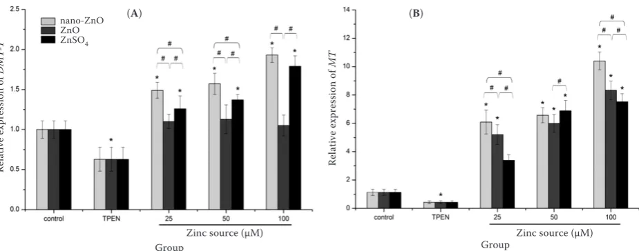
[image:6.595.66.530.322.673.2]Effects of three zinc sources on DMT-1 and MT mRNA expressions in IEC-6 cells. We further investigated the mRNA expressions of DMT-1 and
Figure 4. Effects of TPEN (Zn-deficient medium) or a combination with nano-ZnO, feed-grade ZnO, and ZnSO4 on mRNA expression of ZnT-1 (A), ZnT-4 (B), ZnT-5 (C), ZnT-7 (D) in IEC-6 cells
cells propagated on transwell filters were incubated with or without (control) experimental agents for 24 h *significant difference when compared with control (P < 0.05)
#significant difference when comparing Zn sources within the same Zn concentration (P < 0.05)
data are means ± SD (n = 8)
Re
la
tive e
xpr
ession of
ZnT
1
Zinc source (μM)
nano-ZnO ZnO ZnSO4
(A)
Re
la
tive e
xpr
ession of
ZnT
4
Zinc source (μM) (B)
Group Zinc source (μM)
Re
la
tive e
xpr
ession of
ZnT
5
(C)
Group Zinc source (μM)
Re
la
tive e
xpr
ession of
ZnT
7
MT with the incubation of different zinc sources (Figure 5). A down-regulation of DMT-1 and MT gene expressions with TPEN treatment (P < 0.05) was registered. DMT-1 mRNA expression was increased significantly by supplementing with
nano-ZnO and ZnSO4 (P < 0.05), and no differences
were observed between ZnO and control group (P > 0.05). Moreover, when 25 µM and 50 µM nano-ZnO were added, the mRNA expressions of DMT-1 were significantly higher than those of ZnO and
ZnSO4 groups (P < 0.05). At the concentration of
100 µM , the DMT-1 mRNA expressions of
nano-ZnO and ZnSO4 were significantly higher (P < 0.05)
than that of ZnO. The MT mRNA expression was up-regulated by the three zinc sources at all con-centrations (P < 0.05). Furthermore, the results showed that the mRNA expressions of MT with 25 µM and 100 µM nano-ZnO treatments were
higher than those of ZnO and ZnSO4 (P < 0.05).
DISCUSSION
The IEC-6 cell derived from the rat intestinal epithelial cell is a good model system for study-ing nutrient uptake and transportation because it is difficult to investigate specific environmental parameters in a whole animal. It has been suc-cessfully and widely used in numerous
experi-mental studies for trace elements. In the current study, IEC-6 cell was used as a model to assess
the effect of nano-ZnO, ZnO, ZnSO4, and TPEN
on cell viability, zinc uptake efficiency, and zinc transporters gene expression. The cell viability inhibition by TPEN has already been suggested (Shen et al. 2008). Furthermore, a more recent study reported that TPEN addition noticeably lowered cell viability in HT-29 cells (Gurusamy et al. 2011). In the present study the effects of TPEN on cell viability were similar. Our results showed that viability of cells exposed to TPEN significantly decreased, and indicated that TPEN, a kind of special zinc-chelating reagent, chelated intercellular or intracellular zinc. TPEN thereby impacts the activities of key enzymes that need zinc as a co-factor.
However, no precise figure has been available up to date about the effects of nano-ZnO and ZnO on cell viability and comparison of cell viability between different kinds of inorganic zinc sources in IEC-6 cells. In the present study, viability of cells exposed to nano-ZnO increased significantly and was higher than of those exposed to ZnO at lower concentrations. Furthermore, medium
treatment of ZnSO4 had no significant protective
effect on cell viability, and the three zinc sources at higher concentrations could even decrease it.
The increase of intracellular free Zn2+ which is
Figure 5. Effects of TPEN (Zn-deficient medium) and nano-ZnO, feed-grade ZnO, and ZnSO4 on mRNA expression of divalent metal transporter (DMT-1) (A) and metallothionein (MT) (B) in IEC-6 cells
cells propagated on transwell filters were incubated with or without (control) experimental agents for 24 h *significant difference when compared with control (P < 0.05)
#significant difference when comparing Zn sources within the same concentration (P < 0.05)
data are means ± SD (n = 8)
Re
la
tive e
xpr
ession of
D
M
T-1
Zinc source (μM)
nano-ZnO ZnO ZnSO4
(A)
Group
Re
la
tive e
xpr
ession of
MT
Zinc source (μM) Group
deleterious could eventually lead to the decrease of cell viability and apoptosis (Sakabe et al. 1998),
particularly in ZnSO4 treatment. While ZnSO4 is
totally water-soluble and Zn2+is released in large
quantity, nano-ZnO and ZnO dissolution in water
is very limited. Thus dissolved Zn2+ may gradually
release from cell to meet the need of tissue, even though the dissolution of nano-ZnO would be changed under acidic conditions as well as at the presence of biological components such as amino acids and peptides (Moreau et al. 2007; Xia et al. 2008). The latter study also showed that
cytotoxic-ity of ZnSO4 was still significantly higher than of
nano-ZnO (Xia et al. 2008). The above results also demonstrated that in the IEC-6 cells, nano-ZnO at lower concentration was more bioavailable.
In our experiments, the uptake efficiency of nano-ZnO we observed was significantly higher
than that of ZnO and ZnSO4 groups at all
concen-trations. As mentioned above, the uptake mecha-nism of three zinc sources differs, and the study confirmed that of nano-ZnO was more effecive. Xia et al. (2008) investigated the uptake of ZnO nanoparticle in macrophages (RAW 264.7) and human bronchial epithelial cells (BEAS-2B) using inductively coupled plasma-mass spectrometry (ICP-MS) and fluorescent-labelled labelling ZnO, and it was found out that dissolution of ZnO could happen in the culture medium and endosomes.
In addition, we found that the cellular uptake of nano-ZnO reached a maximum level 3 h post-incubation and decreased with increasing incuba-tion time. This is in line with Yu and Baek (2011) who reported that after human lung epithelial cells had been exposed to the concentration of 125 μg/ml nano-ZnO for a given time, the uptake efficiency peaked at 4 h of nano-ZnO administra-tion and then it decreased (Yu and Baek 2011). In
contrast, the uptake efficiency of ZnSO4 seemed
to peak later. Moreover, no significant differences were observed in the uptake efficiency of ZnO. The explanation of the time response of nano-ZnO may have many aspects. Intrinsically, when the con-centration of nano-ZnO is below solubility, cells are mainly exposed to zinc ions, whereas when the concentration exceeds, cells are confronted with molecular nanoparticles (Cousins et al. 2006). Zinc ions of nano-ZnO were released gradually instead of rapidly. Thus, it was the reason for the higher uptake efficiency of nano-ZnO during the whole incubation time. Secondly, a self-protection
mechanism of cells could be provoked to prevent intracellular zinc excess while being exposed to high Zn levels for a long time. Furthermore, the physicochemical parameters (e.g. size, shape, sur-face area, electrostatic charge, materials composi-tion, hydrophobicity) might be important factors affecting the uptake efficiency, which remains to be tested.
From a physiological perspective, the cell state is
optimal with an appropriate intracellular free Zn2+
level. However, it can be regulated via different transport proteins involved in zinc uptake, storage, and excretion that maintain the zinc homeostasis. In the current study, we examined the effects of different zinc sources on the transcriptional
re-sponse of ZnT-1, ZnT-4, ZnT-5, ZnT-7, DMT-1,
and MT by real-time PCR. The results show that the zinc-deficient status (TPEN addition) leads to significant down-regulation of the mRNA expres-sions of ZnT-1 and ZnT-4, and the absent accounts of ZnT-1 lead to an increased sensitivity of cells to zinc toxicity (Palmiter 2004).
All zinc sources treatments exhibited huge up-regulations of ZnT-1, ZnT-5, and ZnT-7. Conversely, ZnT-4 gene expression did not change in any zinc treatment. This is consistent with an in vitro study which found that ZnT-1 expression is markedly regulated by the dietary zinc supply (Liuzzi et al. 2001), and moreover showed that variation in zinc status does not affect ZnT-4 mRNA levels in small intestine of rat. ZnT-1 plays an important role in zinc efflux of cells. Thus, cells could be up-regulated at high intracellular zinc concentra-tions and down-regulated at low level to maintain zinc homeostasis in IEC-6 cells. Our finding also showed that there were no significant variations
of mRNA expressions of ZnT-1, ZnT-4, ZnT-5,
and ZnT-7 between the three zinc treatments. ZnT-1 was sensitive to zinc ions, and even though nano-ZnO can only release small amount of free
Zn2+, the gradually released zinc could also keep
increases in ZnT-7 and ZnT-5 genes expression in IEC-6 cells upon different zinc sources or TPEN treatment and reduction at high zinc concentra-tion. Our results also suggested that ZnT-5 and ZnT-7 played different roles both at absorption and storage stages.
DMT-1 is predominantly an iron transporter
such as zinc. The present study found a reduc-tion of DMT-1 mRNA expression after addireduc-tion of TPEN, but its increase with the nano-ZnO and
ZnSO4 supplementation. This result is in agreement
with Shen et al. (2008), who reported that mRNA expression of DMT-1 is susceptible to low zinc concentration and resistant to TPEN treatment (Stark 2011). But the transporting mechanism of zinc regulated by DMT-1 has not been investigated yet, thus more detailed exploration is needed. Davis et al. (1998) reported that intestinal zinc absorption was more intense in MT knockout mice than wild, and this result was also further supported by that MT was a vital component for zinc homeostasis regulation. Our results showed that compared with DMT-1 mRNA expression, MT was more sensitive to zinc concentration. This was similar to a previous study, which reported that MT expression was down-regulated during zinc depletion (Cao et al. 2001). These findings are in line with the fact that MT expression was up-regulated at high zinc concentrations to boost zinc efflux, and down-regulated at low concentra-tions to enhance zinc shortage.
CONCLUSION
In conclusion, the current study showed that the effect of zinc in IEC-6 cells varied with different chemical and physical forms (nano-ZnO, ZnO, and
ZnSO4) and the differences concerned cell activity,
uptake, and transport. Compared with the control, supplementation with the three zinc sources en-hanced cell activity and uptake efficiency. Lower concentration of nano-ZnO was more bioavailable for IEC-6 cells viability. The study compared the effects of the three zinc sources on cell viability and confirmed a biphasic effect of zinc with its differ-ent chemical forms. This study also presdiffer-ented that supplementations with the three Zn forms could significantly increase ZnT-1, ZnT-5, MT, and DMT-1,
and that ZnT-5, MT, and DMT-1 expressions in
nano-ZnO treatment were greater than those in
ZnO and ZnSO4 group. In addition, the uptake
and transport of nano-ZnO across the IEC-6 cell monolayers partly via zinc related transporters were induced by a released zinc ion.
REFERENCES
Cao J., Bobo J.A., Liuzzi J.P., Cousins R.J. (2001): Effects of intracellular zinc depletion on metallothionein and ZIP2 transporter expression and apoptosis. Journal of Leukocyte Biology, 70, 559–566.
Case C.L., Carlson M.S. (2002): Effect of feeding organic and inorganic sources of additional zinc on growth per-formance and zinc balance in nursery pigs. Journal of Animal Science, 80, 1917–1924.
Cherian M.G., Howell S.B., Imura N., Klaassen C.D., Koro-patnick J., Lazo J.S., Waalkes M.P. (1994): Contemporary issues in toxicology: role of metallothionein in carcino-genesis. Toxicology and Applied Pharmacology, 126, 1–5. Cousins R.J., McMahon R.J. (2000): Integrative aspects of
zinc transporters. The Journal of Nutrition, 130, 1384– 1387.
Cousins R.J., Liuzzi J.P., Lichten L.A. (2006): Mammalian zinc transport, trafficking, and signals. Journal of Biologi-cal Chemistry, 281, 24085–24089.
Cragg R.A., Phillips S.R., Piper J.M., Varma J.S., Campbell F.C., Mathers J.C., Ford D. (2005): Homeostatic regula-tion of zinc transporters in the human small intestine by dietary zinc supplementation. Gut, 54, 469–478. Davis S.R., McMahon R.J., Cousins R.J. (1998):
Metallothio-nein knockout and transgenic mice exhibit altered intestinal processing of zinc with uniform zinc-dependent zinc trans-porter-1 expression. The Journal of Nutrition, 128, 825–831. De Berardis B., Civitelli G., Condello M., Lista P., Pozzi R.,
Arancia G., Meschini S. (2010): Exposure to ZnO na-noparticles induces oxidative stress and cytotoxicity in human colon carcinoma cells. Toxicology and Applied Pharmacology, 246, 116–127.
Gurusamy K.S., Farooqui N., Loizidou M., Dijk S., Taanman J.W., Whiting S., Farquharson M.J., Fuller B.J., Davidson B.R. (2011): Influence of zinc and zinc chelator on HT-29 colorectal cell line. Biometals, 24, 143–151.
Hanley C., Layne J., Punnoose A., Reddy K.M., Coombs I., Coombs A., Feris K., Wingett D. (2008): Preferential kill-ing of cancer cells and activated human T cells uskill-ing ZnO nanoparticles. Nanotechnology, 19: 295103.
Hojberg O., Canibe N., Poulsen H.D., Hedemann M.S., Jensen B.B. (2005): Influence of dietary zinc oxide and copper sulfate on the gastrointestinal ecosystem in newly weaned piglets. Applied and Environmental Microbiol-ogy, 71, 2267–2277.
Huang L., Tepaamorndech S. (2013): The SLC30 family of zinc transporters – a review of current understanding of their biological and pathophysiological roles. Molecular Aspects of Medicine, 34, 548–560.
Kambe T., Narita H., Yamaguchi-Iwai Y., Hirose J., Ama-no T., Sugiura N., Sasaki R., Mori K., Iwanaga T., Na-gao M. (2002): Cloning and characterization of a novel mammalian zinc transporter, zinc transporter 5, abun-dantly expressed in pancreatic β cells. Journal of Biologi-cal Chemistry, 277, 19049–19055.
Kirschke C.P., Huang L. (2003): ZnT-7, a novel mammalian zinc transporter, accumulates zinc in the Golgi apparatus. Journal of Biological Chemistry, 278, 4096–4102. Liuzzi J.P., Blanchard R.K., Cousins R.J. (2001): Differential
regulation of zinc transporter 1, 2, and 4 mRNA expres-sion by dietary zinc in rats. The Journal of Nutrition, 131, 46–52.
Mavromichalis I., Peter C.M., Parr T.M., Ganessunker D., Baker D.H. (2000): Growth-promoting efficacy in young pigs of two sources of zinc oxide having either a high or a low bioavailability of zinc. Journal of Animal Science, 78, 2896–2902.
Miller H.M., Toplis P., Slade R.D. (2009): Can outdoor rearing and increased weaning age compensate for the removal of in-feed antibiotic growth promoters and zinc oxide? Livestock Science, 125, 121–131.
Moreau J.W., Weber P.K., Martin M.C., Gilbert B., Hutch-eon I.D., Banfield J.F. (2007): Extracellular proteins limit the dispersal of biogenic nanoparticles. Science, 316, 1600–1603.
Owusu-Asiedu A., Nyachoti C.M., Marquardt R.R. (2003): Response of early-weaned pigs to an enterotoxigenic
Es-cherichia coli (K88) challenge when fed diets containing spray-dried porcine plasma or pea protein isolate plus egg yolk antibody, zinc oxide, fumaric acid, or antibiotic. Journal of Animal Science, 81, 1790–1798.
Palmiter R.D. (2004): Protection against zinc toxicity by metallothionein and zinc transporter 1. Proceedings of the National Academy of Sciences of the United States of America, 101, 4918–4923.
Palmiter R.D., Huang L. (2004): Efflux and compartmen-talization of zinc by members of the SLC30 family of solute carriers. Pflügers Archiv – European Journal of Physiology, 447, 744–751.
Qin Y., Thomas D., Fontaine C.P., Colvin R.A. (2009): Si-lencing of ZnT1 reduces Zn2+ efflux in cultured cortical
neurons. Neuroscience Letters, 450, 206–210.
Sakabe I., Paul S., Dansithong W., Shinozawa T. (1998): In-duction of apoptosis in Neuro-2A cells by Zn2+ chelating.
Cell Structure and Function, 23, 95–99.
Sandoval M., Henry P.R., Ammerman C.B., Miles R.D., Lit-tell R.C. (1997): Relative bioavailability of supplemental inorganic zinc sources for chicks. Journal of Animal Sci-ence, 75, 3195–3205.
Shen H., Qin H., Guo J. (2008): Cooperation of metallothio-nein and zinc transporters for regulating zinc homeosta-sis in human intestinal Caco-2 cells. Nutrition Research, 28, 406–413.
Stark W.J. (2011): Nanoparticles in biological systems. An-gewandte Chemie International Edition, 50, 1242–1258. Xia T., Kovochich M., Liong M., Madler L., Gilbert B.,
Shi H., Yeh J.I., Zink J.I., Nel A.E. (2008): Comparison of the mechanism of toxicity of zinc oxide and cerium oxide nanoparticles based on dissolution and oxidative stress properties. ACS Nano, 2, 2121–2134.
Yu J., Baek M. (2011): Effects of physicochemical properties of zinc oxide nanoparticles on cellular uptake. Journal of Physics: Conference Series, 34, 301.