3-(4-Nitrobenzyl)-4
H
-chromen-4-one
Kaalin Gopaul,aNeil Anthony Koorbanally,a*
Mahidansha M. Shaikh,aDeresh Ramjugernathband Hong Suc
aSchool of Chemistry and Physics, University of KwaZulu-Natal, Durban 4000, South Africa,bSchool of Chemical Engineering, University of KwaZulu-Natal, Durban, South Africa, andcChemistry Department, University of Cape Town, Rondebosch 7701, South Africa
Correspondence e-mail: [email protected]
Received 20 February 2013; accepted 4 March 2013
Key indicators: single-crystal X-ray study;T= 173 K; mean(C–C) = 0.003 A˚; Rfactor = 0.050;wRfactor = 0.154; data-to-parameter ratio = 17.3.
In the title compound, C16H11NO4, the dihedral angle between
the ten-membered chromen-4-one ring system (r.m.s. devia-tion = 0.0095 A˚ ) and the benzene ring is 86.16 (5). In the crystal, molecules are linked into a three-dimensional network by weak C—H O hydrogen bonds. The crystal studied was a non-merohedral twin, with the minor twin component refining to 0.093 (1).
Related literature
For the preparation, see: Desideriet al.(2011); Valkonenet al. (2012). For related structures, see: Valkonen et al. (2012); Gopaul et al.(2013). For the biological activity of homoiso-flavonoids, see: Abegazet al.(2007).
Experimental
Crystal data
C16H11NO4 Mr= 281.26 Monoclinic,P21=n a= 4.9246 (9) A˚ b= 10.0160 (19) A˚
c= 25.907 (5) A˚
= 92.845 (4)
V= 1276.3 (4) A˚3 Z= 4
MoKradiation
= 0.11 mm1 T= 173 K
0.350.090.08 mm
Data collection
Bruker Kappa DUO APEXII diffractometer
Absorption correction: multi-scan (TWINABS; Sheldrick, 1997) Tmin= 0.964,Tmax= 0.992
87638 measured reflections 3303 independent reflections 2887 reflections withI> 2(I) Rint= 0.063
Refinement
R[F2> 2(F2)] = 0.050 wR(F2) = 0.154 S= 1.09 3303 reflections
191 parameters
H-atom parameters constrained
max= 0.25 e A˚
3
min=0.23 e A˚3
Table 1
Hydrogen-bond geometry (A˚ ,).
D—H A D—H H A D A D—H A
C2—H2 O2i
0.95 2.38 3.322 (3) 170
C7—H7 O3ii 0.95 2.56 3.491 (3) 166
C9—H9 O4iii
0.95 2.42 3.355 (3) 167
C13—H13 O3iv
0.95 2.53 3.382 (3) 149
C16—H16 O2v 0.95 2.46 3.378 (3) 163
Symmetry codes: (i) xþ3
2;y12;zþ12; (ii) x12;yþ32;z12; (iii)
xþ5
2;y12;zþ12; (iv)xþ1;yþ1;zþ1; (v)xþ1;y;z.
Data collection:APEX2(Bruker, 2006); cell refinement:SAINT
(Bruker, 2006); data reduction:SAINT; program(s) used to solve structure:SHELXS97(Sheldrick, 2008); program(s) used to refine structure: SHELXL97 (Sheldrick, 2008); molecular graphics:
ORTEP-3 for Windows(Farrugia, 2012); software used to prepare material for publication:SHELXL97.
We thank the University of KwaZulu-Natal and the South Africa Research Chairs initiative of the Department of Science and Technology for financial support and the National Research Foundation of South Africa for a bursary for KG.
Supplementary data and figures for this paper are available from the IUCr electronic archives (Reference: FJ2618).
References
Abegaz, B. M., Mutanyatta-Comar, J. & Nindi, M. (2007).Nat. Prod. Commun. 2, 475–498.
Bruker (2006).APEX2andSAINT. Bruker AXS Inc., Madison, Wisconsin, USA.
Desideri, N., Bolasco, A., Fioravanti, R., Proietti Monaco, L., Orallo, F., Ya´n˜ez, M., Ortuso, F. & Alcaro, S. (2011).J. Med. Chem.54, 2155–2164. Farrugia, L. J. (2012).J. Appl. Cryst.45, 849–854.
Gopaul, K., Koorbanally, N. A., Shaikh, M., Su, H. & Ramjugernath, D. (2013). Acta Cryst.E69, o364.
Sheldrick, G. M. (1997).TWINABS. University of Go¨ttingen, Germany. Sheldrick, G. M. (2008).Acta Cryst.A64, 112–122.
Valkonen, A., Laihia, K., Kolehmainen, E., Kauppinen, R. & Perje´si, P. (2012). Struct. Chem.23, 209–217.
Acta Crystallographica Section E Structure Reports Online
supporting information
Acta Cryst. (2013). E69, o513 [doi:10.1107/S1600536813006119]
3-(4-Nitrobenzyl)-4
H
-chromen-4-one
Kaalin Gopaul, Neil Anthony Koorbanally, Mahidansha M. Shaikh, Deresh Ramjugernath and
Hong Su
S1. Comment
Homoisoflavonoids are a group of naturally occurring oxygen heterocyclic compounds, related to the flavonoids, which
consists of a chromone skeleton with a benzyl or benzylidene group at C-3. In the 3-benzyl-4-chromonanone class of
homoisoflavonoid, an extra methylene group exists between the phenyl group and the chromone skeleton. They are
commonly synthesized by either the acid or base catalysed condensation of an aromatic aldehyde with chromanone
(Desideri et al., 2011, Valkonen et al., 2012). Naturally occurring homoisoflavonoids are normally oxygenated and have
shown a wide range of biological activities (Abegaz et al., 2007).
The molecular structure of the title compound is shown in Fig. 1. The dihedral angle between the 10-membered
co-planar chromone ring and the nitrated phenyl ring is 86.16 (5)°. In the crystal, the inter-moclecular weak hydrogen bonds
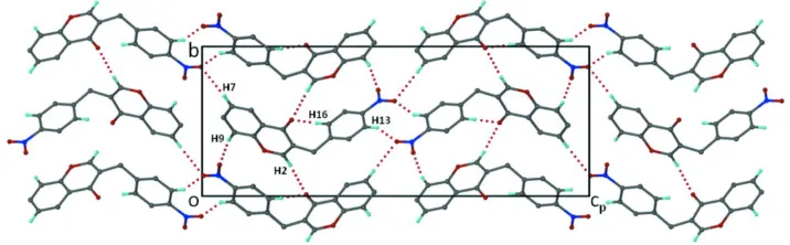
C—H···O link the molecules into a three dimentional network, as shown in Fig.2. The details of the hydrogen bonds are
shown in Table 1.
S2. Experimental
A mixture of chroman-4-one (1.02 g, 6.749 mmol), 4-nitrobenzaldehyde (1.22 g, 8.099 mmol) and 10–15 drops of
piperidine was heated at 80°C for 12 hrs. The reaction mixture was monitored for completion by thin layer
chromatography. Upon completion, the reaction mixture was cooled, diluted with water and neutralized using 10% HCl.
To the viscous reaction mixture, 20 ml of ethyl acetate was added. Upon the addition of hexane to the reaction mixture,
the homoisoflavonoid precipitated out. The powdered product was filtered, washed with hexane and dried under vacuum.
Upon slow evaporation of chloroform, the crystals of the homoisoflavonoid were obtained. (m.p. of 179–180 °C).
1H NMR (400 MHz, CDCl
3) δ: 3.87 (2H, s, H-9), 7.38 (1H, t, J=7.54 Hz, H-6), 7.41 (1H, d, J=8.60 Hz, H-8), 7.45 (1H,
d, J=8.48 Hz, H-2′/6′), 7.65 (1H, td, J=8.32, 1.10 Hz, H-7), 7.79 (1H, s, H-2), 8.12 (1H, d, J=8.56 Hz, H-3′/5′), 8.18 (1H,
dd, J=7.98, 0.66 Hz, H-5).
13C NMR (100 MHz, CDCl
3) δ: 31.89 (C-9), 118.15 (C-8), 123.06 (C-3/1′), 123.79 (C-3′/5′), 123.83 (C-4a), 125.32
(C-6), 125.92 (C-5), 129.65 (C-2′/6′), 133.84 (C-7), 146.76 (C-4′), 153.14 (C-2), 156.52 (C-8a), 177.18 (C-4).
S3. Refinement
The crystal was a non-merohedral twin. Two domains were indexed using CELL_NOW1 and the intensity data for each
domain was then integrated, reduced using the program SAINT. The combined data were scaled and absorption correction
performed using TWINABS. The structure was solved by direct methods using SHELXS97 and refined by full-matrix
least-squares methods based on F2 using SHELXL97. All non-hydrogen atoms were refined anisotropically. All hydrogen
atoms were placed in idealized positions and refined with geometrical constraints. The structure was refined to R factor =
Figure 1
A view of the molecule with displacement ellipsoids drawn at the 50% probability level and H atoms drawn as circles of
arbitary size.
Figure 2
Projection viewed along the a axis, showing the inter-molecular C—H···O hydrogen bonding network. The H-bond
involving H atoms on the molecule of the asymmetric unit were marked for identification. The other H atoms were
omitted for clarity. The H-bonds are shown as dotted lines.
3-(4-Nitrobenzyl)-4H-chromen-4-one
Crystal data
C16H11NO4 Mr = 281.26
Monoclinic, P21/n
Hall symbol: -P 2yn
a = 4.9246 (9) Å
b = 10.0160 (19) Å
c = 25.907 (5) Å
β = 92.845 (4)°
V = 1276.3 (4) Å3 Z = 4
F(000) = 584
Dx = 1.464 Mg m−3
Mo Kα radiation, λ = 0.71073 Å Cell parameters from 3303 reflections
θ = 1.6–27.4°
µ = 0.11 mm−1 T = 173 K Needle, colourless 0.35 × 0.09 × 0.08 mm
Data collection
Bruker Kappa DUO APEXII diffractometer
Radiation source: fine-focus sealed tube Graphite monochromator
0.5° φ scans and ω scans
Absorption correction: multi-scan (TWINABS; Sheldrick, 1997)
[image:3.610.126.484.289.400.2]2887 reflections with I > 2σ(I)
Rint = 0.063
θmax = 27.4°, θmin = 1.6°
h = −6→6
k = 0→12
l = 0→33
Refinement
Refinement on F2
Least-squares matrix: full
R[F2 > 2σ(F2)] = 0.050 wR(F2) = 0.154 S = 1.09 3303 reflections 191 parameters 0 restraints
Primary atom site location: structure-invariant direct methods
Secondary atom site location: difference Fourier map
Hydrogen site location: inferred from neighbouring sites
H-atom parameters constrained
w = 1/[σ2(Fo2) + (0.0779P)2 + 0.7278P]
where P = (Fo2 + 2Fc2)/3
(Δ/σ)max < 0.001
Δρmax = 0.25 e Å−3
Δρmin = −0.23 e Å−3
Special details
Geometry. All e.s.d.'s (except the e.s.d. in the dihedral angle between two l.s. planes) are estimated using the full covariance matrix. The cell e.s.d.'s are taken into account individually in the estimation of e.s.d.'s in distances, angles and torsion angles; correlations between e.s.d.'s in cell parameters are only used when they are defined by crystal symmetry. An approximate (isotropic) treatment of cell e.s.d.'s is used for estimating e.s.d.'s involving l.s. planes.
Refinement. Refinement of F2 against ALL reflections. The weighted R-factor wR and goodness of fit S are based on F2,
conventional R-factors R are based on F, with F set to zero for negative F2. The threshold expression of F2 > σ(F2) is used
only for calculating R-factors(gt) etc. and is not relevant to the choice of reflections for refinement. R-factors based on F2
are statistically about twice as large as those based on F, and R- factors based on ALL data will be even larger.
Fractional atomic coordinates and isotropic or equivalent isotropic displacement parameters (Å2)
x y z Uiso*/Ueq
O1 0.9485 (3) 0.30392 (15) 0.16095 (6) 0.0339 (4) O2 0.3498 (3) 0.50967 (15) 0.23136 (6) 0.0355 (4) O3 0.8175 (4) 0.63590 (18) 0.49727 (6) 0.0449 (4) O4 1.1513 (4) 0.7098 (2) 0.45470 (7) 0.0522 (5) N1 0.9499 (4) 0.63878 (18) 0.45839 (7) 0.0328 (4) C1 0.8059 (4) 0.4022 (2) 0.13399 (8) 0.0282 (4) C2 0.8774 (5) 0.2756 (2) 0.20963 (8) 0.0323 (4)
H2 0.9747 0.2063 0.2275 0.039*
C3 0.6800 (4) 0.33749 (19) 0.23490 (7) 0.0281 (4) C4 0.5276 (4) 0.44552 (19) 0.20968 (7) 0.0264 (4) C5 0.5991 (4) 0.47384 (19) 0.15623 (7) 0.0260 (4) C6 0.4653 (5) 0.5734 (2) 0.12658 (8) 0.0333 (5)
H6 0.3235 0.6233 0.1410 0.040*
C7 0.5368 (5) 0.6000 (2) 0.07681 (9) 0.0383 (5)
H7 0.4457 0.6681 0.0572 0.046*
C8 0.7441 (5) 0.5262 (3) 0.05552 (8) 0.0402 (5)
H8 0.7927 0.5445 0.0212 0.048*
C9 0.8801 (5) 0.4268 (2) 0.08354 (8) 0.0364 (5)
H9 1.0205 0.3767 0.0688 0.044*
C10 0.6125 (5) 0.2953 (2) 0.28877 (8) 0.0343 (5)
H10A 0.6971 0.2072 0.2960 0.041*
C11 0.7026 (4) 0.39038 (19) 0.33199 (7) 0.0273 (4) C12 0.5763 (5) 0.3825 (2) 0.37937 (8) 0.0347 (5)
H12 0.4330 0.3203 0.3831 0.042*
C13 0.6553 (5) 0.4630 (2) 0.42049 (8) 0.0341 (5)
H13 0.5682 0.4569 0.4523 0.041*
C14 0.8652 (4) 0.55301 (19) 0.41435 (7) 0.0273 (4) C15 0.9971 (4) 0.5640 (2) 0.36858 (8) 0.0313 (4)
H15 1.1411 0.6261 0.3653 0.038*
C16 0.9136 (4) 0.4816 (2) 0.32739 (8) 0.0309 (4)
H16 1.0018 0.4879 0.2957 0.037*
Atomic displacement parameters (Å2)
U11 U22 U33 U12 U13 U23
O1 0.0369 (8) 0.0348 (7) 0.0303 (7) 0.0077 (7) 0.0045 (6) −0.0026 (6) O2 0.0400 (8) 0.0371 (8) 0.0303 (8) 0.0037 (7) 0.0105 (6) −0.0019 (6) O3 0.0529 (10) 0.0535 (10) 0.0290 (8) −0.0090 (8) 0.0089 (7) −0.0110 (7) O4 0.0546 (11) 0.0583 (11) 0.0440 (10) −0.0286 (9) 0.0047 (8) −0.0118 (8) N1 0.0365 (9) 0.0334 (9) 0.0284 (9) −0.0030 (8) 0.0001 (7) −0.0015 (7) C1 0.0308 (9) 0.0287 (9) 0.0250 (9) −0.0015 (8) 0.0003 (8) −0.0025 (7) C2 0.0409 (11) 0.0274 (9) 0.0282 (10) 0.0017 (9) −0.0023 (9) −0.0006 (8) C3 0.0368 (10) 0.0251 (9) 0.0222 (9) −0.0056 (8) 0.0003 (8) −0.0022 (7) C4 0.0315 (9) 0.0242 (8) 0.0237 (9) −0.0051 (8) 0.0034 (8) −0.0030 (7) C5 0.0282 (9) 0.0256 (9) 0.0243 (9) −0.0035 (7) 0.0019 (8) −0.0025 (7) C6 0.0362 (10) 0.0321 (10) 0.0316 (10) 0.0035 (9) 0.0018 (9) 0.0024 (8) C7 0.0425 (12) 0.0404 (11) 0.0318 (11) 0.0005 (10) −0.0014 (9) 0.0089 (9) C8 0.0433 (12) 0.0524 (13) 0.0251 (10) −0.0064 (11) 0.0037 (9) 0.0058 (9) C9 0.0358 (11) 0.0461 (12) 0.0278 (10) −0.0012 (10) 0.0067 (9) −0.0044 (9) C10 0.0513 (13) 0.0276 (9) 0.0239 (10) −0.0063 (9) 0.0009 (9) 0.0014 (8) C11 0.0334 (10) 0.0260 (9) 0.0222 (9) 0.0002 (8) −0.0005 (8) 0.0026 (7) C12 0.0384 (11) 0.0375 (11) 0.0286 (10) −0.0126 (9) 0.0042 (9) 0.0005 (8) C13 0.0365 (11) 0.0414 (11) 0.0249 (9) −0.0062 (9) 0.0065 (9) −0.0004 (8) C14 0.0290 (9) 0.0290 (9) 0.0238 (9) 0.0007 (8) −0.0001 (7) −0.0001 (7) C15 0.0320 (10) 0.0334 (10) 0.0285 (10) −0.0060 (8) 0.0017 (8) 0.0022 (8) C16 0.0337 (10) 0.0340 (10) 0.0253 (9) −0.0029 (9) 0.0054 (8) 0.0017 (8)
Geometric parameters (Å, º)
O1—C2 1.356 (3) C7—H7 0.9500
O1—C1 1.379 (3) C8—C9 1.385 (3)
O2—C4 1.243 (2) C8—H8 0.9500
O3—N1 1.227 (2) C9—H9 0.9500
O4—N1 1.228 (2) C10—C11 1.519 (3)
N1—C14 1.472 (3) C10—H10A 0.9900
C1—C5 1.394 (3) C10—H10B 0.9900
C1—C9 1.397 (3) C11—C16 1.393 (3)
C2—C3 1.350 (3) C11—C12 1.406 (3)
C3—C4 1.453 (3) C12—H12 0.9500
C3—C10 1.511 (3) C13—C14 1.387 (3)
C4—C5 1.473 (3) C13—H13 0.9500
C5—C6 1.403 (3) C14—C15 1.385 (3)
C6—C7 1.379 (3) C15—C16 1.394 (3)
C6—H6 0.9500 C15—H15 0.9500
C7—C8 1.396 (3) C16—H16 0.9500
C2—O1—C1 118.18 (16) C8—C9—C1 118.4 (2)
O3—N1—O4 122.80 (18) C8—C9—H9 120.8
O3—N1—C14 118.74 (17) C1—C9—H9 120.8
O4—N1—C14 118.46 (18) C3—C10—C11 115.84 (17)
O1—C1—C5 121.51 (18) C3—C10—H10A 108.3
O1—C1—C9 116.75 (18) C11—C10—H10A 108.3
C5—C1—C9 121.74 (19) C3—C10—H10B 108.3
C3—C2—O1 125.48 (19) C11—C10—H10B 108.3
C3—C2—H2 117.3 H10A—C10—H10B 107.4
O1—C2—H2 117.3 C16—C11—C12 118.41 (18)
C2—C3—C4 119.50 (18) C16—C11—C10 122.68 (18)
C2—C3—C10 121.08 (19) C12—C11—C10 118.86 (18)
C4—C3—C10 119.42 (18) C13—C12—C11 121.51 (19)
O2—C4—C3 122.79 (18) C13—C12—H12 119.2
O2—C4—C5 122.13 (18) C11—C12—H12 119.2
C3—C4—C5 115.08 (17) C12—C13—C14 118.41 (18)
C1—C5—C6 118.21 (18) C12—C13—H13 120.8
C1—C5—C4 120.18 (18) C14—C13—H13 120.8
C6—C5—C4 121.60 (18) C15—C14—C13 122.27 (19)
C7—C6—C5 121.0 (2) C15—C14—N1 119.37 (18)
C7—C6—H6 119.5 C13—C14—N1 118.35 (17)
C5—C6—H6 119.5 C14—C15—C16 118.37 (19)
C6—C7—C8 119.5 (2) C14—C15—H15 120.8
C6—C7—H7 120.2 C16—C15—H15 120.8
C8—C7—H7 120.2 C11—C16—C15 121.03 (18)
C9—C8—C7 121.2 (2) C11—C16—H16 119.5
C9—C8—H8 119.4 C15—C16—H16 119.5
C7—C8—H8 119.4
C2—O1—C1—C5 1.8 (3) C7—C8—C9—C1 −0.3 (4)
C2—O1—C1—C9 −178.58 (19) O1—C1—C9—C8 −179.1 (2)
C1—O1—C2—C3 −1.4 (3) C5—C1—C9—C8 0.5 (3)
O1—C2—C3—C4 −0.9 (3) C2—C3—C10—C11 107.3 (2)
O1—C2—C3—C10 178.39 (19) C4—C3—C10—C11 −73.4 (3)
C2—C3—C4—O2 −177.5 (2) C3—C10—C11—C16 −23.1 (3)
C10—C3—C4—O2 3.2 (3) C3—C10—C11—C12 159.7 (2)
C2—C3—C4—C5 2.6 (3) C16—C11—C12—C13 0.4 (3)
C10—C3—C4—C5 −176.76 (17) C10—C11—C12—C13 177.8 (2)
O1—C1—C5—C6 179.24 (18) C11—C12—C13—C14 −0.1 (3)
O1—C1—C5—C4 −0.1 (3) C12—C13—C14—N1 −179.9 (2)
C9—C1—C5—C4 −179.63 (19) O3—N1—C14—C15 174.2 (2)
O2—C4—C5—C1 177.96 (19) O4—N1—C14—C15 −6.2 (3)
C3—C4—C5—C1 −2.1 (3) O3—N1—C14—C13 −6.2 (3)
O2—C4—C5—C6 −1.3 (3) O4—N1—C14—C13 173.4 (2)
C3—C4—C5—C6 178.63 (18) C13—C14—C15—C16 0.4 (3)
C1—C5—C6—C7 −0.1 (3) N1—C14—C15—C16 179.96 (19)
C4—C5—C6—C7 179.1 (2) C12—C11—C16—C15 −0.3 (3)
C5—C6—C7—C8 0.4 (3) C10—C11—C16—C15 −177.6 (2)
C6—C7—C8—C9 −0.2 (4) C14—C15—C16—C11 0.0 (3)
Hydrogen-bond geometry (Å, º)
D—H···A D—H H···A D···A D—H···A
C2—H2···O2i 0.95 2.38 3.322 (3) 170
C7—H7···O3ii 0.95 2.56 3.491 (3) 166
C9—H9···O4iii 0.95 2.42 3.355 (3) 167
C13—H13···O3iv 0.95 2.53 3.382 (3) 149
C16—H16···O2v 0.95 2.46 3.378 (3) 163