Received 9 January 2019 Accepted 23 January 2019
Edited by S. Parkin, University of Kentucky, USA
Keywords:crystal structure;
2-amino-6-bromopyridine; tetrafluoro-benzoic acid; 2-amino-6-pyridinium 2,3,5,6-tetrafluorobenzoate; hydrogen bond.
CCDC reference:1893117
Supporting information:this article has supporting information at journals.iucr.org/e
Crystal structure of the co-crystal salt
2-amino-6-bromopyridinium 2,3,5,6-tetrafluorobenzoate
Eric Bosch*
Department of Chemistry, Missouri State University, Springfield, MO 65897, USA. *Correspondence e-mail: ericbosch@missouristate.edu
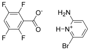
The asymmetric unit of the co-crystal salt 2-amino-6-bromopyridinium 2,3,5,6-tetrafluorobenzoate, C5H6BrN2
+
C7HF4O2
, contains one pyridinium cation and one benzoate anion. In the crystal, the aminopyridinium cationic unit forms two hydrogen bonds to the benzoate oxygen atoms in an R2
2
(8) motif. Two pyridinium benzoate units are hydrogen bonded through self-complementary hydrogen bonds between the second amine hydrogen and a carboxylate O with a secondR22(8) motif to form a discrete hydrogen-bonded complex containing two
2-amino-6-bromopyridinium moieties and two 2,3,5,6-tetrafluorobenzoate moieties. The 2-amino-6-bromopyridinium moieties -stack in a head-to-tail mode with a centroid–centroid separation of 3.7227 (12) A˚ and adjacent tetrafluorobenzoates also -stack in a head-to-tail mode with a centroid– centroid separation of 3.6537 (13) A˚ .
1. Chemical context
The fields of crystal engineering and supramolecular chemistry rely on the identification and application of versatile synthons to guide the construction of molecular solids (Desiraju, 1995, 2013). For example carboxylic acids are known to form a centrosymmetric dimer through self-complementary O— H O hydrogen bonds (Fig. 1a) in addition to hydrogen-bonded catemer chains and rings. It has been shown that these hydrogen bonds can be diverted by O—H N hydrogen bonding to pyridines, often supported by a non-conventional pyridine C—H O hydrogen bond (Fig. 1b). The interaction of the more basic pyridines, for example 4-(N,N -dimethyl-amino)pyridine, with carboxylic acids most often yields charge-assisted hydrogen-bonded salts (Fig. 1c). Similarly, the combination of 2-aminopyridines and benzoic acids has been
ISSN 2056-9890
Figure 1
demonstrated to be a reliable supramolecular synthon resulting in the formation of charge-assisted hydrogen-bonded complexes shown in Fig. 1d (Bis & Zaworotko, 2005). The formation of hydrogen-bonded co-crystals or salts of amines and acids has potential in the pharmaceutical field where the physicochemical properties of active pharmaceuticals, including aqueous solubility and physical and chemical stability, may be modulated and tailored by co-crystal or salt formation (Schultheiss & Newman, 2009). For example a study involving the non-steroidal anti-inflammatory drug piroxicam reported the formation of 19 pyridine based co-crystals (Wales
et al., 2012). The present study presents the first co-crystal/salt formed between a substituted pyridine and 2,3,5,6-tetra-fluorobenzoic acid.
2. Structural commentary
The asymmetric unit of the co-crystal salt 2-amino-6-bromo-pyridinium 2,3,5,6-tetrafluorobenzoate (I), contains one pyri-dinium cation and one benzoate anion that are held together by two charge-assisted hydrogen bonds (Table 1, first two entries) to form an R2
2(8) motif (Fig. 2). The bond distance
C12—O2 is slightly shorter than C12—O1, with distances of 1.236 (2) and 1.267 (2) A˚ respectively. The atoms that form this R2
2(8) motif (Fig. 1) are almost coplanar, with the
maximum deviation above and below the least-squares plane calculated through all of these atoms being 0.169 (7) and 0.147 (8) A˚ , respectively, for O2 and O1. The angle between the planes defined by the benzene and pyridine rings is 67.04 (7)and the carboxylate anion is twisted out of the plane
of the benzene ring, with C12 0.103 (3) A˚ above the plane of the benzene ring and O1 1.043 (3) A˚ above, and O2 0.713 (4) A˚ below the plane defined by the benzene ring.
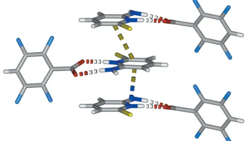
3. Supramolecular features
In the co-crystal salt (I), adjacent amino pyridinium benzoate salt units are linked into dimeric salt complexes with self-complementary hydrogen bonds (Table 1, entry 3) from the second amine hydrogen atom and carboxylate oxygen atom O2 in a secondR2
4(8) motif (Fig. 3). The two components are
relatively well separated within the crystal structure into zones parallel to thecaxis.
There are two interactions that involve the tetrafluoro-benzoate (Fig. 4). Adjacent tetrafluorotetrafluoro-benzoates-stack in a head-to-tail mode with aCg1 Cg1idistance of 3.6537 (13) A˚
research communications
Acta Cryst.(2019). E75, 284–287 Eric Bosch C
5H6BrN2+C7HF4O2
285
Table 1Hydrogen-bond geometry (A˚ ,).
D—H A D—H H A D A D—H A
N1—H1 O1 0.91 (2) 1.68 (2) 2.585 (2) 177 (2)
N2—H2A O2 0.87 (2) 1.98 (2) 2.845 (2) 175 (2)
N2—H2B O2i 0.87 (2) 2.02 (2) 2.854 (2) 162 (2)
C9—H9 Br1ii 0.95 2.94 3.867 (2) 166
[image:2.610.92.248.253.347.2] [image:2.610.315.565.347.523.2]Symmetry codes: (i)xþ1;yþ1;zþ1; (ii)x;yþ1;zþ1.
Figure 2
The molecular structure of the co-crystal salt (I) showing the atom-labeling scheme. Displacement ellipsoids drawn at the 50% probability level and hydrogen bonds (Table 1) are shown as dotted lines.
Figure 3
[image:2.610.316.564.573.713.2]Part of the crystal structure of (I) viewed along b, highlighting the hydrogen-bonded dimeric salt unit.
Figure 4
[image:2.610.46.295.594.710.2][symmetry code: (i)x, 1y,z; Cg1 is the centroid of the benzene ring C6–C11] and there is a close C—F inter-action with a Cg1 F3iidistance of 3.1640 (17) A˚ [symmetry code: (ii)x,y1
2, 1 2z].
The 2-aminopyridinium groups form offset alternating head-to-tail -stacks parallel to the b axis (Fig. 5) with a
Cg2 Cg2iii distance of 3.7227 (12) A˚ and a shortest perpen-dicular interplanar distance of 3.2547 (8) A˚ [symmetry code: (iii) 1x,y1
2, 3
2z;Cg2 is the centroid of the pyridine ring].
There is one short contact to the bromine with a C9 Br1iv distance of 3.867 (2) A˚ [symmetry code: (iv) 1 x, 1 y, 2z].
4. Database survey
A search of the Cambridge Crystallographic Database (Version 5.39, update of August 2018; Groom et al., 2016) usingConquest(Brunoet al., 2002) for structures including the neutral carboxylic acid dimer synthon as shown in Fig. 1a
revealed 6,016 hits, while a search for neutral pyridine carb-oxylic acid interactions where the distance between the acid proton and the pyridine N is equal to or less than the sum of the van der Waals radii revealed 2189 hits. In 966 of the 2189 structures the distance between the carbonyl O and the pyri-dine H is also equal to or less than the sum of the van der Waals radii, corresponding to the synthon shown in Fig. 1b. A related search of the Cambridge Crystallographic Database for co-crystals with 4-(N,N-dimethylamino)pyridine and carboxylic acids revealed only four neutral co-crystals and 54 structures corresponding to the pyridinium carboxylate as shown in Fig. 1c. A similar search for co-crystals formed between 2-aminopyridines with benzoic acids yielded 41 hits, of which 40 feature charge-assisted aminopyridinium carboxylate hydrogen-bonded co-crystals as the result of proton transfer shown in Fig. 1d. The structure that is reported to form a neutral hydrogen-bonded complex corresponds to the co-crystal formed between 2-aminopyridine and 4-aminobenzoic acid [refcode WOPCOV; Chandrasekaran & Babu, 2014]. Finally there is only one reported co-crystal of tetrafluorobenzoic acid, or the corresponding
2,3,5,6-tetrafluorobenzoate, with an organic base. In that example theophylline forms a neutral hydrogen-bonded complex (Corpinotet al., 2016).
5. Synthesis and crystallization
2-Amino-6-bromopyridine and 2,3,5,6-tetrafluorobenzoic acid were used as supplied. An equimolar amount (0.1 mmol) of each component were added to a screw-capped vial and 3 mL of ethanol added to effect a clear colorless solution that was allowed to slowly concentrate over two weeks. A homo-geneous mass of crystals was obtained.
6. Refinement
Crystal data, data collection and structure refinement details are summarized in Table 2. All hydrogen atoms were located in Fourier-difference maps. Hydrogen atoms involved in hydrogen-bonding interactions were restrained in the refine-ment with N—H = 0.87 (2) A˚ and withUiso(H) = 1.2Ueq(N).
The aromatic H atoms were included in the refinement at calculated positions with C—H = 0.95 A˚ and Uiso(H) =
[image:3.610.43.296.74.220.2]1.2Ueq(C).
Table 2
Experimental details.
Crystal data
Chemical formula C5H6BrN2+C7HF4O2
Mr 367.11
Crystal system, space group Monoclinic,P21/c
Temperature (K) 100
a,b,c(A˚ ) 13.7230 (9), 6.5757 (4),
15.3224 (10)
(
) 111.841 (1)
V(A˚3) 1283.42 (14)
Z 4
Radiation type MoK
(mm1) 3.26
Crystal size (mm) 0.250.200.03
Data collection
Diffractometer Bruker APEXII CCD
Absorption correction Multi-scan (SADABS; Bruker,
2014)
Tmin,Tmax 0.788, 1.000
No. of measured, independent and observed [I> 2(I)] reflections
16099, 2847, 2395
Rint 0.045
(sin/)max(A˚1) 0.641
Refinement
R[F2> 2(F2)],wR(F2),S 0.026, 0.059, 1.04
No. of reflections 2847
No. of parameters 199
No. of restraints 3
H-atom treatment H atoms treated by a mixture of
independent and constrained refinement
max, min(e A˚
3) 0.41,0.32
[image:3.610.313.562.87.395.2]Computer programs:SMARTandSAINT(Bruker, 2014),SHELXT2018/2(Sheldrick, 2015a),SHELXL2018/3(Sheldrick, 2015b) andX-SEED(Barbour, 2001).
Figure 5
Acknowledgements
We thank the Missouri State University Provost Incentive Fund that funded the purchase of the X-ray diffractometer.
References
Barbour, L. J. (2001).J. Supramol. Chem.1, 189–191.
Bis, J. A. & Zaworotko, M. J. (2005).Cryst. Growth Des.5, 1169–1179. Bruker (2014).SMART, SAINTand SADABS. Bruker AXS Inc.,
Madison, Wisconsin, USA.
Bruno, I. J., Cole, J. C., Edgington, P. R., Kessler, M., Macrae, C. F., McCabe, P., Pearson, J. & Taylor, R. (2002).Acta Cryst.B58, 389– 397.
Chandrasekaran, J. & Babu, B. (2014). Private communication (Refcode WOPCOV). CCDC, Cambridge, England.
Corpinot, M. K., Stratford, S. A., Arhangelskis, M., Anka-Lufford, J., Halasz, I., Judasˇ, N., Jones, W. & Bucˇar, D.-K. (2016). CrystEng-Comm,18, 5434–5439.
Desiraju, G. R. (1995).Angew. Chem. Int. Ed. Engl.34, 2311–2327. Desiraju, G. R. (2013).J. Am. Chem. Soc.135, 9952–9967.
Groom, C. R., Bruno, I. J., Lightfoot, M. P. & Ward, S. C. (2016).Acta Cryst.B72, 171–179.
Schultheiss, N. & Newman, A. (2009).Cryst. Growth Des. 9, 2950– 2967.
Sheldrick, G. M. (2015a).Acta Cryst.A71, 3–8. Sheldrick, G. M. (2015b).Acta Cryst.C71, 3–8.
Wales, C., Thomas, L. H. & Wilson, C. C. (2012).CrystEngComm,14, 7264–7274.
research communications
Acta Cryst.(2019). E75, 284–287 Eric Bosch C
sup-1 Acta Cryst. (2019). E75, 284-287
supporting information
Acta Cryst. (2019). E75, 284-287 [https://doi.org/10.1107/S2056989019001294]
Crystal structure of the co-crystal salt 2-amino-6-bromopyridinium
2,3,5,6-tetrafluorobenzoate
Eric Bosch
Computing details
Data collection: SMART (Bruker, 2014); cell refinement: SMART (Bruker, 2014); data reduction: SAINT (Bruker, 2014);
program(s) used to solve structure: SHELXT2018/2 (Sheldrick, 2015a); program(s) used to refine structure:
SHELXL2018/3 (Sheldrick, 2015b); molecular graphics: X-SEED (Barbour, 2001); software used to prepare material for
publication: X-SEED (Barbour, 2001).
2-Amino-6-bromopyridinium 2,3,5,6-tetrafluorobenzoate
Crystal data
C5H6BrN2+·C7HF4O2−
Mr = 367.11 Monoclinic, P21/c
a = 13.7230 (9) Å b = 6.5757 (4) Å c = 15.3224 (10) Å β = 111.841 (1)° V = 1283.42 (14) Å3
Z = 4
F(000) = 720 Dx = 1.900 Mg m−3
Mo Kα radiation, λ = 0.71073 Å Cell parameters from 3850 reflections θ = 2.7–25.7°
µ = 3.26 mm−1
T = 100 K
Cut block, colourless 0.25 × 0.20 × 0.03 mm
Data collection
Bruker APEXII CCD diffractometer
Radiation source: fine-focus sealed tube Graphite monochromator
Detector resolution: 8.3660 pixels mm-1
phi and ω scans
Absorption correction: multi-scan (SADABS; Bruker, 2014) Tmin = 0.788, Tmax = 1.000
16099 measured reflections 2847 independent reflections 2395 reflections with I > 2σ(I) Rint = 0.045
θmax = 27.1°, θmin = 1.6°
h = −17→17 k = −8→8 l = −19→19
Refinement
Refinement on F2
Least-squares matrix: full R[F2 > 2σ(F2)] = 0.026
wR(F2) = 0.059
S = 1.04 2847 reflections 199 parameters 3 restraints
Hydrogen site location: mixed
H atoms treated by a mixture of independent and constrained refinement
w = 1/[σ2(F
o2) + (0.0271P)2 + 0.3891P]
where P = (Fo2 + 2Fc2)/3
(Δ/σ)max = 0.002
Δρmax = 0.41 e Å−3
supporting information
sup-2 Acta Cryst. (2019). E75, 284-287
Special details
Geometry. All esds (except the esd in the dihedral angle between two l.s. planes) are estimated using the full covariance matrix. The cell esds are taken into account individually in the estimation of esds in distances, angles and torsion angles; correlations between esds in cell parameters are only used when they are defined by crystal symmetry. An approximate (isotropic) treatment of cell esds is used for estimating esds involving l.s. planes.
Fractional atomic coordinates and isotropic or equivalent isotropic displacement parameters (Å2)
x y z Uiso*/Ueq
Br1 0.35106 (2) 0.41134 (3) 0.84202 (2) 0.01733 (7)
F1 0.10223 (10) 0.15160 (19) 0.49369 (9) 0.0248 (3)
F4 0.17441 (10) 0.8060 (2) 0.40322 (9) 0.0275 (3)
F2 −0.10146 (10) 0.2028 (2) 0.38905 (10) 0.0321 (3)
F3 −0.03037 (12) 0.8562 (2) 0.30158 (10) 0.0365 (4)
O1 0.27555 (11) 0.4079 (2) 0.59997 (10) 0.0188 (3)
O2 0.32636 (12) 0.4894 (3) 0.48172 (11) 0.0238 (4)
N1 0.46382 (13) 0.4100 (3) 0.72562 (12) 0.0130 (4)
H1 0.3977 (13) 0.404 (3) 0.6815 (14) 0.016*
N2 0.53368 (14) 0.4068 (3) 0.61009 (13) 0.0171 (4)
H2A 0.4701 (14) 0.424 (3) 0.5700 (14) 0.020*
H2B 0.5859 (15) 0.422 (3) 0.5919 (16) 0.020*
C4 0.57133 (17) 0.4158 (3) 0.88912 (15) 0.0170 (4)
H4 0.577544 0.416389 0.952978 0.020*
C2 0.65046 (17) 0.4148 (3) 0.77289 (15) 0.0172 (4)
H2 0.711166 0.414715 0.757094 0.021*
C12 0.25906 (16) 0.4564 (3) 0.51564 (15) 0.0155 (5)
C5 0.47591 (16) 0.4124 (3) 0.81748 (14) 0.0147 (4)
C1 0.54936 (16) 0.4114 (3) 0.70135 (15) 0.0136 (4)
C7 0.07144 (17) 0.3268 (3) 0.44649 (15) 0.0173 (5)
C3 0.66028 (17) 0.4183 (3) 0.86458 (15) 0.0182 (5)
H3 0.728259 0.422448 0.912677 0.022*
C11 0.10738 (17) 0.6527 (3) 0.40039 (15) 0.0183 (5)
C10 0.00175 (18) 0.6802 (4) 0.34810 (15) 0.0228 (5)
C8 −0.03368 (17) 0.3535 (4) 0.39179 (16) 0.0206 (5)
C6 0.14445 (16) 0.4777 (3) 0.45227 (14) 0.0157 (5)
C9 −0.06994 (18) 0.5308 (4) 0.34355 (15) 0.0241 (5)
H9 −0.142601 0.549773 0.307970 0.029*
Atomic displacement parameters (Å2)
U11 U22 U33 U12 U13 U23
Br1 0.01626 (12) 0.02221 (12) 0.01623 (12) −0.00014 (9) 0.00917 (8) 0.00008 (9)
F1 0.0203 (7) 0.0188 (7) 0.0320 (8) −0.0013 (5) 0.0061 (6) 0.0036 (6)
F4 0.0277 (8) 0.0257 (8) 0.0320 (8) −0.0027 (6) 0.0144 (6) 0.0081 (6)
F2 0.0175 (7) 0.0384 (9) 0.0383 (9) −0.0100 (6) 0.0081 (6) −0.0041 (7)
F3 0.0341 (8) 0.0443 (9) 0.0320 (9) 0.0155 (7) 0.0133 (7) 0.0215 (7)
O1 0.0134 (7) 0.0282 (9) 0.0142 (8) 0.0001 (7) 0.0045 (6) 0.0028 (7)
sup-3 Acta Cryst. (2019). E75, 284-287
N1 0.0101 (8) 0.0137 (9) 0.0154 (9) 0.0021 (7) 0.0051 (7) 0.0015 (7)
N2 0.0115 (9) 0.0234 (10) 0.0184 (10) 0.0008 (8) 0.0079 (8) 0.0027 (8)
C4 0.0196 (11) 0.0169 (11) 0.0134 (10) 0.0032 (9) 0.0049 (9) −0.0012 (9)
C2 0.0135 (10) 0.0150 (11) 0.0231 (12) 0.0021 (9) 0.0070 (9) 0.0004 (9)
C12 0.0145 (11) 0.0167 (11) 0.0159 (11) −0.0007 (8) 0.0062 (9) −0.0057 (8)
C5 0.0153 (10) 0.0129 (10) 0.0173 (11) 0.0005 (9) 0.0077 (9) 0.0003 (9)
C1 0.0137 (10) 0.0093 (10) 0.0195 (11) 0.0009 (8) 0.0083 (8) 0.0018 (8)
C7 0.0174 (11) 0.0193 (11) 0.0154 (11) 0.0014 (9) 0.0062 (9) −0.0003 (9)
C3 0.0143 (11) 0.0169 (11) 0.0191 (11) 0.0022 (9) 0.0011 (9) −0.0012 (9)
C11 0.0192 (11) 0.0240 (12) 0.0152 (11) −0.0010 (9) 0.0103 (9) 0.0006 (9)
C10 0.0226 (12) 0.0313 (14) 0.0159 (11) 0.0111 (11) 0.0088 (10) 0.0098 (10)
C8 0.0132 (11) 0.0312 (13) 0.0191 (12) −0.0056 (10) 0.0080 (9) −0.0057 (10)
C6 0.0150 (11) 0.0225 (12) 0.0118 (10) 0.0018 (9) 0.0075 (9) −0.0036 (9)
C9 0.0129 (11) 0.0439 (16) 0.0144 (12) 0.0052 (10) 0.0037 (9) 0.0002 (10)
Geometric parameters (Å, º)
Br1—C5 1.886 (2) C4—C3 1.405 (3)
F1—C7 1.342 (2) C4—H4 0.9500
F4—C11 1.355 (3) C2—C3 1.361 (3)
F2—C8 1.349 (3) C2—C1 1.413 (3)
F3—C10 1.345 (3) C2—H2 0.9500
O1—C12 1.267 (2) C12—C6 1.517 (3)
O2—C12 1.236 (2) C7—C8 1.384 (3)
N1—C5 1.355 (3) C7—C6 1.389 (3)
N1—C1 1.357 (3) C3—H3 0.9500
N1—H1 0.909 (16) C11—C10 1.383 (3)
N2—C1 1.334 (3) C11—C6 1.383 (3)
N2—H2A 0.868 (16) C10—C9 1.374 (3)
N2—H2B 0.866 (16) C8—C9 1.371 (3)
C4—C5 1.360 (3) C9—H9 0.9500
C5—N1—C1 120.04 (18) F1—C7—C8 118.9 (2)
C5—N1—H1 118.4 (15) F1—C7—C6 120.24 (19)
C1—N1—H1 121.5 (15) C8—C7—C6 120.8 (2)
C1—N2—H2A 117.8 (16) C2—C3—C4 120.9 (2)
C1—N2—H2B 120.3 (16) C2—C3—H3 119.5
H2A—N2—H2B 119 (2) C4—C3—H3 119.5
C5—C4—C3 117.10 (19) F4—C11—C10 118.3 (2)
C5—C4—H4 121.4 F4—C11—C6 120.04 (19)
C3—C4—H4 121.4 C10—C11—C6 121.6 (2)
C3—C2—C1 119.5 (2) F3—C10—C9 120.1 (2)
C3—C2—H2 120.2 F3—C10—C11 119.1 (2)
C1—C2—H2 120.2 C9—C10—C11 120.8 (2)
O2—C12—O1 126.5 (2) F2—C8—C9 120.0 (2)
O2—C12—C6 118.28 (19) F2—C8—C7 118.5 (2)
O1—C12—C6 115.18 (18) C9—C8—C7 121.5 (2)
supporting information
sup-4 Acta Cryst. (2019). E75, 284-287
N1—C5—Br1 115.97 (15) C11—C6—C12 121.12 (19)
C4—C5—Br1 120.82 (16) C7—C6—C12 121.8 (2)
N2—C1—N1 117.94 (19) C8—C9—C10 118.1 (2)
N2—C1—C2 122.87 (19) C8—C9—H9 120.9
N1—C1—C2 119.18 (19) C10—C9—H9 120.9
Hydrogen-bond geometry (Å, º)
D—H···A D—H H···A D···A D—H···A
N1—H1···O1 0.91 (2) 1.68 (2) 2.585 (2) 177 (2)
N2—H2A···O2 0.87 (2) 1.98 (2) 2.845 (2) 175 (2)
N2—H2B···O2i 0.87 (2) 2.02 (2) 2.854 (2) 162 (2)
C4—H4···Br1ii 0.95 3.13 4.017 (2) 155
C9—H9···Br1iii 0.95 2.94 3.867 (2) 166