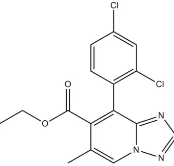
Ethyl
8-(2,4-dichlorophenyl)-6-methyl-1,2,4-triazolo[1,5-
a
]pyridine-7-carboxyl-ate
Yang Li,* Chen Sun and Ran Zhang
School of Chemical Engineering, Taishan Medical University, Taian 271016, People’s Republic of China
Correspondence e-mail: chemyangli@gmail.com
Received 29 October 2013; accepted 6 November 2013
Key indicators: single-crystal X-ray study;T= 298 K; mean(C–C) = 0.004 A˚;
Rfactor = 0.055;wRfactor = 0.170; data-to-parameter ratio = 13.6.
In the title compound, C16H13Cl2N3O2, the carboxylate group and the benzene ring attached to the central 1,2,4-triazolo-[1,5-a]pyridine bicycle are twisted from its mean plane by 55.6 (1) and 72.6 (1), respectively. In the crystal, weak C— H O interactions link the molecules into zigzag chains propagating in [100].
Related literature
For applications of [1,2,4]triazolo[1,5-a]pyridine derivatives, see: Luo & Hu (2006); Liu & Hu (2002). For details of the synthesis, see: Jones & Sliskovic (1983); Wanget al.(2003); Ge
et al.(2009); Jiaet al.(2010). For standard bond lengths, see: Allenet al.(1987).
Experimental
Crystal data
C16H13Cl2N3O2 Mr= 350.19
Orthorhombic,Pbca a= 14.693 (2) A˚ b= 13.531 (2) A˚ c= 16.347 (2) A˚
V= 3250.0 (8) A˚3 Z= 8
MoKradiation
= 0.41 mm1 T= 298 K
0.330.260.21 mm
Data collection
Brucker SMART APEXII CCD area-detector diffractometer Absorption correction: multi-scan
(SADABS; Bruker, 1999) Tmin= 0.876,Tmax= 0.919
15766 measured reflections 2860 independent reflections 2206 reflections withI> 2(I) Rint= 0.090
Refinement
R[F2> 2(F2)] = 0.055 wR(F2) = 0.170 S= 1.07 2860 reflections
210 parameters
H-atom parameters constrained
max= 0.42 e A˚
3
min=0.33 e A˚
[image:1.610.84.255.510.673.2]3
Table 1
Hydrogen-bond geometry (A˚ ,).
D—H A D—H H A D A D—H A
C7—H7 O1i
0.93 2.55 3.296 (4) 137
Symmetry code: (i)xþ1 2;y;zþ
1 2.
Data collection:SMART(Bruker, 1998); cell refinement:SAINT (Bruker, 1999); data reduction:SAINT; program(s) used to solve structure:SHELXS97(Sheldrick, 2008); program(s) used to refine structure: SHELXL97 (Sheldrick, 2008); molecular graphics: SHELXTL(Sheldrick, 2008); software used to prepare material for publication:SHELXTL.
Supplementary data and figures for this paper are available from the IUCr electronic archives (Reference: CV5435).
References
Allen, F. H., Kennard, O., Watson, D. G., Brammer, L., Orpen, A. G. & Taylor, R. (1987).J. Chem. Soc. Perkin Trans. 2, pp. S1–19.
Bruker (1998).SMART. Bruker AXS Inc., Madison, Wisconsin, USA. Bruker (1999).SADABSandSAINT. Bruker AXS Inc., Madison, Wisconsin,
USA.
Ge, Y. Q., Jia, J., Yang, H., Zhao, G. L., Zhan, F. X. & Wang, J. W. (2009). Heterocycles,78, 725–736.
Jia, J., Ge, Y. Q., Tao, X. T. & Wang, J. W. (2010).Heterocycles,81, 185–194. Jones, G. & Sliskovic, D. R. (1983).Adv. Heterocycl. Chem.34, 79–143. Liu, T. & Hu, Y. (2002).Bioorg. Med. Chem. Lett.12, 2411–2413. Luo, Y. & Hu, Y. (2006).Arch. Pharm. Chem. Life Sci.339, 262–266. Sheldrick, G. M. (2008).Acta Cryst.A64, 112–122.
Wang, J. W., Jia, J., Hou, D. J., li, H. M. & Yin, J. (2003).Chin. J. Org. Chem.23, 173–175.
Acta Crystallographica Section E Structure Reports
Online
supporting information
Acta Cryst. (2013). E69, o1796 [doi:10.1107/S160053681303033X]
Ethyl 8-(2,4-dichlorophenyl)-6-methyl-1,2,4-triazolo[1,5-
a
]pyridine-7-carboxyl-ate
Yang Li, Chen Sun and Ran Zhang
S1. Comment
The [1,2,4]triazolo[1,5-a]pyridine derivatives exhibit antifungal, anticancer and anti-inflammatory activities (Liu & Hu,
2002; Luo & Hu, 2006). However, only small number of [1,2,4]triazolo[1,5-a]pyridines is known. The commonly used
synthetic methods are the annulation of 1,2,4-triazole ring starting with amino substituted pyridines by a multistep
procedure (Jones & Sliskovic, 1983). Recently, imidazo[1,5-a]pyridines, pyrazolo[1,5-a]pyridines,
imidazo[1,2-a]pyridines and indolizines have been synthesized in our group wityh the use of a novel tandem reaction (Wang et al.,
2003; Ge et al., 2009; Jia et al., 2010). We tried to extend this reaction to synthesize the [1,2,4]triazolo[1,5-a]pyridine
heterocycles and obtained the title compound (I). Herewith we present its crystal structure.
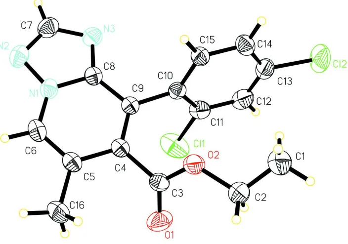
In (I) (Fig. 1), all bond lengths and angles are normal (Allen et al., 1987). The carboxylate group and benzene ring
attached to the central [1,2,4]triazolo[1,5-a]pyridine bicycle are twisted from its mean plane at 55.6 (1) and 72.6 (1)°,
respectively. In the crystal, weak intermolecular C—H···O interactions (Table 1) link molecules into zigzag chains
propagated in [100].
S2. Experimental
(2,4-Dichlorophenyl)(1H-1,2,4-triazol-5-yl)methanone (6 mmol), ethyl 4-bromo-3-methylbut-2-enoate (12 mmol),
potassium carbonate (1.8 g, 13.2 mmol) and DMF (30 ml) were added to a 100 ml round-bottomed flask. The reaction
system was stirred for 8 h. Then the mixture was poured into water (200 ml) and extracted with dichloromethane (3 x 50
ml). Organic layers were combined and dried over anhydrous Na2SO4, then filtered. By rotary evaporation, the mixture
was concentrated. After that, these crude products were depurated by using column chromatography in 76% isolated
yield. Crystals suitable for X-ray diffraction analysis were obtained by slow evaporation of a solution of the title
compound in a hexane/ethyl acetate mixture (3:1 v/v) at room temperature over a period of one week.
S3. Refinement
All H atoms were found on difference maps, but placed in idealized positions (C—H = 0.93–0.97 Å), and included in the
Figure 1
View of (I) with displacement ellipsoids drawn at the 30% probability level.
Ethyl 8-(2,4-dichlorophenyl)-6-methyl-1,2,4-triazolo[1,5-a]pyridine-7-carboxylate
Crystal data
C16H13Cl2N3O2 Mr = 350.19
Orthorhombic, Pbca Hall symbol: -P 2ac 2ab a = 14.693 (2) Å b = 13.531 (2) Å c = 16.347 (2) Å V = 3250.0 (8) Å3 Z = 8
F(000) = 1440 Dx = 1.431 Mg m−3
Mo Kα radiation, λ = 0.71073 Å Cell parameters from 5439 reflections θ = 2.4–26.6°
µ = 0.41 mm−1 T = 298 K Block, colourless 0.33 × 0.26 × 0.21 mm
Data collection
Brucker SMART APEXII CCD area-detector diffractometer
Radiation source: fine-focus sealed tube Graphite monochromator
phi and ω scans
Absorption correction: multi-scan (SADABS; Bruker, 1999) Tmin = 0.876, Tmax = 0.919
15766 measured reflections 2860 independent reflections 2206 reflections with I > 2σ(I) Rint = 0.090
θmax = 25.0°, θmin = 2.4° h = −15→17
k = −14→16 l = −19→19
Refinement
Refinement on F2 Least-squares matrix: full R[F2 > 2σ(F2)] = 0.055 wR(F2) = 0.170
2860 reflections 210 parameters 0 restraints
Secondary atom site location: difference Fourier map
Hydrogen site location: inferred from neighbouring sites
H-atom parameters constrained
w = 1/[σ2(F
o2) + (0.081P)2 + 1.792P] where P = (Fo2 + 2Fc2)/3
(Δ/σ)max < 0.001 Δρmax = 0.42 e Å−3 Δρmin = −0.33 e Å−3
Special details
Geometry. All e.s.d.'s (except the e.s.d. in the dihedral angle between two l.s. planes) are estimated using the full covariance matrix. The cell e.s.d.'s are taken into account individually in the estimation of e.s.d.'s in distances, angles and torsion angles; correlations between e.s.d.'s in cell parameters are only used when they are defined by crystal symmetry. An approximate (isotropic) treatment of cell e.s.d.'s is used for estimating e.s.d.'s involving l.s. planes.
Refinement. Refinement of F2 against ALL reflections. The weighted R-factor wR and goodness of fit S are based on F2, conventional R-factors R are based on F, with F set to zero for negative F2. The threshold expression of F2 > σ(F2) is used only for calculating R-factors(gt) etc. and is not relevant to the choice of reflections for refinement. R-factors based on F2 are statistically about twice as large as those based on F, and R- factors based on ALL data will be even larger.
Fractional atomic coordinates and isotropic or equivalent isotropic displacement parameters (Å2)
x y z Uiso*/Ueq
Cl1 0.05484 (8) 0.40274 (8) 0.16648 (8) 0.1076 (5) Cl2 0.19892 (9) 0.61061 (7) −0.08078 (8) 0.1131 (5) N1 0.26136 (16) 0.13047 (17) 0.22123 (12) 0.0520 (6) N2 0.32941 (18) 0.1240 (2) 0.27788 (15) 0.0667 (7) N3 0.33128 (17) 0.27522 (19) 0.21631 (14) 0.0597 (6) O1 −0.01914 (15) 0.1842 (2) 0.05658 (15) 0.0854 (8) O2 0.09633 (14) 0.21635 (17) −0.02602 (11) 0.0703 (6) C1 0.0817 (4) 0.3050 (5) −0.1491 (3) 0.138 (2) H1A 0.1140 0.3576 −0.1224 0.207* H1B 0.0391 0.3323 −0.1874 0.207* H1C 0.1242 0.2634 −0.1775 0.207* C2 0.0347 (3) 0.2488 (4) −0.0903 (2) 0.0953 (13) H2A 0.0074 0.1916 −0.1164 0.114* H2B −0.0137 0.2883 −0.0667 0.114* C3 0.06111 (18) 0.19142 (19) 0.04424 (16) 0.0498 (6) C4 0.13260 (17) 0.17205 (19) 0.10725 (15) 0.0451 (6) C5 0.13144 (18) 0.07882 (19) 0.14927 (16) 0.0494 (6) C6 0.19780 (19) 0.0596 (2) 0.20462 (17) 0.0558 (7)
H6 0.2000 −0.0012 0.2310 0.067*
C7 0.3669 (2) 0.2128 (3) 0.27135 (19) 0.0687 (9)
H7 0.4161 0.2311 0.3038 0.082*
C8 0.26346 (18) 0.22155 (19) 0.18455 (15) 0.0478 (6) C9 0.19725 (16) 0.24312 (18) 0.12376 (15) 0.0439 (6) C10 0.20186 (17) 0.33997 (18) 0.08023 (15) 0.0460 (6) C11 0.1376 (2) 0.4141 (2) 0.09152 (19) 0.0597 (7) C12 0.1379 (2) 0.4988 (2) 0.0427 (2) 0.0717 (9)
H12 0.0942 0.5477 0.0500 0.086*
C15 0.26961 (18) 0.3567 (2) 0.02229 (16) 0.0521 (7)
H15 0.3154 0.3099 0.0158 0.062*
C16 0.0598 (2) 0.0018 (2) 0.1339 (2) 0.0667 (8) H16A 0.0770 −0.0588 0.1604 0.100* H16B 0.0542 −0.0093 0.0761 0.100* H16C 0.0026 0.0241 0.1554 0.100*
Atomic displacement parameters (Å2)
U11 U22 U33 U12 U13 U23
Cl1 0.0972 (8) 0.0803 (7) 0.1453 (10) 0.0092 (5) 0.0583 (7) −0.0185 (6) Cl2 0.1464 (10) 0.0607 (6) 0.1323 (10) −0.0150 (6) −0.0448 (8) 0.0396 (6) N1 0.0569 (13) 0.0554 (13) 0.0438 (11) 0.0063 (11) 0.0013 (10) 0.0036 (10) N2 0.0705 (16) 0.0752 (18) 0.0544 (14) 0.0091 (14) −0.0085 (12) 0.0073 (12) N3 0.0583 (14) 0.0654 (15) 0.0555 (13) −0.0044 (11) −0.0075 (11) −0.0013 (11) O1 0.0490 (13) 0.130 (2) 0.0777 (15) −0.0008 (13) 0.0030 (11) 0.0062 (15) O2 0.0600 (12) 0.1014 (17) 0.0494 (11) −0.0223 (11) −0.0043 (9) 0.0141 (10) C1 0.120 (4) 0.192 (6) 0.102 (3) −0.071 (4) −0.037 (3) 0.068 (4) C2 0.091 (3) 0.118 (3) 0.077 (2) −0.042 (2) −0.033 (2) 0.035 (2) C3 0.0508 (16) 0.0464 (14) 0.0523 (14) −0.0042 (11) 0.0019 (12) −0.0053 (11) C4 0.0476 (14) 0.0442 (14) 0.0436 (13) 0.0007 (11) 0.0073 (11) −0.0030 (10) C5 0.0534 (15) 0.0442 (14) 0.0507 (14) −0.0001 (11) 0.0123 (12) −0.0022 (11) C6 0.0678 (18) 0.0448 (14) 0.0549 (15) 0.0036 (13) 0.0116 (14) 0.0056 (12) C7 0.0627 (19) 0.086 (2) 0.0576 (18) 0.0003 (16) −0.0104 (15) −0.0001 (16) C8 0.0521 (15) 0.0480 (15) 0.0435 (13) 0.0012 (11) 0.0037 (11) −0.0013 (11) C9 0.0460 (14) 0.0429 (13) 0.0428 (13) 0.0010 (11) 0.0041 (10) −0.0021 (10) C10 0.0474 (14) 0.0434 (14) 0.0473 (14) −0.0026 (11) −0.0062 (11) −0.0045 (11) C11 0.0558 (16) 0.0466 (16) 0.0765 (19) 0.0008 (12) 0.0009 (14) −0.0113 (13) C12 0.067 (2) 0.0407 (16) 0.107 (3) 0.0075 (13) −0.0178 (19) −0.0072 (16) C13 0.077 (2) 0.0452 (16) 0.074 (2) −0.0100 (15) −0.0236 (17) 0.0054 (14) C14 0.0716 (19) 0.0550 (17) 0.0559 (16) −0.0131 (14) −0.0018 (14) 0.0030 (13) C15 0.0542 (16) 0.0476 (15) 0.0545 (15) −0.0030 (12) −0.0005 (12) 0.0026 (12) C16 0.0708 (19) 0.0487 (16) 0.081 (2) −0.0097 (14) 0.0069 (16) −0.0006 (15)
Geometric parameters (Å, º)
Cl1—C11 1.733 (3) C4—C5 1.437 (4)
Cl2—C13 1.743 (3) C5—C6 1.355 (4)
N1—C6 1.365 (4) C5—C16 1.502 (4)
N1—N2 1.366 (3) C6—H6 0.9300
N1—C8 1.371 (3) C7—H7 0.9300
N2—C7 1.326 (4) C8—C9 1.421 (4)
N3—C8 1.338 (3) C9—C10 1.493 (4)
N3—C7 1.341 (4) C10—C11 1.389 (4)
O1—C3 1.200 (3) C10—C15 1.393 (4)
O2—C3 1.304 (3) C11—C12 1.398 (4)
C1—H1A 0.9600 C13—C14 1.371 (4)
C1—H1B 0.9600 C14—C15 1.376 (4)
C1—H1C 0.9600 C14—H14 0.9300
C2—H2A 0.9700 C15—H15 0.9300
C2—H2B 0.9700 C16—H16A 0.9600
C3—C4 1.494 (4) C16—H16B 0.9600
C4—C9 1.378 (4) C16—H16C 0.9600
C6—N1—N2 126.2 (2) N3—C7—H7 121.2
C6—N1—C8 124.0 (2) N3—C8—N1 109.6 (2) N2—N1—C8 109.7 (2) N3—C8—C9 132.1 (2) C7—N2—N1 101.1 (2) N1—C8—C9 118.4 (2) C8—N3—C7 102.1 (3) C4—C9—C8 117.7 (2) C3—O2—C2 117.9 (2) C4—C9—C10 123.4 (2) C2—C1—H1A 109.5 C8—C9—C10 118.9 (2) C2—C1—H1B 109.5 C11—C10—C15 117.3 (3) H1A—C1—H1B 109.5 C11—C10—C9 122.6 (2) C2—C1—H1C 109.5 C15—C10—C9 120.0 (2) H1A—C1—H1C 109.5 C10—C11—C12 120.9 (3) H1B—C1—H1C 109.5 C10—C11—Cl1 120.5 (2) C1—C2—O2 110.6 (3) C12—C11—Cl1 118.6 (2) C1—C2—H2A 109.5 C13—C12—C11 119.0 (3)
O2—C2—H2A 109.5 C13—C12—H12 120.5
C1—C2—H2B 109.5 C11—C12—H12 120.5
O2—C2—H2B 109.5 C12—C13—C14 122.0 (3) H2A—C2—H2B 108.1 C12—C13—Cl2 118.9 (3) O1—C3—O2 124.0 (3) C14—C13—Cl2 119.1 (3) O1—C3—C4 124.1 (3) C13—C14—C15 118.5 (3) O2—C3—C4 111.9 (2) C13—C14—H14 120.8 C9—C4—C5 121.8 (2) C15—C14—H14 120.8 C9—C4—C3 119.8 (2) C14—C15—C10 122.1 (3) C5—C4—C3 118.4 (2) C14—C15—H15 119.0 C6—C5—C4 118.6 (2) C10—C15—H15 119.0 C6—C5—C16 118.8 (3) C5—C16—H16A 109.5 C4—C5—C16 122.5 (3) C5—C16—H16B 109.5 C5—C6—N1 119.4 (2) H16A—C16—H16B 109.5
C5—C6—H6 120.3 C5—C16—H16C 109.5
N1—C6—H6 120.3 H16A—C16—H16C 109.5 N2—C7—N3 117.6 (3) H16B—C16—H16C 109.5 N2—C7—H7 121.2
Hydrogen-bond geometry (Å, º)
D—H···A D—H H···A D···A D—H···A
C7—H7···O1i 0.93 2.55 3.296 (4) 137