1-[2-Hydroxy-4-(prop-2-yn-1-yloxy)-phenyl]ethanone
V. Selvarani,aM. A. Neelakantan,a* T. Srinivasanband D. Velmuruganb
aChemistry Research Centre, National Engineering College, K.R. Nagar, Kovilpatti
628 503, India, andbCentre of Advanced Study in Crystallography and Biophysics,
University of Madras, Guindy Campus, Chennai 600 025, India Correspondence e-mail: [email protected]
Received 18 November 2013; accepted 30 November 2013
Key indicators: single-crystal X-ray study;T= 293 K; mean(C–C) = 0.002 A˚; Rfactor = 0.043;wRfactor = 0.130; data-to-parameter ratio = 17.8.
In the title compound, C11H10O3, there is an intramolecular
O—H O hydrogen bond generating anS(6) ring motif. The O atom of the hydroxy group deviates by 0.0200 (1) A˚ from the benzene ring to which it is attached. The propyne group is almost linear, the C—C C angle being 177.83 (15), and is
almost coplanar with the benzene ring; the C—C—O—C torsion angle being only 1.1 (2). In the crystal, molecules
are linked via C—H O hydrogen bonds, forming infinite
C(11) chains running parallel to [103]. These chains are linked by a pair of C—H O hydrogen bonds, enclosing R2
2
(8) inversion dimers, forming a corrugated two-dimensional network lying parallel to (103).
Related literature
For the biological activity of benzaldehyde derivatives, see: Zhaoet al.(2007); Ley & Bertram (2001); Deloguet al.(2010). For a related structure, see: Esakkiammal et al. (2012). For graph-set notation, see: Bernsteinet al.(1995).
Experimental
Crystal data
C11H10O3 Mr= 190.19
a= 4.9975 (2) A˚
b= 10.4305 (4) A˚
= 97.257 (2)
V= 974.54 (7) A˚3 Z= 4
MoKradiation
= 0.10 mm1 T= 293 K
0.350.300.20 mm
Data collection
Bruker SMART APEXII area-detector diffractometer Absorption correction: multi-scan
(SADABS; Bruker, 2008)
Tmin= 0.968,Tmax= 0.981
9316 measured reflections 2451 independent reflections 1898 reflections withI> 2(I)
Rint= 0.021
Refinement
R[F2> 2(F2)] = 0.043 wR(F2) = 0.130 S= 1.05 2451 reflections 138 parameters 3 restraints
H atoms treated by a mixture of independent and constrained refinement
max= 0.17 e A˚
3
min=0.21 e A˚
3
Table 1
Hydrogen-bond geometry (A˚ ,).
D—H A D—H H A D A D—H A
O1—H1 O2 0.82 1.83 2.5551 (14) 146 C4—H4 O3i 0.93 2.54 3.4609 (16) 172 C11—H11 O2ii
0.93 2.32 3.2337 (18) 168
Symmetry codes: (i)xþ3;yþ2;z; (ii)xþ3 2;yþ
3 2;z
1 2.
Data collection:APEX2(Bruker, 2008); cell refinement:SAINT
(Bruker, 2008); data reduction:SAINT; program(s) used to solve structure:SHELXS97(Sheldrick, 2008); program(s) used to refine structure: SHELXL97 (Sheldrick, 2008); molecular graphics:
ORTEP-3 for Windows(Farrugia, 2012) andMercury(Macraeet al., 2008); software used to prepare material for publication:SHELXL97
andPLATON(Spek, 2009).
The authors thank the TBI X-ray facility and the UGC (SAP) CAS in Crystallography and Biophysics, University of Madras, India, for the data collection and other facilities. TS thanks the DST for an Inspire Fellowship.
Supplementary data and figures for this paper are available from the IUCr electronic archives (Reference: SU2668).
References
Bernstein, J., Davis, R. E., Shimoni, L. & Chang, N.-L. (1995).Angew. Chem. Int. Ed. Engl.34, 1555–1573.
Bruker (2008).APEX2,SAINTandSADABS. Bruker AXS Inc., Madison, Wisconsin, USA.
Delogu, G., Podda, G., Corda, M., Fadda, M. B., Fais, A. & Era, B. (2010).
Bioorg. Med. Chem. Lett.20, 6138–6140.
Esakkiammal, M., Selvarani, V., Neelakantan, M. A., Silambarasan, V. & Velmurugan, D. (2012).Acta Cryst.E68, o2465.
Farrugia, L. J. (2012).J. Appl. Cryst.45, 849–854.
Ley, J. P. & Bertram, H. J. (2001).Bioorg. Med. Chem. Lett.9, 1879–1885. Macrae, C. F., Bruno, I. J., Chisholm, J. A., Edgington, P. R., McCabe, P.,
Pidcock, E., Rodriguez-Monge, L., Taylor, R., van de Streek, J. & Wood, P. A. (2008).J. Appl. Cryst.41, 466–470.
Sheldrick, G. M. (2008).Acta Cryst.A64, 112–122. Spek, A. L. (2009).Acta Cryst.D65, 148–155.
Zhao, X., Song, D. K., Radbil, A. B. & Radbil, B. A. (2007).Russ. J. Appl. Chem.80, 1373–1375.
Acta Crystallographica Section E
Structure Reports
Online
supporting information
Acta Cryst. (2014). E70, o24 [https://doi.org/10.1107/S1600536813032613]
1-[2-Hydroxy-4-(prop-2-yn-1-yloxy)phenyl]ethanone
V. Selvarani, M. A. Neelakantan, T. Srinivasan and D. Velmurugan
S1. Comment
Schiff bases derived from amines and substituted benzaldehydes exhibit antibacterial, anticancer and antitumour activities
(Zhao et al., 2007). Several benzaldoximes, benzaldehyde-O-ethyloximes and acetophenone oximes were synthesized and
evaluated as tyrosinase inhibitors (Ley & Bertram, 2001). Bis-salicylaldehydes has been shown to exhibited greater
inhibitory activity than salicylaldehyde (Delogu et al., 2010). In view of these potential applications and in continuation
of our work on the crystal structures of benzaldehyde derivatives, we synthesized the title compound and report herein on
its crystal structure.
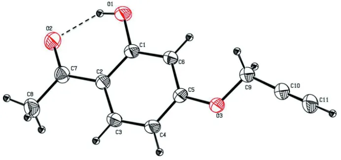
The molecular structure of the title compound is stabilized by an O—H···O intramolecular hydrogen bond (Fig. 1 and
Table 1), which forms an S(6) graph-set motif (Bernstein et al., 1995). The hydroxyl O atom, O1, deviates by 0.0200 (1)
Å from the benzene ring (C1-C6) to which it is attached. The oxygen atom substituted propyne group is slightly twisted
from the benzene ring (C1–C6) to which it is attached as evidenced by the torsion angle C6–C5–O3–C9 = -1.1 (2) °. The
propyne group is almost linear, the C9–C10≡C11 angle being 177.83 (15)°, and it is also in the flagpole position on atom
O3. The mean plane of the acetaldehyde group makes a dihedral angle of 0.39 (9)° with the benzene ring (C1–C6),
indicating that they are almost coplanar.
In the crystal, molecules are linked via C—H···O hydrogen bonds forming infinite C(11) chains running parallel to
direction [103]. These chains are linked via a pair of C—H···O hydrogen bonds, enclosing R22(8) inversion dimers,
forming wave-like two-dimensional networks lying parallel to (103); see Table 1 and Fig. 2.
S2. Experimental
Equimolar amounts of 3-bromopropyne (10 mmol), 2,4-dihydroxyacetophenone (10 mmol) and potassium carbonate (15
mmol) were suspended in dried acetone (30 ml) and refluxed for 5 h. The reaction mixture was filtered while hot to
remove insoluble impurities, neutralized with water and then extracted with ethyl acetate and dried with Na2SO4. The
extracts were concentrated to obtain a brown solid which was then purified by column chromatography over SiO2 by
eluting with a mixture of 5% ethyl acetate in n-hexane. Evaporation of the purified extract yielded the title compound in
the form of a pure white solid [Yield: 83%]. Colourless block-like crystals, suitable for X-ray diffraction analysis, were
obtained by the slow evaporation of a solution in ethyl acetate.
S3. Refinement
All H atoms could be located in difference Fourier maps. The methyl H atoms were refined with Uiso(H) = 1.5Ueq(C). The
OH and other C-bound H atoms were included in calculated positions are refined as riding atoms: O-H = 0.82 Å, C-H =
Figure 1
The molecular structure of the title molecule, with atom labelling. Displacement ellipsoids are drawn at the 30%
[image:3.610.132.480.291.405.2]probability level. The intramolecular O-H···O hydrogen bond is shown as a dashed line (see Table 1 for details)
Figure 2
A view along the a axis of the crystal packing of the title compound. The hydrogen bonds are shown as dashed lines (see
Table 1 for details; H atoms not involved in hydrogen bonding have been excluded for clarity).
1-[2-Hydroxy-4-(prop-2-yn-1-yloxy)phenyl]ethan-one
Crystal data
C11H10O3
Mr = 190.19 Monoclinic, P21/n
Hall symbol: -P 2yn
a = 4.9975 (2) Å
b = 10.4305 (4) Å
c = 18.8467 (7) Å
β = 97.257 (2)°
V = 974.54 (7) Å3
Z = 4
F(000) = 400
Dx = 1.296 Mg m−3
Mo Kα radiation, λ = 0.71073 Å Cell parameters from 2451 reflections
θ = 2.2–28.4°
µ = 0.10 mm−1
T = 293 K Block, colourless 0.35 × 0.30 × 0.20 mm
Data collection
Bruker SMART APEXII area-detector diffractometer
Radiation source: fine-focus sealed tube Graphite monochromator
ω and φ scans
Absorption correction: multi-scan (SADABS; Bruker, 2008)
Tmin = 0.968, Tmax = 0.981
Rint = 0.021
θmax = 28.4°, θmin = 2.2°
h = −6→6
k = −13→13
l = −25→25
Refinement
Refinement on F2
Least-squares matrix: full
R[F2 > 2σ(F2)] = 0.043
wR(F2) = 0.130
S = 1.05 2451 reflections 138 parameters 3 restraints
Primary atom site location: structure-invariant direct methods
Secondary atom site location: difference Fourier map
Hydrogen site location: inferred from neighbouring sites
H atoms treated by a mixture of independent and constrained refinement
w = 1/[σ2(F
o2) + (0.0637P)2 + 0.1394P]
where P = (Fo2 + 2Fc2)/3
(Δ/σ)max < 0.001
Δρmax = 0.17 e Å−3
Δρmin = −0.21 e Å−3
Extinction correction: SHELXL97 (Sheldrick, 2008), Fc*=kFc[1+0.001xFc2λ3/sin(2θ)]-1/4
Extinction coefficient: 0.020 (4)
Special details
Geometry. All esds (except the esd in the dihedral angle between two l.s. planes) are estimated using the full covariance matrix. The cell esds are taken into account individually in the estimation of esds in distances, angles and torsion angles; correlations between esds in cell parameters are only used when they are defined by crystal symmetry. An approximate (isotropic) treatment of cell esds is used for estimating esds involving l.s. planes.
Refinement. Refinement of F2 against ALL reflections. The weighted R-factor wR and goodness of fit S are based on F2,
conventional R-factors R are based on F, with F set to zero for negative F2. The threshold expression of F2 > 2sigma(F2) is
used only for calculating R-factors(gt) etc. and is not relevant to the choice of reflections for refinement. R-factors based on F2 are statistically about twice as large as those based on F, and R- factors based on ALL data will be even larger.
Fractional atomic coordinates and isotropic or equivalent isotropic displacement parameters (Å2)
x y z Uiso*/Ueq
C1 0.9425 (3) 0.69719 (11) 0.08613 (7) 0.0455 (3)
C2 0.9120 (2) 0.81055 (10) 0.12463 (6) 0.0421 (3)
C3 1.0579 (3) 0.91806 (11) 0.10720 (6) 0.0475 (3)
H3 1.0417 0.9941 0.1320 0.057*
C4 1.2229 (3) 0.91474 (11) 0.05494 (7) 0.0506 (3)
H4 1.3186 0.9874 0.0446 0.061*
C5 1.2467 (3) 0.80135 (11) 0.01728 (6) 0.0446 (3)
C6 1.1094 (3) 0.69245 (11) 0.03266 (6) 0.0468 (3)
H6 1.1283 0.6169 0.0076 0.056*
C7 0.7340 (3) 0.81464 (12) 0.18017 (6) 0.0485 (3)
C8 0.7041 (4) 0.93600 (15) 0.22030 (9) 0.0632 (4)
H8A 0.595 (4) 0.9292 (18) 0.2554 (11) 0.095*
H8B 0.648 (4) 1.005 (2) 0.1896 (10) 0.095*
H8C 0.872 (4) 0.9636 (19) 0.2446 (10) 0.095*
C9 1.4462 (3) 0.69606 (12) −0.07557 (7) 0.0527 (3)
H9A 1.2727 0.6639 −0.0972 0.063*
H9B 1.5342 0.6298 −0.0449 0.063*
C10 1.6120 (3) 0.73056 (13) −0.13064 (7) 0.0546 (3)
C11 1.7405 (3) 0.75594 (16) −0.17657 (8) 0.0680 (4)
O1 0.8072 (2) 0.58946 (8) 0.09853 (6) 0.0691 (4)
H1 0.7187 0.6017 0.1317 0.104*
O2 0.6074 (2) 0.71799 (10) 0.19459 (5) 0.0624 (3)
O3 1.4107 (2) 0.80891 (8) −0.03494 (5) 0.0574 (3)
Atomic displacement parameters (Å2)
U11 U22 U33 U12 U13 U23
C1 0.0506 (7) 0.0372 (5) 0.0508 (6) −0.0075 (5) 0.0149 (5) 0.0001 (4)
C2 0.0460 (6) 0.0395 (5) 0.0418 (5) −0.0015 (5) 0.0098 (5) 0.0009 (4)
C3 0.0570 (7) 0.0372 (5) 0.0501 (6) −0.0056 (5) 0.0140 (5) −0.0060 (4)
C4 0.0595 (8) 0.0385 (6) 0.0569 (7) −0.0126 (5) 0.0196 (6) −0.0035 (5)
C5 0.0457 (7) 0.0431 (6) 0.0472 (6) −0.0071 (5) 0.0149 (5) −0.0022 (4)
C6 0.0531 (7) 0.0370 (5) 0.0531 (6) −0.0072 (5) 0.0177 (6) −0.0073 (4)
C7 0.0549 (8) 0.0484 (6) 0.0438 (6) 0.0027 (5) 0.0127 (5) 0.0033 (5)
C8 0.0781 (11) 0.0589 (8) 0.0572 (8) 0.0039 (8) 0.0270 (8) −0.0065 (6)
C9 0.0567 (8) 0.0491 (7) 0.0558 (7) −0.0084 (6) 0.0200 (6) −0.0096 (5)
C10 0.0569 (8) 0.0544 (7) 0.0553 (7) −0.0037 (6) 0.0179 (6) −0.0085 (6)
C11 0.0790 (11) 0.0666 (9) 0.0649 (8) −0.0040 (8) 0.0339 (8) −0.0095 (7)
O1 0.0912 (8) 0.0423 (5) 0.0835 (7) −0.0214 (5) 0.0488 (6) −0.0082 (4)
O2 0.0751 (7) 0.0571 (6) 0.0612 (6) −0.0077 (5) 0.0334 (5) 0.0043 (4)
O3 0.0673 (6) 0.0475 (5) 0.0644 (6) −0.0165 (4) 0.0354 (5) −0.0110 (4)
Geometric parameters (Å, º)
C1—O1 1.3469 (14) C7—O2 1.2384 (15)
C1—C6 1.3879 (16) C7—C8 1.4918 (18)
C1—C2 1.4056 (16) C8—H8A 0.91 (2)
C2—C3 1.3991 (16) C8—H8B 0.94 (2)
C2—C7 1.4573 (16) C8—H8C 0.95 (2)
C3—C4 1.3627 (17) C9—O3 1.4275 (14)
C3—H3 0.9300 C9—C10 1.4529 (18)
C4—C5 1.3920 (16) C9—H9A 0.9700
C4—H4 0.9300 C9—H9B 0.9700
C5—O3 1.3602 (14) C10—C11 1.1712 (19)
C5—C6 1.3765 (16) C11—H11 0.9300
C6—H6 0.9300 O1—H1 0.8200
O1—C1—C6 117.20 (10) O2—C7—C8 119.53 (12)
O1—C1—C2 121.51 (11) C2—C7—C8 119.89 (11)
C6—C1—C2 121.27 (10) C7—C8—H8A 114.1 (12)
C3—C2—C1 117.34 (11) C7—C8—H8B 112.2 (12)
C3—C2—C7 121.92 (10) H8A—C8—H8B 110.2 (17)
C1—C2—C7 120.75 (10) C7—C8—H8C 111.3 (12)
C4—C3—C2 122.03 (10) H8A—C8—H8C 104.4 (16)
C4—C3—H3 119.0 H8B—C8—H8C 103.9 (17)
C2—C3—H3 119.0 O3—C9—C10 107.45 (10)
C3—C4—H4 120.4 C10—C9—H9A 110.2
C5—C4—H4 120.4 O3—C9—H9B 110.2
O3—C5—C6 124.19 (10) C10—C9—H9B 110.2
O3—C5—C4 114.68 (10) H9A—C9—H9B 108.5
C6—C5—C4 121.13 (10) C11—C10—C9 177.83 (15)
C5—C6—C1 118.99 (10) C10—C11—H11 180.0
C5—C6—H6 120.5 C1—O1—H1 109.5
C1—C6—H6 120.5 C5—O3—C9 117.87 (9)
O2—C7—C2 120.59 (11)
O1—C1—C2—C3 −179.06 (12) C4—C5—C6—C1 0.8 (2)
C6—C1—C2—C3 −0.40 (19) O1—C1—C6—C5 178.60 (12)
O1—C1—C2—C7 0.7 (2) C2—C1—C6—C5 −0.1 (2)
C6—C1—C2—C7 179.39 (12) C3—C2—C7—O2 −179.90 (12)
C1—C2—C3—C4 0.3 (2) C1—C2—C7—O2 0.3 (2)
C7—C2—C3—C4 −179.52 (12) C3—C2—C7—C8 −0.1 (2)
C2—C3—C4—C5 0.4 (2) C1—C2—C7—C8 −179.89 (13)
C3—C4—C5—O3 178.37 (12) C6—C5—O3—C9 −1.1 (2)
C3—C4—C5—C6 −0.9 (2) C4—C5—O3—C9 179.62 (12)
O3—C5—C6—C1 −178.44 (12) C10—C9—O3—C5 175.97 (12)
Hydrogen-bond geometry (Å, º)
D—H···A D—H H···A D···A D—H···A
O1—H1···O2 0.82 1.83 2.5551 (14) 146
C4—H4···O3i 0.93 2.54 3.4609 (16) 172
C11—H11···O2ii 0.93 2.32 3.2337 (18) 168