• No results found

Crystal structure of 5 [4 (di­methyl­amino)­phen­yl] 3 (4 methyl­phen­yl) 4,5 di­hydro 1H pyrazole 1 carbaldehyde

N/A
N/A
Protected

Academic year: 2020

Share "Crystal structure of 5 [4 (di­methyl­amino)­phen­yl] 3 (4 methyl­phen­yl) 4,5 di­hydro 1H pyrazole 1 carbaldehyde"

Copied!
9
0
0

Loading.... (view fulltext now)

Full text

(1)

Crystal structure of

5-[4-(dimethyl-

amino)phenyl]-3-(4-methylphenyl)-4,5-dihydro-1

H

-pyrazole-1-carbaldehyde

Farook Adam,a* Seranthimata Samshuddin,bNadiah Ameram,aSubramayaband Laxminarayana Samarthab

aSchool of Chemical Sciences, Universiti Sains Malaysia, 11800 Pulau Pinang,

Malaysia, andbDepartment of PG Studies in Chemistry, Alva’s College, Moodbidri,

Karnataka 574 227, India. *Correspondence e-mail: [email protected]

Received 1 December 2015; accepted 4 December 2015

Edited by E. R. T. Tiekink, University of Malaya, Malaysia

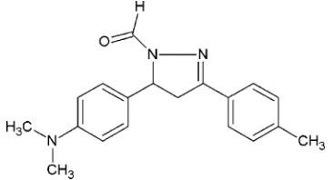
The title compound, C19H21N3O, comprises a central pyrazole

ring which is N-connected to an aldehyde group and C-connected twice to substituted benzene rings. The pyrazole ring is twisted on the C—C single bond, and the least-squares plane through this ring forms dihedral angles of 82.44 (5) and 4.52 (5) with the (dimethylamino)benzene and p-tolyl rings,

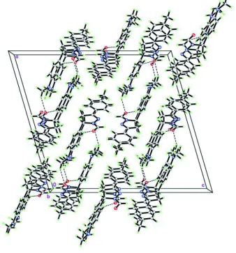
respectively. In the crystal, weak C—H O hydrogen bonds link molecules into supramolecular tubes along thebaxis.

Keywords:crystal structure; substituted pyrazole; pyrazole derivatives; pharmacological properties.

CCDC reference:1440601

1. Related literature

For pharmacological properties of pyrazole derivatives, see: Sarojini et al. (2010); Samshuddin et al. (2012). For their industrial applications, see: Wileyet al.(1958); Luet al.(1999). For related structures, see Funet al.(2010); Baktıret al.(2011).

2. Experimental

2.1. Crystal data

C19H21N3O Mr= 307.39

Monoclinic,C2=c a= 21.9524 (15) A˚ b= 6.2511 (4) A˚ c= 24.1521 (16) A˚

= 106.3069 (9)

V= 3181.0 (4) A˚3 Z= 8

MoKradiation

= 0.08 mm1 T= 100 K

0.450.260.15 mm

2.2. Data collection

Bruker APEX DUO CCD area-detector diffractometer Absorption correction: multi-scan

(SADABS; Sheldrick, 1996) Tmin= 0.921,Tmax= 0.962

27492 measured reflections 4750 independent reflections 4090 reflections withI> 2(I) Rint= 0.028

2.3. Refinement

R[F2> 2(F2)] = 0.043 wR(F2) = 0.122 S= 1.04 4750 reflections

211 parameters

H-atom parameters constrained

max= 0.37 e A˚ 3

[image:1.610.80.263.608.707.2]

min=0.22 e A˚ 3

Table 1

Hydrogen-bond geometry (A˚ ,).

D—H A D—H H A D A D—H A

C1—H1A O1i

0.95 2.52 3.4175 (12) 158 C19—H19A O1ii

0.98 2.45 3.3902 (15) 161

Symmetry codes: (i)x;y1;z; (ii)xþ1 2;y

1 2;zþ

1 2.

Data collection: APEX2 (Bruker, 2014); cell refinement: SAINT (Bruker, 2014); data reduction:SAINT; program(s) used to solve structure:SHELXS2013(Sheldrick, 2008); program(s) used to refine structure: SHELXL2014 (Sheldrick, 2015); molecular graphics: SHELXTL(Sheldrick, 2008); software used to prepare material for publication:PLATON(Spek, 2009).

Acknowledgements

SS thanks Alva’s Education Foundation, Moodbidri, for providing research facilities. The authors would like to thank Universiti Malaysia Kelantan, SLAI, the Malaysian Ministry of Higher Education and the Universiti Sains Malaysia for RU research grants (Nos. PKIMIA/846017 and 1001/PKIMIA/ 811269), which partly supported this work.

Supporting information for this paper is available from the IUCr electronic archives (Reference: TK5413).

References

Baktır, Z., Akkurt, M., Samshuddin, S., Narayana, B. & Yathirajan, H. S. (2011).Acta Cryst.E67, o1292–o1293.

Bruker (2014).APEX2,SAINTandSADABS. Bruker AXS Inc., Madison, Wisconsin, USA.

Fun, H.-K., Hemamalini, M., Samshuddin, S., Narayana, B. & Yathirajan, H. S. (2010).Acta Cryst.E66, o582–o583.

data reports

(2)

Lu, Z. Y., Zhu, W. G., Jiang, Q. & Xie, M. G. (1999).Chin. Chem. Lett.10, 679– 682.

Samshuddin, S., Narayana, B., Sarojini, B. K., Khan, M. T. H., Yathirajan, H. S., Raj, C. G. D. & Raghavendra, R. (2012).Med. Chem. Res.21, 2012–2022. Sarojini, B. K., Vidyagayatri, M., Darshanraj, C. G., Bharath, B. R. &

Manjunatha, H. (2010).Lett. Drug. Des. Discov.7, 214–224.

Sheldrick, G. M. (1996).SADABS. University of Go¨ttingen, Germany. Sheldrick, G. M. (2008).Acta Cryst.A64, 112–122.

Sheldrick, G. M. (2015).Acta Cryst.C71, 3–8. Spek, A. L. (2009).Acta Cryst.D65, 148–155.

(3)

supporting information

sup-1 Acta Cryst. (2015). E71, o1031–o1032

supporting information

Acta Cryst. (2015). E71, o1031–o1032 [https://doi.org/10.1107/S2056989015023294]

Crystal structure of

5-[4-(dimethylamino)phenyl]-3-(4-methylphenyl)-4,5-di-hydro-1

H

-pyrazole-1-carbaldehyde

Farook Adam, Seranthimata Samshuddin, Nadiah Ameram, Subramaya and Laxminarayana

Samartha

S1. Introduction

Pyrazolyl derivatives are well-known for their versatile pharmacological activities (Sarojini et al., 2010; Samshuddin et

al., 2012). In addition, many 1,3,5-triaryl-2-pyrazolyls have a variety of industrial applications such as functioning as

scintillation solutes (Wiley et al., 1958) and fluorescent agents (Lu et al., 1999). The crystal structures of some pyrazolyls

containing a N-alkyl chain viz., 3,5-bis(4-fluorophenyl)-4,5-dihydro-1H-pyrazole-1-carbaldehyde (Baktır et al., 2011) and

1-[3,5-bis(4-fluorophenyl)-4,5-dihydro-1H-pyrazol-1-yl]ethanone (Fun et al., 2010) have been reported. In view of the

importance of pyrazolines, the title compound (I) was prepared and its crystal structure reported.

S2. Supramolecular features

The asymmetric unit of (I) consists of a single crystallographic independent molecule as shown in Fig. 1. The pyrazoline

ring (N1/N2/C7/C8/C9) is twisted about the C8—C7 bond [Q2 = 0.0964 (10) Å and φ2 = 133.5 (6)°] with maximum

deviations of 0.057 (1) and -0.053 (1) Å from its mean plane for atoms C7 and C8, respectively. The methyl-substituted

phenyl ring (C10–C15) and dimethylamino-substituted phenyl ring (C1–C6) make dihedral angles of 4.52 (5) and

82.44 (5)°, respectively, with the pyrazoline ring. In crystal, molecules are connected by weak C—H···O hydrogen bonds

into one-dimensional spiral-like chains (Fig. 2), propagating along the crystallographic b-axis.

S3. Synthesis and crystallization

A mixture of (2E)-3-[4-(dimethylamino)phenyl]-1-(4-methylphenyl)prop-2-en-1-one (2.65 g, 0.01 mol) and hydrazine

hydrate (1 ml) in 30 ml formic acid was refluxed for 6 h. The reaction mixture was cooled and poured into 250 ml

ice-cold water. The precipitate formed was collected by filtration and purified by recrystallization from ethanol. Single

crystals were grown from ethyl acetate by slow evaporation (m.p 473–476 K; yield: 68%).

S4. Refinement

The carbon-bound H-atoms were placed in calculated positions (C—H = 0.95–1.00 Å) and were included in the

refinement in the riding model approximation, with Uiso(H) set to 1.2–1.5Uequiv(C) A rotating group model was applied to

(4)
[image:4.610.141.476.72.249.2]

Figure 1

The molecular structure of (I), showing the atom labels and 50% probability displacement ellipsoids.

Figure 2

[image:4.610.136.474.295.658.2]
(5)

supporting information

sup-3 Acta Cryst. (2015). E71, o1031–o1032

5-[4-(Dimethylamino)phenyl]-3-(4-methylphenyl)-4,5-dihydro-1H-pyrazole-1-carbaldehyde

Crystal data

C19H21N3O

Mr = 307.39 Monoclinic, C2/c a = 21.9524 (15) Å b = 6.2511 (4) Å c = 24.1521 (16) Å β = 106.3069 (9)° V = 3181.0 (4) Å3

Z = 8

F(000) = 1312 Dx = 1.284 Mg m−3

Mo radiation, λ = 0.71073 Å Cell parameters from 9179 reflections θ = 3.0–30.3°

µ = 0.08 mm−1

T = 100 K Block, colourless 0.45 × 0.26 × 0.15 mm

Data collection

Bruker APEX DUO CCD area-detector diffractometer

Radiation source: fine-focus sealed tube Graphite monochromator

φ and ω scans

Absorption correction: multi-scan (SADABS; Sheldrick, 1996) Tmin = 0.921, Tmax = 0.962

27492 measured reflections 4750 independent reflections 4090 reflections with I > 2σ(I) Rint = 0.028

θmax = 30.3°, θmin = 1.8°

h = −30→29 k = −8→8 l = −34→34

Refinement

Refinement on F2

Least-squares matrix: full R[F2 > 2σ(F2)] = 0.043

wR(F2) = 0.122

S = 1.04 4750 reflections 211 parameters 0 restraints

Hydrogen site location: inferred from neighbouring sites

H-atom parameters constrained w = 1/[σ2(F

o2) + (0.0683P)2 + 1.9935P]

where P = (Fo2 + 2Fc2)/3

(Δ/σ)max < 0.001

Δρmax = 0.37 e Å−3

Δρmin = −0.22 e Å−3

Special details

Geometry. All esds (except the esd in the dihedral angle between two l.s. planes) are estimated using the full covariance matrix. The cell esds are taken into account individually in the estimation of esds in distances, angles and torsion angles; correlations between esds in cell parameters are only used when they are defined by crystal symmetry. An approximate (isotropic) treatment of cell esds is used for estimating esds involving l.s. planes.

Fractional atomic coordinates and isotropic or equivalent isotropic displacement parameters (Å2)

x y z Uiso*/Ueq

(6)

H4A 0.1847 0.1467 0.3062 0.019* C5 0.10411 (5) 0.07238 (15) 0.24333 (4) 0.01551 (18) H5A 0.0858 0.2108 0.2418 0.019* C6 0.07085 (5) −0.08737 (15) 0.20693 (4) 0.01555 (18) C7 0.00689 (5) −0.04206 (15) 0.16474 (4) 0.01606 (18) H7A −0.0115 0.0911 0.1764 0.019* C8 −0.04155 (5) −0.22678 (17) 0.15670 (4) 0.01855 (19) H8A −0.0255 −0.3427 0.1850 0.022* H8B −0.0828 −0.1755 0.1607 0.022* C9 −0.04769 (4) −0.30150 (15) 0.09584 (4) 0.01509 (18) C10 −0.08377 (4) −0.48998 (15) 0.06946 (4) 0.01530 (18) C11 −0.08705 (5) −0.55364 (16) 0.01292 (4) 0.01714 (19) H11A −0.0660 −0.4718 −0.0093 0.021* C12 −0.12078 (5) −0.73494 (16) −0.01069 (4) 0.01826 (19) H12A −0.1226 −0.7754 −0.0490 0.022* C13 −0.15214 (5) −0.85946 (16) 0.02091 (4) 0.0189 (2) C14 −0.14841 (5) −0.79645 (18) 0.07700 (5) 0.0228 (2) H14A −0.1690 −0.8797 0.0993 0.027* C15 −0.11508 (5) −0.61392 (17) 0.10118 (4) 0.0203 (2) H15A −0.1136 −0.5733 0.1394 0.024* C16 0.03751 (4) 0.14662 (16) 0.08404 (4) 0.01661 (18) H16A 0.0363 0.1457 0.0444 0.020* C17 −0.18897 (6) −1.05622 (18) −0.00455 (5) 0.0265 (2) H17A −0.1673 −1.1291 −0.0295 0.040* H17B −0.2317 −1.0148 −0.0273 0.040* H17C −0.1919 −1.1528 0.0266 0.040* C18 0.27313 (5) −0.42547 (17) 0.33548 (5) 0.0208 (2) H18A 0.3182 −0.4282 0.3570 0.031* H18B 0.2665 −0.5105 0.3002 0.031* H18C 0.2479 −0.4857 0.3594 0.031* C19 0.28414 (5) −0.04748 (17) 0.36345 (5) 0.0205 (2) H19A 0.3290 −0.0845 0.3794 0.031* H19B 0.2634 −0.0456 0.3945 0.031* H19C 0.2806 0.0941 0.3454 0.031*

Atomic displacement parameters (Å2)

U11 U22 U33 U12 U13 U23

(7)

supporting information

sup-5 Acta Cryst. (2015). E71, o1031–o1032

C7 0.0182 (4) 0.0175 (4) 0.0121 (4) −0.0028 (3) 0.0037 (3) −0.0018 (3) C8 0.0199 (4) 0.0220 (5) 0.0145 (4) −0.0060 (4) 0.0060 (3) −0.0034 (3) C9 0.0143 (4) 0.0165 (4) 0.0138 (4) −0.0010 (3) 0.0030 (3) −0.0012 (3) C10 0.0143 (4) 0.0157 (4) 0.0150 (4) −0.0014 (3) 0.0026 (3) −0.0002 (3) C11 0.0198 (4) 0.0162 (4) 0.0154 (4) −0.0026 (3) 0.0051 (3) 0.0004 (3) C12 0.0212 (4) 0.0175 (4) 0.0148 (4) −0.0018 (3) 0.0029 (3) −0.0011 (3) C13 0.0182 (4) 0.0171 (4) 0.0187 (4) −0.0039 (3) 0.0007 (3) 0.0004 (3) C14 0.0236 (5) 0.0257 (5) 0.0194 (5) −0.0110 (4) 0.0063 (4) 0.0002 (4) C15 0.0214 (5) 0.0238 (5) 0.0160 (4) −0.0073 (4) 0.0057 (4) −0.0021 (4) C16 0.0160 (4) 0.0167 (4) 0.0158 (4) −0.0017 (3) 0.0024 (3) 0.0018 (3) C17 0.0296 (5) 0.0218 (5) 0.0240 (5) −0.0107 (4) 0.0010 (4) −0.0018 (4) C18 0.0230 (5) 0.0185 (5) 0.0214 (5) 0.0047 (4) 0.0069 (4) 0.0027 (4) C19 0.0177 (4) 0.0208 (5) 0.0213 (5) 0.0008 (4) 0.0026 (4) −0.0018 (4)

Geometric parameters (Å, º)

O1—C16 1.2256 (12) C8—H8B 0.9900 N1—C16 1.3459 (12) C9—C10 1.4622 (13) N1—N2 1.3892 (11) C10—C15 1.3982 (13) N1—C7 1.4901 (12) C10—C11 1.4049 (13) N2—C9 1.2889 (12) C11—C12 1.3858 (13) N3—C3 1.3908 (12) C11—H11A 0.9500 N3—C19 1.4550 (13) C12—C13 1.3991 (14) N3—C18 1.4556 (13) C12—H12A 0.9500 C1—C2 1.3884 (14) C13—C14 1.3910 (15) C1—C6 1.3986 (14) C13—C17 1.5051 (14) C1—H1A 0.9500 C14—C15 1.3922 (14) C2—C3 1.4094 (13) C14—H14A 0.9500 C2—H2A 0.9500 C15—H15A 0.9500 C3—C4 1.4112 (13) C16—H16A 0.9500 C4—C5 1.3923 (13) C17—H17A 0.9800 C4—H4A 0.9500 C17—H17B 0.9800 C5—C6 1.3938 (13) C17—H17C 0.9800 C5—H5A 0.9500 C18—H18A 0.9800 C6—C7 1.5121 (13) C18—H18B 0.9800 C7—C8 1.5446 (14) C18—H18C 0.9800 C7—H7A 1.0000 C19—H19A 0.9800 C8—C9 1.5118 (13) C19—H19B 0.9800 C8—H8A 0.9900 C19—H19C 0.9800

(8)

C2—C1—H1A 119.2 C13—C12—H12A 119.4 C6—C1—H1A 119.2 C14—C13—C12 118.05 (9) C1—C2—C3 121.18 (9) C14—C13—C17 120.48 (9) C1—C2—H2A 119.4 C12—C13—C17 121.47 (9) C3—C2—H2A 119.4 C13—C14—C15 121.39 (9) N3—C3—C2 120.99 (9) C13—C14—H14A 119.3 N3—C3—C4 121.77 (9) C15—C14—H14A 119.3 C2—C3—C4 117.14 (9) C14—C15—C10 120.38 (9) C5—C4—C3 120.88 (9) C14—C15—H15A 119.8 C5—C4—H4A 119.6 C10—C15—H15A 119.8 C3—C4—H4A 119.6 O1—C16—N1 123.54 (9) C4—C5—C6 121.73 (9) O1—C16—H16A 118.2 C4—C5—H5A 119.1 N1—C16—H16A 118.2 C6—C5—H5A 119.1 C13—C17—H17A 109.5 C5—C6—C1 117.53 (9) C13—C17—H17B 109.5 C5—C6—C7 120.91 (8) H17A—C17—H17B 109.5 C1—C6—C7 121.55 (8) C13—C17—H17C 109.5 N1—C7—C6 111.69 (8) H17A—C17—H17C 109.5 N1—C7—C8 100.43 (7) H17B—C17—H17C 109.5 C6—C7—C8 114.89 (8) N3—C18—H18A 109.5 N1—C7—H7A 109.8 N3—C18—H18B 109.5 C6—C7—H7A 109.8 H18A—C18—H18B 109.5 C8—C7—H7A 109.8 N3—C18—H18C 109.5 C9—C8—C7 102.92 (8) H18A—C18—H18C 109.5 C9—C8—H8A 111.2 H18B—C18—H18C 109.5 C7—C8—H8A 111.2 N3—C19—H19A 109.5 C9—C8—H8B 111.2 N3—C19—H19B 109.5 C7—C8—H8B 111.2 H19A—C19—H19B 109.5 H8A—C8—H8B 109.1 N3—C19—H19C 109.5 N2—C9—C10 121.69 (9) H19A—C19—H19C 109.5 N2—C9—C8 114.10 (8) H19B—C19—H19C 109.5 C10—C9—C8 124.20 (8)

(9)

supporting information

sup-7 Acta Cryst. (2015). E71, o1031–o1032

C2—C1—C6—C7 179.72 (9) C11—C12—C13—C17 179.86 (9) C16—N1—C7—C6 −72.07 (12) C12—C13—C14—C15 0.62 (16) N2—N1—C7—C6 113.18 (9) C17—C13—C14—C15 −179.41 (10) C16—N1—C7—C8 165.67 (9) C13—C14—C15—C10 −0.67 (17) N2—N1—C7—C8 −9.08 (10) C11—C10—C15—C14 0.25 (15) C5—C6—C7—N1 103.78 (10) C9—C10—C15—C14 −178.56 (9) C1—C6—C7—N1 −75.15 (11) N2—N1—C16—O1 176.82 (9) C5—C6—C7—C8 −142.69 (9) C7—N1—C16—O1 2.38 (16)

Hydrogen-bond geometry (Å, º)

D—H···A D—H H···A D···A D—H···A

C1—H1A···O1i 0.95 2.52 3.4175 (12) 158

C19—H19A···O1ii 0.98 2.45 3.3902 (15) 161

Figure

Table 1
Figure 1The molecular structure of (I), showing the atom labels and 50% probability displacement ellipsoids

References

Related documents