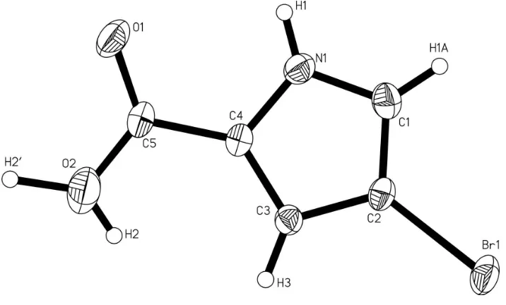
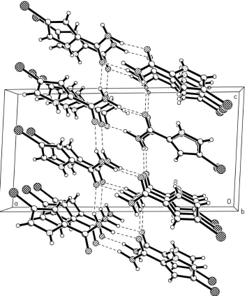
• No results found

4 Bromo 1H pyrrole 2 carb­­oxy­lic acid

N/A
N/A
Protected

Academic year: 2020

Share "4 Bromo 1H pyrrole 2 carb­­oxy­lic acid"

Copied!
7
0
0

Loading.... (view fulltext now)

Full text

(1)

4-Bromo-1

H

-pyrrole-2-carboxylic acid

Le Zheng, Fang Hu, Xiang Chao Zeng* and Kai Ping Li

Department of Chemistry, Jinan University, Guangzhou, Guangdong 510632, People’s Republic of China

Correspondence e-mail: xczeng@126.com

Received 26 June 2012; accepted 6 August 2012

Key indicators: single-crystal X-ray study;T= 293 K; mean(C–C) = 0.005 A˚;

Rfactor = 0.034;wRfactor = 0.081; data-to-parameter ratio = 16.3.

In the title compound, C5H4BrNO2, the non-H atoms of the pyrrole ring and the Br atom are approximately coplanar, with an r.m.s. deviation from the best fit plane of 0.025 (6) A˚ ;. The dihedral angle between the plane of the carboxy group and this plane is 14.1 (2). In the crystal, O—H O hydrogen

bonds link the molecules together, forming corrugated sheets parallel to thebcplane.

Related literature

For pyrrole compounds obtained from marine organisms, see: Liuet al.(2005); Faulkner (2002). For the bioactivity of pyrrole derivatives, see: Banwellet al.(2006); Sosaet al.(2002). For related structures, see: Zenget al.(2007); Tanget al.(2008).

Experimental

MoKradiation

= 6.29 mm1

0.240.200.14 mm

Data collection

Oxford Gemini S Ultra area-detector diffractometer Absorption correction: multi-scan

(CrysAlis PRO; Oxford Diffraction, 2010)

Tmin= 0.314,Tmax= 0.473

2436 measured reflections 1387 independent reflections 1081 reflections withI> 2(I)

Rint= 0.021

Refinement

R[F2> 2(F2)] = 0.034

wR(F2) = 0.081

S= 1.10 1387 reflections

85 parameters

H-atom parameters constrained max= 0.39 e A˚

3

min=0.45 e A˚ 3

Table 1

Hydrogen-bond geometry (A˚ ,).

D—H A D—H H A D A D—H A

O2—H20

O1i

1.07 1.86 2.914 (4) 166 O2—H2 O1ii

0.82 2.28 3.030 (4) 153

Symmetry codes: (i)xþ1;yþ1 2;zþ

1

2; (ii)x;yþ 3 2;z

1 2.

Data collection: CrysAlis PRO(Oxford Diffraction, 2010); cell refinement: CrysAlis PRO; data reduction: CrysAlis PRO; program(s) used to solve structure: SHELXS97 (Sheldrick, 2008); program(s) used to refine structure:SHELXL97(Sheldrick, 2008); molecular graphics:SHELXTL(Sheldrick, 2008); software used to prepare material for publication:SHELXTL.

We thank the Natural Science Foundation of Guangdong Province, China (No. 06300581) for generously supporting this study.

Supplementary data and figures for this paper are available from the IUCr electronic archives (Reference: FJ2579).

References

Banwell, M. G., Hamel, E., Hockless, D. C. R., Verdier-Pinard, P., Willis, A. C. & Wong, D. J. (2006).Bioorg. Med. Chem.14, 4627–4638.

Faulkner, D. J. (2002).Nat. Prod. Rep.18, 1–48.

Liu, J. F., Guo, S. P. & Jiang, B. (2005).Chin. J. Org. Chem.25, 788–799. Oxford Diffraction (2010).CrysAlis PRO. Oxford Diffraction Ltd, Yarnton,

England.

Sheldrick, G. M. (2008).Acta Cryst.A , 112–122.

Structure Reports

Online

(2)

supporting information

supporting information

Acta Cryst. (2012). E68, o2689 [doi:10.1107/S1600536812034800]

4-Bromo-1

H

-pyrrole-2-carboxylic acid

Le Zheng, Fang Hu, Xiang Chao Zeng and Kai Ping Li

S1. Comment

Pyrrole derivatives are well known in many marine organisms (Faulkner, 2002), some show important bioactivities, such

as antitumor activity (Banwell et al., 2006) and protein kinase inhibiting activity (Sosa et al., 2002). These are the reasons

why they have attracted our interest. This study is relevant to our previous studies on Methyl 2-(4,5-dibromo-1H

-pyrrole-2-carboxamido)propionate (Zeng et al., 2007) and 1H-Pyrrole-2-carboxylic acid (Tang et al., 2008).

In the title molecule, bond lengths and angles are unexceptional. The non-H atoms of the pyrrole ring and Br atom are

approximately coplanar (plane 1), with r.m.s. deviation from the best fit plane of 0.025 (6)°, the dihedral angle between

the carboxy plane and Plane 1 is 14.1 (2)°. The OH hydrogen atom is disordered over two positions (shown as in Fig. 1),

which form weak intermolecular O2—H2···O1 hydrogen bonds respectively (Table 1). In the crystal, the above hydrogen

bonds link the molecules into corrugated sheets parallel to the bc plane (shown as in Fig. 2 and Fig. 3).

S2. Experimental

The methyl 4-bromopyrrole-2-carbonylaminoacetate (1.30 g, 5 mmol) and potassium carbonate (1.38 g, 10 mmol) were

added to acetonitrile (20 ml), the mixture was stirred at reflux for 24 h. After the reaction mixture was cooled and filtered,

the filtrate was evaporated in vacuo, and then the residue was chromatographed over Si gel 60 using EtOAc-petroleum

ether (1:2.5) as eluting solvent and the title compound (I) was obtained (55.2% yield). Light yellow monoclinic crystals

suitable for X-ray analysis (m.p. 424 K) grew over a period of one week when the EtOH solution of I was exposed to the

air at room temperature.

S3. Refinement

All non-H atoms were refined with anisotropic displacement parameters. The H atoms except the H(OH) were positioned

geometrically[C—H = 0.93 Å for CH, and N—H = 0.86 Å] and refined using a riding model, with Uiso = 1.2Ueq of the

(3)
[image:3.610.128.486.72.288.2]

Figure 1

The molecular structure of the title compound, with the atom-numbering scheme. Displacement ellipsoids are drawn at

(4)
[image:4.610.131.485.71.495.2]

supporting information

Figure 2

(5)
[image:5.610.127.486.70.372.2]

Figure 3

sheet formed by weak hydrogen bonds (dashed lines).

4-Bromo-1H-pyrrole-2-carboxylic acid

Crystal data

C5H4BrNO2

Mr = 190.00

Monoclinic, P21/c

Hall symbol: -P 2ybc

a = 16.0028 (13) Å

b = 4.9046 (6) Å

c = 8.2367 (7) Å

β = 93.199 (7)°

V = 645.47 (11) Å3

Z = 4

F(000) = 368

Dx = 1.955 Mg m−3

Melting point < 424 K

Mo radiation, λ = 0.71073 Å Cell parameters from 950 reflections

θ = 3.5–29.1°

µ = 6.29 mm−1

T = 293 K

(6)

supporting information

Refinement

Refinement on F2

Least-squares matrix: full

R[F2 > 2σ(F2)] = 0.034

wR(F2) = 0.081

S = 1.10 1387 reflections 85 parameters 0 restraints

Primary atom site location: structure-invariant direct methods

Secondary atom site location: difference Fourier map

Hydrogen site location: inferred from neighbouring sites

H-atom parameters constrained

w = 1/[σ2(F

o2) + (0.034P)2 + 0.2705P]

where P = (Fo2 + 2Fc2)/3

(Δ/σ)max < 0.001

Δρmax = 0.39 e Å−3

Δρmin = −0.45 e Å−3

Special details

Geometry. All e.s.d.'s (except the e.s.d. in the dihedral angle between two l.s. planes) are estimated using the full covariance matrix. The cell e.s.d.'s are taken into account individually in the estimation of e.s.d.'s in distances, angles and torsion angles; correlations between e.s.d.'s in cell parameters are only used when they are defined by crystal symmetry. An approximate (isotropic) treatment of cell e.s.d.'s is used for estimating e.s.d.'s involving l.s. planes.

Refinement. Refinement of F2 against ALL reflections. The weighted R-factor wR and goodness of fit S are based on F2,

conventional R-factors R are based on F, with F set to zero for negative F2. The threshold expression of F2 > σ(F2) is used

only for calculating R-factors(gt) etc. and is not relevant to the choice of reflections for refinement. R-factors based on F2

are statistically about twice as large as those based on F, and R- factors based on ALL data will be even larger.

Fractional atomic coordinates and isotropic or equivalent isotropic displacement parameters (Å2)

x y z Uiso*/Ueq Occ. (<1)

Br1 0.07690 (2) 0.68153 (9) −0.16395 (4) 0.04763 (17) N1 0.24925 (19) 0.3185 (6) 0.1354 (4) 0.0400 (7)

H1 0.2653 0.1983 0.2065 0.048*

C3 0.2505 (2) 0.6756 (8) −0.0297 (4) 0.0321 (7)

H3 0.2674 0.8285 −0.0861 0.039*

O1 0.40382 (15) 0.4281 (6) 0.2816 (3) 0.0514 (7) C4 0.29799 (19) 0.5248 (7) 0.0817 (4) 0.0321 (8) C2 0.17094 (19) 0.5519 (8) −0.0414 (4) 0.0327 (8) O2 0.4311 (2) 0.7498 (8) 0.0940 (4) 0.0731 (10)

H2 0.4079 0.8099 0.0102 0.013 (15)* 0.50

H2′ 0.4898 0.7973 0.1562 0.009 (13)* 0.50

C1 0.1716 (2) 0.3316 (8) 0.0598 (5) 0.0421 (9)

H1A 0.1274 0.2128 0.0743 0.050*

C5 0.3821 (2) 0.5608 (9) 0.1583 (4) 0.0373 (8)

Atomic displacement parameters (Å2)

U11 U22 U33 U12 U13 U23

(7)

C5 0.0278 (17) 0.048 (2) 0.0357 (17) 0.0020 (18) −0.0038 (13) −0.0053 (19)

Geometric parameters (Å, º)

Br1—C2 1.876 (3) O1—C5 1.239 (4)

N1—C1 1.360 (5) C4—C5 1.465 (4)

N1—C4 1.366 (4) C2—C1 1.364 (5)

N1—H1 0.8600 O2—C5 1.342 (5)

C3—C4 1.375 (5) O2—H2 0.8200

C3—C2 1.408 (4) O2—H2′ 1.0702

C3—H3 0.9300 C1—H1A 0.9300

C1—N1—C4 109.9 (3) C3—C2—Br1 125.8 (3)

C1—N1—H1 125.1 C5—O2—H2 109.5

C4—N1—H1 125.1 C5—O2—H2′ 118.5

C4—C3—C2 106.1 (3) H2—O2—H2′ 132.0

C4—C3—H3 126.9 N1—C1—C2 107.1 (3)

C2—C3—H3 126.9 N1—C1—H1A 126.5

N1—C4—C3 108.1 (3) C2—C1—H1A 126.5

N1—C4—C5 118.5 (3) O1—C5—O2 122.9 (3)

C3—C4—C5 133.1 (3) O1—C5—C4 119.9 (3)

C1—C2—C3 108.8 (3) O2—C5—C4 117.1 (3)

C1—C2—Br1 125.2 (3)

C1—N1—C4—C3 0.6 (4) C3—C2—C1—N1 0.8 (4)

C1—N1—C4—C5 175.0 (3) Br1—C2—C1—N1 −175.1 (2)

C2—C3—C4—N1 −0.1 (4) N1—C4—C5—O1 −8.7 (5)

C2—C3—C4—C5 −173.3 (3) C3—C4—C5—O1 163.9 (4)

C4—C3—C2—C1 −0.5 (4) N1—C4—C5—O2 174.5 (3)

C4—C3—C2—Br1 175.4 (2) C3—C4—C5—O2 −12.8 (6)

C4—N1—C1—C2 −0.9 (4)

Hydrogen-bond geometry (Å, º)

D—H···A D—H H···A D···A D—H···A

Figure

Figure 1The molecular structure of the title compound, with the atom-numbering scheme
Figure 2
Figure 3

References

Related documents

In this study, we identified 9 protein markers for predicting time to recurrence using the protein expression data on 222 TCGA pri- marily high-grade serous ovarian cancers

For the purpose of analyzing the impurities in the water samples coming from different roofs, four building within the KCAET campus viz location 1(library -

To overcome the problems and weakness, this project need to do some research and studying to develop better technology. There are list of the objectives to be conduct

The above block diagram shows the SPV fed to Dc/Dc Converter for different dc applications, To analysis the performance of dc-dc converters(Buck, Boost,

22 subjects showing low or undetectable activities of BAT were randomly divided into 2 groups: one was exposed to cold at 17°C for 2 hours every day for 6 weeks (cold group; n

Foxo deletion on osteoblast differentiation in both bone marrow and calvaria cells suggests that the increases in ALP activity and mineralization observed in the bone

Histologically, the lesion is composed of fibrous connective tissue trabeculae (top quarter of image) and adipose connective tissue (bottom three quarters of image); within

• Data shows credit using and rationing of risk averts, risk neutrals and risk lovers respectively. As to risk averts, the credit is mainly used to pay children’s tuition, medical